UWSA1-4 Flashcards
(137 cards)
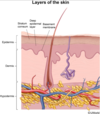
A 5-year-old boy comes to the office with periorbital edema and rust-colored urine for 2 days. His oral intake has been normal, but his parents are concerned that he is voiding less frequently than usual. Three weeks ago, he had a pustule on his face that subsequently developed an overlying, adherent, honey-crusted layer and resolved with antibiotics. The patient’s blood pressure is mildly elevated. Urinalysis is positive for protein and red blood cell casts. Which of the following characteristics best describes the bacteria involved in this patient’s condition?
This patient’s most likely diagnosis is acute poststreptococcal glomerulonephritis (PSGN). PSGN is an immune complex-mediated disease that occurs 1-3 weeks after a pharyngeal or skin infection (eg, impetigo) by Streptococcus pyogenes (group A Streptococcus or GAS). Deposition of streptococcal nephritogenic antigens within the glomerulus causes immune complex formation, which in turn triggers complement activation and inflammation.
GAS is a gram-positive, beta-hemolytic, bacitracin-sensitive bacterium that grows in chains. Gram-positive bacteria retain the crystal violet stain and appear purple. When GAS is plated on blood agar culture, GAS causes beta hemolysis (complete red cell lysis) and transparency in the normally red agar media. Bacitracin can be used to distinguish GAS from other streptococcal strains, with GAS being sensitive to bacitracin and most non-group A strains resistant.
Symptoms of PSGN typically include edema, gross hematuria, hypertension, and oliguria. The edema is commonly periorbital and can become generalized. Gross hematuria often presents as tea- or cola-colored urine, and urinalysis is positive for protein and blood. Serum abnormalities include a low C3 and mildly elevated creatinine. Antibody titers reflecting a previous GAS infection are helpful in diagnosis (Table). A biopsy is usually not indicated for diagnosis when the presentation is classic.
Strep Pyogenes features
GAS is a gram-positive, beta-hemolytic, bacitracin-sensitive bacterium that grows in chains. Gram-positive bacteria retain the crystal violet stain and appear purple.
When GAS is plated on blood agar culture, GAS causes beta hemolysis (complete red cell lysis) and transparency in the normally red agar media.
Bacitracin can be used to distinguish GAS from other streptococcal strains, with GAS being sensitive to bacitracin and most non-group A strains resistant.
Acute poststreptococcal Glomerulonephritits pathophysiology
PSGN is an immune complex-mediated disease that occurs 1-3 weeks after a pharyngeal or skin infection (eg, impetigo) by Streptococcus pyogenes (group A Streptococcus or GAS)
Deposition of streptococcal nephritogenic antigens within the glomerulus causes immune complex formation, which in turn triggers complement activation and inflammation.
Acute poststreptococcal Glomerulonephritits clinical features
Can be asymptomatic
Symptoms of PSGN typically include edema, gross hematuria, hypertension, and oliguria. The edema is commonly periorbital and can become generalized. Gross hematuria often presents as tea- or cola-colored urine, and urinalysis is positive for protein and blood. Serum abnormalities include a low C3 and mildly elevated creatinine. Antibody titers reflecting a previous GAS infection are helpful in diagnosis (Table). A biopsy is usually not indicated for diagnosis when the presentation is classic.
If symptomatic:
Gross hematuria (tea- or cola-colored urine)
Edema (periorbital, generalized)
Hypertension
Acute poststreptococcal Glomerulonephritits lab findings
Urinalysis: + protein, + blood, ± red blood cell casts
Serum:
↓ C3 & possible ↓ C4
↑ Serum creatinine
↑ Anti-DNase B (antideoxyribonuclease-B) & ↑ AHase (antihyaluronidase)
↑ ASO (antistreptolysin O) & ↑ anti-NAD (from preceding pharyngitis) (antinicotinamide-adenine dinucleotidase)
Staphylococcus aureus features
Staphylococcus aureus is a gram-positive, catalase-positive, coagulase-positive bacterium that grows in clusters.
Differentiating staph from strep
the catalase test is a particularly important test used to determine whether the Gram + cocci is a staphylococci or a streptococci.
Staph is catalase Positive
Strep is catalase Negative
Quelling Reaction
The Quellung reaction refers to capsule swelling that is evident on microscopy after a bacterium is exposed to its capsular antigens. Streptococcus pneumoniae, the most common cause of community-acquired pneumonia, is an example of a Quellung-reaction-positive bacterium.
Preceeding infections of Reactive Arthritis
Genitourinary infection: Chlamydia trachomatis
Enteritis: Salmonella, Shigella, Yersinia, Campylobacter, Clostridioides (formerly Clostridium) difficile
Reactive Arthritis symptoms
Asymmetric oligoarthritis
Enthesitis
Dactylitis
Ocular: conjunctivitis, anterior uveitis
Genital: urethritis, cervicitis, prostatitis
Dermal: keratoderma blennorrhagicum, circinate balanitis
Oral ulcers
Reactive arthritis risk factor
HLA B27
Reactive arthritis Pathophysiology
Genitourinary or gastrointestinal infection typically precedes the onset of symptoms by 2-6 weeks. The manifestations are caused by immune-complexes involving bacterial antigens. However, it does not represent disseminated infection, and joint aspirates are sterile (ie, it is a reactive, not infectious, arthritis).
Ankylosing Spondylitis description
Ankylosing spondylitis is a chronic disease affecting the axial skeleton and causes progressive stiffness and pain.
It is associated with HLA B27, but extra-articular symptoms are not prominent.
Disseminated gonococcal infection description
Disseminated gonococcal infection presents as
purulent arthritis (eg, knee, wrist, ankle)
or
as the triad of tenosynovitis, dermatitis, and non-purulent polyarthralgias.
Hypersensitivity Vasculitis
Hypersensitivity vasculitis is a drug reaction characterized by a purpuric rash.

Viral Arthritis description
Viral arthritis causes symmetric, polyarticular disease. It is most common following parvovirus, hepatitis B, hepatitis C, rubella, and alphavirus infection.
Chromosome 15 genomic imprinting disorders
- Prader-Willi Syndrome
- Angelman Syndrome
Pathophysiology of Prader-Willi Syndrome
This child has Prader-Willi syndrome (PWS), a genomic imprinting disorder caused by the loss of paternally inherited alleles on chromosome 15q11-13. Within this region, certain genes are differentially imprinted and silenced depending on the parent of origin. Alleles that are maternally imprinted cannot be actively expressed unless there is a functional paternal copy. PWS can be caused by any genetic defect that results in the absence of this paternal contribution, most commonly microdeletions of the paternal 15q11-13 region or maternal uniparental disomy (ie, both chromosome 15s are inherited from the mother). This patient’s normal fluorescence in-situ hybridization (FISH) findings indicate that his condition is most likely due to maternal uniparental disomy.
Causes of prader-willi syndrome
Loss of paternally inherited allele from chromosome 15:
Paternal microdeletion
or
Maternal uniparental disomy
Clinical Features of Prader-Willi syndrome
Neonatal hypotonia
Hyperphagia/obesity
Short stature
Small hands & feet
Hypogonadism
Dysmorphic facies
Intellectual disability
Causes of Angelman Syndrome
Loss of maternally inherited allele from chromosome 15
Maternal microdeletion
Paternal uniparental disomy
S&S of Angelman syndrome
Epilepsy/seizures
Ataxic gait/tremors
Poor motor & language development
Inappropriate laughter/smiling
Intellectual disability
Celiac disease genetic implications
Celiac disease has a genetic component and is associated with the human leukocyte antigen
(HLA) DQ2 and DQ8 genotypes.
Celiac Disease Pathophysiology
Celiac disease (celiac sprue, gluten-sensitive enteropathy) is caused by dietary exposure to gliadins (a component of gluten) in wheat and related grains in susceptible individuals. This sensitivity to gluten can trigger autoimmune reactions in the gastrointestinal tract. In the small bowel, celiac disease causes marked villous atrophy in a background of diffuse enteritis. CD8+ T-cell lymphocytes infiltrate the small intestine surface epithelium, and a wide range of inflammatory cells (CD4+ T-cell lymphocytes, plasma cells, macrophages, eosinophils, and mast cells) take up residence in the lamina propria. Celiac disease has a genetic component and is associated with the human leukocyte antigen (HLA) DQ2 and DQ8 genotypes.