W10 Skeletal System Flashcards
(52 cards)
Describe the functions of the skeletons and bone tissues?
The skeletal system has a number of important functions:
Support
Protection
Assistance in moving
Mineral homeostasis
Blood cell production
Triglyceride storage
Describe support in the skeletal system
Provides a structural framework for the soft tissues of the body
Describe protection in the skeletal system
The skeleton protects internal organs
Describe assistance in moving
Movement of the skeletal muscle attaches to bones, assist in contraction
Describe mineral homeostasis
Osseous tissue stores severval minerals (calcium & phosphorous, bones can release minerals to maintain homeostatic levels
Describe Blood cell production
Blood cells are produced by red bone marrow, which is found in certain bones
Describe Triglyceride storage
Yellow bone arrow consists mainly of adipocytes that store triglycerides
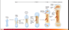
Describe the gross anatomy of a typical long bones
Epiphysis (bone end) ↓ Diaphysis (bone shaft) ↓ Epiphysis (bone end)
What are the 6 classifications of bones?
Long bone
Short bone
Flat bone
Irregular bone
Sesamoid bone
Sutural bones

Describe bone tissue
A unique form of hard connective tissue. Collagen fibres and mineral salt crystals (Ca & phosphate) = hydroxyapatite. Bones arr composed of two structures compact (cortical) & spongy (cancellous) bone (Spongy bones contain myeloid tissue)
Describe the gross anatomy of a typical long bone of the Epiphysis region
Articular cartilage - thin layer of cartilage that covers the surface of the epiphysis that articulates with other bones
Metaphysis- Between diaphysis & epiphysis.
Contains the epiphyseal growth plate in growing bones

Describe the gross anatomy of a typical long bone in the Diaphysis reigon
Endosteum - a thin connective tissue layer that lines the surface of the medullary cavity
Medullary cavity - A hallow clindrical space within the middle of the diaphysis. Contains yellow bone marrow in adults
Periosteum - a connective tissue layer attached to the outside of the bone

Describe the structure of a flat bone
Structure of a flat bone similar to a long bone but simpler

Describe the blood supply in bone
Bone is a living tissue so it contains an extensive network of blood vessels. These vessels overlap so if one becomes damaged the bone will not die because it receives blood from another vessel
Describe the chemical composition of the bone
Osseous tissue is a type of connective tissue, it contains cells that are surrounded and embedded within a extracellular matrix. Composed of 25% water 24% collagen fibers 50% crystallized mineral salts
What are the cells of osseous tissue?
Osteoprogenitor (osteogenic) Osteoblasts Osteocytes
Define Osteoprogenitor cells
They are unspecialized stem cells, they are the only bone cells that can divide. Important in fracture repair

Define Osteoblasts
Synthesis and secrete collagen fibres and other components of the extracellular matrix.
They initiate calcification - bone formation also know as ossification

Define Osteocytes
Mature bone cells and most numerous cells. Responsible for day to day function of metabolism of osseous tissue.
Exchange aided by cytoplasmic projections in canaliculi (little canals, caneloni) FORMED WHEN OSTEOBLASTS BECOME SURROUNDED WITH BONE MATRIX

Define Osteoclasts
Large multinucleate cells, release enzymes and acids that digest the extracellular matrix. Involved in resorption of osseous tissue (important for development, maintainance and repair)

Describe the histology of a compact &spongy bone
Compact bone: diaphysis of long bones, surface of short, flat and irregular bones
Spongy bone - epiphysis of long bone, most of short flat and irregular bone.
Describe the histology of a compact bone structure
Compact bones contains very few spaces. It provides protection and support and resists stress of weight and movement.

Its ↑ tensile strength is due to the type of materials and how its arranged
What are the components of bone structure?
Osteon - repeating structural unit of compact bone. Consist of a central canal containing blood vessels surrounded by concentric lamellae, osteocytes, lacuna and canaliculi.
Concentric lamellae (thin layer) - rings of hard, calcified extracellular matrix
Lacuna - small space located between the lamellae, these spaces contain osteocytes
Canaliculi - small channels filled with cellular projections & extracellular fluid that radiate from lacuna, provides routes for oxygen and nutrients to each osteocytes

What are the two types of canals in compact bone structure?
Central canals - run though the centre of osteons & connect with perforating canals, surrounded by concentric lamellae
Perforating canals - allow blood vessels, lymphatic vessels and nerves to enter compact bone