W9 Nervous System Flashcards

(75 cards)

1
Q

What type of stimuli do smell and taste detect?

A

Smell and taste are chemical senses, they arise due to the interaction of odorants and tastants (chemicals) with specialised receptors

2
Q

List the 5 primary taste sensations

A

Bitter

Sour

Salty

Sweet

Umami (meat or savoury)

3
Q

Describe the receptors for taste

A

Located in taste buds, mainly located on tongue, they are also present on the soft palate, pharynx(throat), and epiglottis (cartilage lid over the voice box)

4
Q

What is the function of the Olfactory gland?

A

Produce mucus that is secreted onto the olfactory epithelium, the mucus moistens the epithelium and dissolves odorants

5
Q

What is the effect of an odorant binding to an olfactory receptor?

A

The binding of an odorant to the olfactory receptors leads to the generation of a depolarising graded potential. If depolarisation is large enough, action potentials are generated & nerve impulses are sent to the CNS

6
Q

PICWhat is the pathway of olfactory information to the CNS?

A

The axons of the olfactory bulb neurons exit the olfactory bulb and move into the olfactory tract.

Axons travelling in the olfactory tract project to the primary olfactory area of the cerebral cortex.

Other axons of the olfactory tract project to the hypo thalamus and other brain regions involved in memory & emotion

7
Q

What are the 5 primary tastes?

A

Bitter Sour Salty Sweet Umami

8
Q

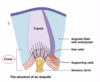
PICWhat is the structure of a taste bud?

A

Consists of three types of epithelial cells:

  1. Supporting cells
  2. Gustatory receptor cells
  3. Basal cells
9
Q

Define a supporting cell

A

Surround the gustatory cells and provide chemical and structural support

10
Q

Define the Gustatory receptor cell

A

Responsible for detecting chemicals, long microvillus called a gustatory hair that projects into the external surface of the taste pore

11
Q

Define Basal cells

A

Stem cells that produce supporting cells, they have a lifespan of about 10 days

12
Q

What is the effect of the binding of a tastant to a taste receptor?

A

A tastant is any chemical that stimulates taste receptor cells, they have to be dissolved into the saliva before they can make contact with gustatory. The result of tastant binding to a receptor is the generation of depolarisng and release a neurotransmitter -> stimulates sensory neurons that project to the CNS

13
Q

What is the pathway of gustatory infomation to the CNS?

A

Gustatory information is carried by 3 cranial nerves Nerve impulses travel down these nerves to the medulla oblongata Neural impulses are then sent to either the limbic regions, the hypothalamus, or the thalamus -> Info from the thalamus is then sent to the cerebral cortex

14
Q

What are the accessory structures of the eye?

A

Eyelids (palpebrae) Eyelashes Eyebrows Lacrimal apparatus Extrinsic eye muscles

15
Q

What is the lacrimal fluid?

A

The lacrimal fluid (tears) is produced in the lacrimal glands and secreted by the lacrimal apparatus, then drains into the lacrimal ducts

16
Q

PICWhat is the function of the lacrimal fluid?

A

Lacrimal fluid (tears) are a water solution containing salts, mucus, and lysozyme ( a material enzyme).

LF protects, cleans, lubricates, and moistens the eye ball

17
Q

PICWhat is the function of the lacrimal fluid?

A

Lacrimal fluid (tears) are a water solution containing salts, mucus, and lysozyme ( a material enzyme). LF protects, cleans, lubricates, and moistens the eye ball

18
Q

What is the function of the extrinsic eye muscle?

A

There are 6: Superior rectus Inferior rectus Lateral rectus Medial rectus Superior oblique Inferior oblique The extrinsic eyeball moves laterally, medially, superiorly and inferiorly

19
Q

What are the 3 layers of the eyeball?

A
  1. The fibrous tunic 2. The vascular tunic 3. The retina
20
Q

What is the outmost later of the eye ball and describe it

A

The fibrous tunic is the superficial layer of the eye consisting of the sclera & cornea: clear coating that covers the iris, curved and helps to focus light onto the retina Sclera: White outer layer of eyeball, made up of dense connective tissue, gives shape, and rigidity to eyeball, also acts as attachment point;

21
Q

What are the three components of the vascular tunic?

A

The vascular tunic is the middle layer of the eyeball. Consists of 3 components 1. The Choriod 2. The cillary body 3. The iris

22
Q

What is the function of the choroid?

A

Is the most posterior portion of the vascular tunic and lines most of the internal surface of the sclera

23
Q

PICWhat is the function of the ciliary body?

A

Consists of the cilary processes: contains capilaries that secrete aqueous humor (provides nutrients for the lens and cornea)

The cilary muscle - alters the shapes of the lens for near or far vision

24
Q

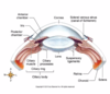
What is the function of the Iris?

A

Is the coloured potion of the eyeball, suspended between the cornea and the lens, it regulates the amount of light entering the eye through the pupil

25
What is the function of the choriod,the cillary body, and the iris? (during light)
In bright light the PNS causes circular muscles to contract - decreasing pupil size In dim light: the SNS activates constrition of muscles that radiate from the eye (radial muscles) - increasing pupil size (dilation)
26
What are the two layers of the retina?
The retina is the innermost layer of the eye ball, composed of: The Pigmented layer Neural layer
27
PICDefine the pigmented layer? (in the retina)
Sheet on melanin - containing cells between the choriod and the neural layer, this layer prevents reflection and scattering of light within the eyeball
28
PIC Define the neural layer of the retina
An outgrowth of the brain. This layer processes visual info before sending it down to the optic nerve
29
PICWhat are the three cell types of the neural layer?
The photoreceptors The bipolar cell layer The ganglion cell layer
30
How are the three cell types of the neural layer arranged?
The neural layer is arranged such that light passes through the ganglion adn the bipolar layers before reaching the photoreceptors
31
Whar are the 2 types of photoreceptors?
The rods; allow us to see dim light, they do no provide colour vision The cones; activated during bright light conditions, and colour, responding to blue, green and red
32
Whar are the 2 types of photoreceptors?
The rods; allow us to see dim light, they do no provide colour vision The cones; activated during bright light conditions, and colour, responding to blue, green and red
33
What is the structure that causes a blind spot?
Pathway of visual info in photoreceptors -\>bipolar cells -\> ganglion cells The axons of the ganglion cells exit the eyeball through the optic disc and form the optic nerve. The disc has no photo receptors, so light that falls on the optic disc is not seen
34
What anatomical and functional features of the central fovea improve vision?
The central fovea is a small depression in the exact centre of the posterior retina. Contains only cones, also bipolar cells and ganglion cells do not cover cones. = high resolution
35
What is the function of the lens?
The lens lies posterior to the pupil and iris, the lens helps to fucus images onto the retina for clear vision
36
What are the 2 divisions of the visual field?
Light rays from the nasal half of the visual field fall on the temporal half of the retina. Light rays from the temporal half of the visual field fall on the nasal half of the retina
37
What is the pathway of visual information to the CNS?
0
38
Identify the anatomy of the outer ear
The external ear: collects sound waves and channels them inwards
39
PICDescribe the anatomy of the external/outer ear
The auricle The external auditory canal The tympanic membrane
40
Identify the anatomy of the middle ear
The oval window The round window
41
Describe the anatomy of the middle ear
The auditory ossicles are the smallest bones in the body and extend to the middle ear known as: The malleus (hammer) The incus (anvil) The stapes (stirrup)
42
What is sound?
Sound is a pressure wave created by a vibrating object. Cycles of compression and decompression determine the frequency and pitch
43
What are the 3 main structures of the ear?
External ear Middle ear Inner ear
44
PICDefine the external ears function
The external ear: collects sound waves and channels them inwards
45
PICDescribe the anatomy of the inner ear
The outer bony labyrinth: contains a fluid similar in composition to CSF called perilymph. The inner membranous labyrinth: contains a fluid called endolymph
46
PICDefine the function of the inner earn
Contains the receptors for hearing and equilibrium (balance)
47
Define the tympanic membrane
Also referred to as the ear drum, it is a thin layer of connective tissue covered by skin on the external side and mucus on the internal. It also separates the external auditory canal from the middle ear
48
What do the ceruminous glands secrete and what is its function?
The ceruminous glands are loacted near the exterior opening of the external canal, they secrete cerumen (earwax), the hair and cerumen in the external canal help to prevent dust etc geting in
49
Where is the middle ear located?
The middle ear is a small air-filled cavity located in the temporal bone, it is separated from the external canal by the tympanic membrane
50
PICWhat membrane does malleus connect to -\> incus -\> staples?
The *handle* of the **malleus** attaches to the internal surface of the tympanic membrane. The *head* of the **malleus** froms a joint (articulates) with the **incus** on one side. On the other side it articulates with the *head* of the **stapes**.
51
What does the auditory tube allow?
The eustachian tube (auditory tube) connects to the middle ear to the nasopharynx. During swallowing and yawning it opens and allows the air pressure in to middle ear to equalise with atmospheric pressure
52
What does the auditory tube allow? (middle ear)
The eustachian tube (auditory tube) connects to the middle ear to the nasopharynx. During swallowing and yawning it opens and allows the air pressure in to middle ear to equalise with atmospheric pressure
53
What is the function of the tensor tympani and the stapedius? (middle ear)
The tensor tympani and the stapedius are tiny skeletal muscles attach to the auditory ossicles. The tensor tympanic limits movement and increases the tension of the tympanic membrane to prevent damage from loud noises. The stapedius muscles acts to protect the oval window, dapening large vibrations of the stapes due to loud noices
54
PICWhat are the three portions of the bony labyrinth?
The semicircular canals The vestibule The cochlea
55
Describe the inner ears cochlea
Resembles a snail shell, it is divided into three channles, the outer two channels are bony labyrinth, sandwiched between is membranous labyrinth
56
What are the three channels of the cochlea?
The cochlear duct: filled with endolymph The scala vestibuli: filled with perilymph The scala tympani: filled with perilymph. Between these we have membranes - **The vestibular membrane**: separates the _scala vestibuli_ from the _cochlea duct_ **The basilar membrane** separates the _cochlea_ duct from the s_cala tympani_
57
What is the spiral organ?
Rests on the basilar membrane, this is the sensory part of the cochlea
58
What are hair cells and hair cell bundles
Hair cells are the receptors for hearing. At the apical tip of each hair cells are hair cell bundles (*40-80 microvilli that extend into the endolympth of the cochlear duct*)
59
What do hair cells snyapse onto?
Snapse onto sensory neurons that *form the cochlear branch* of *the vestibulocochlear nerve*, the cell bodies of these sensory neurons are l_ocated in the spiral ganglion_
60
What is the tectorial membrane?
The Tectorial membrane is a gelatinous membrane that projects over and makes contact with the hair cells of the spiral organ. (activating an action potential)
61
What are the events that lead to auditory nervee stimulation?
1. Sound waves (**compression decompression**, and *vibrate the tympanic membrane*) 2. The *vibration* of the **tympanic membrane** cause displacment of the auditory ossicles. 3. **Movement of the stapes at the oval window** establishes *pressure waves in the perilymph of the vestibular duct* ( moving fluid in and out -----\>) 4. As the pressure waves move up the vestibular duct, they **distort the basilar membrane** on their way to the round window of the tympanic duct. 5. The vibration causes the hair cells to press against the tectorial membrane ---\> 6. Info about the region and the intensity of stimulation is replayed to the ***CNS over the cochlear branch of the cranial nerve VIII***
62
How does the structure of the basilar membrane determine the frequency of sound it responds too?
The basal membrane changes from wider and flexible at one end --\> to narrower and stiffer at the other end - the stucture vibrates at different locations depending on the frequency. ---\> Hair cells are activated depending on which segment of the basilar membrane is vibrating (exposure to frequency)
63
How does the structure of the basilar membrane determine the frequency of sound it responds too?
The basal membrane changes from wider and flexible at one end --\> to narrower and stiffer at the other end - the stucture vibrates at different locations depending on the frequency. ---\> Hair cells are activated depending on which segment of the basilar membrane is vibrating (exposure to frequency)
64
What are the two types of equilibrium?
Static equilibrium: refers to linear acceleration/deceleration Dynamic equilibrium: maintenance of body position in response to (rotation movements)
65
What receptor organ is involved in detecting changes to equilibrium?
Vestibular apparatus, two components of this are - static equilibrium sensed by otoliths. Dynamic equilibrim (rotation movements) sensed by semicircular canals
66
What are the three components of the vestibular apparatus?
The saccule The semivirvular ducts
67
What is the saccule and the utricle ?
Two mechanistically similar sensing apparatus called macula that are orientated perpendicular to each other (**macula utrculi, macula sacculi)**
68
What are the two cell types present in the macula?
**Hair cells:** these are the sensory receptors that dectect changes in head postions *(hair bundles contain cell bundles 40-80 microvilli)* **Supporting cells:** act as insulation conponents and chemical support
69
What are the otiliths and what is their function?
**Otoliths**: are crystals of calcium carbonate that sit as a layer on top of the optlithic membrane. The otoliths add weight to the otholithic membrane and increase the influence of gravity on this membrane during movement
70
What happens when you tilt your head backwards
When tiliting your head either forwards or backwards the otolithic membrane (weighed by the otoliths) slide down hill due to gravity, This bends the microvilli that form hair bundles. Creates depolarising graded potential in the hair cells (-\> CNS)
71
What happens when you tilt your head backwards
When tiliting your head either forwards or backwards the otolithic membrane (weighed by the otoliths) slide down hill due to gravity, this bends the microvilli that form hair bundles. Creates depolarising graded potential in the hair cells (-\> CNS)
72
How are the semicircular ducts arranged in dynamic equilibrium?
Arranged so that each loop lies at right angles to eachother in the three planes of space, this allows the semicircular ducts to detect rotational movement of the head. (anterior, posterior, lateral)
73
What are the ampulla?
The ampulla are dilated portions of each semicircular duct, these contain receptors for rotational movements (hair cells) ## Footnote Within ampulla structures called **crista** lie within. The **cupula** is a gelatinous membrane that extends over the crista
74
What happens when you shake your head from side to side?
When your head is still the cupulla sits directly above the crista, as such, the microvilli of the haircells are not bent. --\> When your head moves the endolymph inside these ducts move, --\> endolymph pushes on cupula --\> bends the microvilli of the hair cells, opening ion channels and causing a depolarising potential --\> release of neurotransmitter to CNS
75
PICWhat nerve sends vestibular information to the CNS?
The sensory neurons innervating hair cells of the utricle saccule and semicircular ducts send axons to the CNS --\> vestibular branch of the vestibulocochlear (VIII) cranial nerve