Week 1 Flashcards

(220 cards)

1
Q

3 types of muscle

How many and where are the nuclei?

A

Sk: multinucleated/periferal

C: One/center

Sm: One/center

2
Q

What tissue give risue to all muscles?

What is an exception?

A

Mesoderm

Exception: iris (derives from ectoderm)

3
Q

What contractile all muscles contain?

A

Actin and Myosin

4
Q

Describe the three types of muscles

A

Skeletal muscle is composed of large, elongated, multinucleated fibers.

Cardiac muscle is composed of irregular branched cells bound together longitudinally by intercalated disks.

Smooth muscle is an agglomerate of fusiform cells. The density of the packing between the cells depends on the amount of extracellular connective tissue present.

5
Q

What cells give rise to muscle cells?

What these cells form to produce muscle cells?

A

Mesenchymal cells -> Myoblasts -> Myotubes -> Mature muscle

6
Q

Muscle fiber vs. myofibril

A

Muscle fiber = muscle cell

Myofibril = made up of the myofilaments actin and myosin

7
Q

What is the purpose of the connective tissue in skeletal muscle?

A

Transmits the forces (muscle cells do not extend the length of the musscle)

Transmitting blood vessels

8
Q

How muscle is organized (subcomponents)?

A
9
Q

When is the number of muscle fibers steady?

A

14 years old (~puberty)

10
Q

What might regenerate skeletal muscle cells in an adult?

A

Satellite cells

11
Q

What is the molecule that regulates number of muscle cells (hormone)?

How does it regulate the number muscle fibers?

A

Myostatin

It suppresses skeletal muscle development.

12
Q

What determines the strength of the muscle?

A

Total number of muscle fibers (not length)

13
Q

What is the difference between hypertrophy and hyperplasia?

A

Hypertrophy = Increase in muscle size

Hyperplasia = Increase in number of muscle cells

14
Q

What is the functional unit of muscle cell?

Where does it extrends from?

What multiple sacromeres from?

A

Sacromere

Z to Z

Myofibrils

15
Q

What skeletal muscle bands can we see?

A

Actin (7nm) makes up the thin I band (isotropic to polarized light)

Myosin (15nm) makes up the A band (anisotropic to polarized light)

16
Q

Where are the T-tubules in skeletal muscle cells?

A

A-I band junction

17
Q

What is triad (skeletal muscle) made of?

What is the function of triad?

A

T tubule + 2 SR (terminal cisterna of sarcoplasmic reticulum)

Calcium for uniform contraction

18
Q

What are three bands in skeletal muscle?

A

A band made up of actin and myosin

I band made up of actin

H band made up of myosin

19
Q

How contraction of muscle affects:

A band?

I band?

H band?

Two adjacent Z disks?

A

the A band stays the same length

the I bands and H bands shorten (sliding filament model)

The Z disks are moving closer to one another

20
Q

What covers neuro-muscular junction?

A

Schwann cell

21
Q

What is external lamina?

A

A structure similar to basal lamina that surrounds the sarcolemma of muscle cells. It is secreted by myocytes and consists primarily of Collagen type IV, laminin and perlecan (heparan sulfate proteoglycan). Nerve cells, including perineurial cells and Schwann cells also have an external lamina-like protective coating.

22
Q

What is another name for neuromuscular junctions?

A

Motor end plates

23
Q

What molecule plays crucial role in muscle contraction?

A

Calcium

24
Q

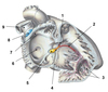
Characteristics of cardiac muscle cells

A

Striations

Intercalated disks

1-2 centrally located nuclei per cell

Bifurcating & anastomosing cells

Highly vascular

Have atrial granules

25
What are atrial granules and where they are found?
The are found in cardiac muscle cells They contain atrial natriuretic peptide (ANP) that acts on kidney to regulate blood pressure through sodium and water reabsorption.
26
What are the three layers in heart?
Endocardium (homologous to tunica intima) Myocardium (homologous to tunica medai) Epicardium (homologous to tunica adventitia)
27
What is a difference between cardiac and skeletal muscle cells in terms of the T-tubules?
Skeletal muscle have and cardiac muscle lack cisternae Skeletal muscle have T-tubule on A/I band border while cardiac muscle have t-tubule on Z-line
28
What is dyad (cardaiac muscle) made of?
T-tubule + sacroplasmic reticulum
29
What surrounds pericardial cavity?
Pericardium (visceral pericardium) Parietal pericardium
30
What is an organelle that is excessively present in cardiac muscle?
Mitochondria
31
What are four types of muscle cells in heart?
**Contractile cardicytes** (myocardiocytes) = **contraction** **Purkinje fibers** (modified myocardiocytes) = found **deep** to **endocardium** lining the **interventricular septum**; **impulse conducting**; **Myoendocrine cardiocytes** = producing **atrial natriuretic factor** **Nodal cardiocytes** = control the **rhytmic** contraction; found in **SA** and **AV node**; found **deep to endocardium** of the **interatrial** and **interventricular septa**
32
How Purkinje fibers can be distinguished from other muscle cells?
Location (deep to endocardium) Size (larger) Staining (lighter because of glycogen content)
33
Cardiac tamponade
Pericardial sac effusion
34
Unique cardiomyocytes that can be seen on microscope
Purkinje fibers Myoendocribe cardiocytes
35
How cardiac muscle cells are connected?
Vertical (transverse): **fascia** (longer strip that goes between cells; wider) **adherens** and **desmosomes** Horizontal (longitudinal): **gap junctions**.
36
What is syncytium? What allows cardiac muscle to act as syncytium?
**Syncytium** is a **multinucleated** cell that can **result** from **multiple cell fusions** of **uninuclear cells**, in contrast to a coenocyte, which can result from multiple nuclear divisions without accompanying cytokinesis. Can because of : fascia adherens, desmosomes and gap junctions
37
Cardiac muscle cells exhibit spontaneous rhythmic contraction What does this contraction depends on? What regulates it?
Dependent upon gap junctions autonomic nervous system (ANS) regulation
38
Smooth Muscle characteristics
elongated fusiform cell appearance (relaxed) one nucleus / centrally located no striations Cytoplasmic dense bodies serve as Z lines Possess caveolae and some SER but not T system Gap junctions in single-unit smooth muscle External basal lamina around each cell Involuntary contraction initiated by several modalities one being the ANS
39
What is the function of caveolae?
Aid in Ca+2 uptake and release.
40
Different types of smooth muscle
**Single unit** (conntected by **gap junction** - syncytium; cannot contract independently of one another; found in the wall of hallow visceras) **Multi-Unit** (has its **own nerve supply** and can contract independetly of one another; en passant) **Vascular** (mix of single and muti unit)
41
Examples of single and mulit unit muscle cells
SU: intestine, uterus, ureters MU: iris
42
En passant ?? | (smooth muscle)
axonal swellings containing synaptic vesciles
43
Heart / Thorax 1
1. Mediastinal part of parietal pleura 2. Axillary vein 3. Horizontal fissure 4. Inferior lobe, right lung 5. Diaphragmatic part of parietal pleura 6. Fibrous pericardium 7. Musculophrenic artery and vein 8. Inferior lob, left lung 9. Left oblique fissure 10. Cardiac notch 11. Costal part of parietal pleura 12. Internal thoracic artery and vein
44
Two potential spaces around lungs
Costomediastinal recess Costodiaphragmatic recess
45
Mediastinum
An **undelineated group of structures in** the **thorax**, surrounded by loose connective tissue. It is the central compartment of the thoracic cavity. It contains the **heart**, the **great vessels** of the heart, the **esophagus**, the **trachea**, the **phrenic nerve,** the **cardiac nerve**, the** thoracic duct**, the **thymus**, and the lymph nodes of the central chest.
46
Heart / Thorax 2
1. Esophagus 2. T-9 Vertebra 3. Thoracic aorta 4. Mediastinal part of parietal pleura 5. Left phrenic serve, left pericardiacophrenic artery and vein 6. Pericardium 7. Central tendom of diaphragm, middle leaflet covered by pericardium 8. Right costomediastinal recess 9. Inferior vena cava
47
Superior mediastinum components
Roots of great vessels Esophagus Trachea Vagal, phrenic, and cardiac nerves
48
Inferior mediastinum
(anterior portion) **branches** of the **interal thoracic artery** and some **thymus** in children (middle) **heart** and **ascending aorta** and **SVC**"**everything in** the **pericardial sac**" (posterior) all other vessels, nerves and visceral structures **anterior** to **vertebrae** and **between** the **parietal pleura** of both lungs
49
Heart / Thorax 3
0. Thymic remanant 1. Internal thoracic artery 2. Rib 1 3. Left phernic nerve, left pericardiacophrenic artery 4. Costal part of parietal pleura 5. Fibrous pericardium 6. Line of fusion of fibrous pericardium to diaphragm 7. Superior epigastric artery 8. Musculophrenic artery 9. Line of fusion for fibrous pericardium with superior vena cava 10. Right axillary artery and vein 11. Right internal thoracic vein
50
Heart / Thorax 4
1. Left Vagus 2. Left subclavian 3. Left axillary artery 4. Left brachiocephalic vein 5. Costal part of parietal pleura 6. Left costocdiaphragmatic recess 7. Fibrous pericardium 8. Right and left phrenic nerves 9. Right costodiaphragmatic recess 10. Mediastinal part of parietal pleura 11. Superior vena cava 12. Arch of aorta 13. Right brachiocephalic vein 14. Right vagus nerve
51
Heart / Thorax 5
1. Left brachiocephalic vein 2. Ligamentum arteriosum and left recurrent laryngeal nerve 3. Left vagus nerve 4. Mediastinal part of parietal pleura 5. Transverse pericardial sinus 6. Fibrous and parietal serous pericardium 7. Left ventricle 8. Auricle of left atrium 9. Right ventricle 10. Right atrium 11. Auricle of right atrium 12. Ascending aorta 13. Pericardial reflections 14. Superior vena cava 15. Right brachiocephalic vein
52
Heart / Thorax 6
1. Ligamentum arteriousm 2. Transverse pericardiac sinus 3. Left superior and inferior pulmonary veins 4. Oblique pericardial sinus 5. Parietal layer of serous pericardium (fused to fibrous) 6. Inferior vena cava 7. Middle cardiac vein 8. Coronary sinus 9. Superior vena cava
53
Heart / Thorax 7
1. Left vagus nerve 2. Pulmonary trunk 3. Transverse pericardial sinus 4. Left phernic nerve and pericardiacophrenic artery and vein 5. Left inferior pulmonary vein 6. Parietal layer of serous pericardium 7. Fibrous pericardium 8. Inferior vena cava 9. Right superior pulmonary vein 10. Superior vena cava 11. Ascending aorta 12. Ligamentum arteriosum 13. Left recurrent laryngeal nerve
54
Heart 1
1. Brachiocephalic artery (trunk) 2. Right atrium 3. Inferior vena cava 4. Coronary sinus 5. Pulmonary veins 6. Auricle of left atrium 7. Right and left pulmonary arteries 8. Left subclavian 9. Left common caroitd
55
Heart 2
1. Left coronary artery 2. Cricumflex artery 3. Left marginal artery (obtuse) 4. Great cardiac vein 5. Anterior interventricular artery (Left anterior descending, LAD) 6. Right marginal artery (acute) 7. Small cardiac vein 8. Right coronary artery 9. Atrial branch of right coronary artery 10. SA artery
56
Heart 3
1. Sinuatrial node artery 2. SA Node 3. Small cardiac vein 4. Right coronary artery 5. Middle cardiac vein 6. Right marginal artery 7. Posterior interventricular artery (posterior descrnding) 8. Left marginal artery 9. Coronary sinus 10. Great cardiac vein 11. Circumflex artery
57
Heart 4
1. Ascending aorta 2. Auricle of right atrium 3. Crista terminalis 4. Pectinate muscles 5. Septal cuscp of tricuspid valve 6. Ostium and valve of coronary sinus 7. Inferior vena cava 8. Fossa ovalis 9. Limbus of fossa ovalis 10. Interatrial septum 11. Superior and inferior right pulmonary veins 12. Right pulmonary artery 13. Superior vena cava
58
Heart 5
1. Pumonary trunk 2. Auricle of left atrium 3. Pulmonary valve 4. Conus arteriosus 5. Supraventricular crest 6. Papillary muscles (septal) 7. Papillary muscles (anterior) 8. Papillary muscles (posterior) 9. Septomarginal trabecula 10. Trabeculae carneae 11. Chordae tendineae 12. Tricuspid valve (posterior cusp) 13. Tricuspid valve (septal cusp) 14. Tricuspid valve (anterior) 15. Ascending aorta
59
Heart 6
1. Ligamentum arteriosum 2. Coronary sinus 3. Inferior vena cava 4. Cordinae tendineae 5. Trabeculae carneae 6. Papillary muscles (anterior/posterior) 7. Epicardium 8. Endocardium 9. Myocardium 10. Bicuspid valve (anterior) 11. Bicuspid valve (posterior) 12. Pulmonary trunk 13. Auricle of left atrium 14. Arhc of aorta
60
Heart 7
1. Aortic valve (left, right, and posterior cusp) 2. Right coronary artery 3. Tricuspid valve (anteior, septal, and posterior cusp) 4. Atrioventricular nodal artery 5. Posterior intraventricular artery 6. Middle cardiac vein 7. Left and right fibrous trigones 8. Bicuspid valve (anterior and posterior cusp) 9. Circumflex artery 10. Left coronary artery 11. Pulmonary valve (left,right, and anterior?)
61
Heart 8 Aortic Valve
1. Ostium of left coronary artery 2. Mitral valve anterior cusp 3. Interventricular septum muscular part 4. Interventricular septum membranous part 5. Nodule 6. Ostium of right coronary artery 7. Sinuses of aortic valve
62
Heart 9 Tricuspid
1. Tricuspid valve (septal, anterior, and posterior) 2. Chordae tendineae 3. Papillary muscle (posteior) 4. Papillary muscle (posteior, septal, and anterior) 5. Interventricular septum membranous part 6. Ostium of coronary sinus 7. Ostium of inferior vena cava
63
Heart 10
1. Left bundle 2. Right bundle 3. Subendocrinal (Purkinje) fibers 4. Atrioventricular bundle (of His) 5. Atrioventricular node 6. Fossa ovalis 7. Crista terminalis 8. Sinu-atrial (SA) node 9. Superior vena cava
64
Heart / Thorax 8
1. Cervical cardiac vagal branches 2. Left vagus nerve 3. Thoracic cardiac vagal branches 4. Left recurrent largyngeal nerve 5. Cardiac plexus 6. Thoracic sympathetic cardiac branches 7. Sympathetic chain ganglion 8. Cervical symapthetic cardiac branches
65
Aortic valve stenosis What happens during it? Would be treated immediately? What this might result in?
Opening of the aortic valve is narrowed Heart hypertrophy to compensate No because heart is getting stroned Heart failure
66
What are the components of innate immunity?
Epithelia (physical barrier) Phagocytic cells (macrophages and neutrophils) Natrual killer cells Blood proteins: complement system
67
What are the components of adaptive immunity?
B cells and plasma cells -- humoral immunity (antibody mediated) T cells - cell-mediated immunity (cellular immunity)
68
3 types of Lymphocytes and their function
B cells - respond to cell free and plasma membrane bound antigens T cells - respond to cell-bound antigens Natural killer cells - non-specific production of perforins and granzymes
69
What is a difference between T cells and NK cells?
NK cells: lack T cell recepotr (TCR) lack CD4 and CD8 do not enter thymus to become immunocompetent
70
What are accessory cells of immune system? Their function?
Macrophages (APC or phagocytic) Dendritic cells (fibroblast-like cells forming the stroma of lymphatic tissue) Epithelial reticular cells - found exclusively in the thymus APCs (express MHCI & MHCII; present antigen; produce cytokines) \* many APCs belong to mononuclear phagocytic system
71
T cells Where do they originate? Where do they become immunocompetent? Where they can be easily found? What "test" do they need to pass?
Bone marrow Thymus **Paracortical** regions of the **lymph nodes** & **Periarterial** sheaths of the **spleen** (vasculature) Ability to differentiate between self and antigen
72
What is Naive T cell?
Immunocompetent T cell that must be activated
73
2 types of mature T cells
Memory T cells Effector T cells
74
Three types of effector T cells
T helper cells (recognition of foreign antigens) Cytotoxic T Cells (responsible for killing foreign cells, tumor cells, and and virus infected cells) Suppressor T cells (suppresses the immune response of other T lymphocytes)
75
B cells General function? Origin? In order to become plasma cells what cell they need to interact? When do B cells proliferate and differentiate? Are they found in all lymphoid tissue?
Humoral immunity Bone marrow T-helper When they encouter antigen In all except thymus (very little)
76
B cells Types? Function? Where B memory cells are found?
Plasma cells (clock face): synthesize / secrete Ab; responsible for primaryresponse B Memory cells: responsible for secondary response Mante layer of lymph node
77
What is important about IgG and IgA?
IgG is most abundant in serum, fetal aquired immunity IgA most abundant in glandular secretions (digestive, respiratory, and integument)
78
What is the name of the protein that is attached to IgA and prevents them from degradation in mucosal linings?
J protein
79
Stroma vs. Parenchyma
**Stroma** **supportive tissue =** connective tissue fiber e.g. reticular fibers in lymph node; could be also dendritic cells; epithelial-reticular cell, but all have tight junctions **Parenchyma** **functional unit** = sits in the stroma = T, B, Ma, hepatocyte (liver)
80
Lymphocytes undergo ____ differentiation in the primary lymphatic organs Lymphocytes undergo \_\_\_\_ activation in the secondary lymphatic organs
antigen-independent antigen-dependent
81
Lymphatic System
Concentrate and eliminate antigens Production and maturation of lymphocytes Addition of antibodies Provides a means for returning tissue fluid Absorption of chylomicrons from the small intestine
82
Lymphoid Organs Primary? Secondary?
**PRIMARY** Bone marrow Fetal liver Thymus **SECONDARY** Diffuse Lymphatic Tissue Tonsils Lymph nodes Spleen
83
What cells/componets (4) are usually found in lymphoid organs?
Reticular fibers Dendritic cells (APC) Macrophages (APC/phagocytic) Lymphatic vessels
84
What captures antigens in intestinal walls? What are these cells are adjacent to? What layer are lymphocytes located in?
M cells (mucosa/microfold) Peyer's patches Lamina propia
85
What is lymphatic nodule Examples? Primary composition? Is it pernament? Connective tissue (capsule)?
basic structural unit of diffuse lymphatic tissue tonsils, lymph nodes, spleen ,GALT (gut associated lymphatic tissue), B (bronchus) ALT and M (mucosa) ALT B cells, lymphoblasts, plasma cells, memory cells Not always pernament; can be transitory
86
Types of lymphatic nodules (differentiation stage) Color?
primary nodule = one that has not seen antigen secondary = one with a germinal center in response to antigen (light cells inside that undergo mitosis -- lymphoblasts)
87
Are there germline centers in fetus?
No, there are none because fetus has not seen any antigen.
88
Waldeyer’s Ring
Anatomical term collectively describing the annular arrangement of lymphoid tissue in the pharynx
89
How infections originating in pharynx is separated from the rest of the body?
Tonsils possess an incomplete capsule of connective tissue which acts as a protective barrier against leakage of antigen into pharyngeal CT spaces
90
Tonsils what is it? what chracteristic cells do they contain?
Aggregate of nodules grouped around crypts They contain M cells and APCs
91
How to distinguish different tonsils?
Palatine – stratified squamous Lingual – stratified squamous Nasopharyngeal - respiratory
92
Lymph nodes Where are they found? Function? Where T cells are found in lymph nodes?
situated in course of lymph vessels allows for lymph to pass through before entering blood stream (filters) ; mantain / produce B and T cells paracortical regions
93
Name of the passage for passage of arterioles, venules and efferent lymphatics in lymph node
Hilum
94
What is the differece between septum and trabeculate?
Septum go from the capsular surface through the organ Trabeculae end in the organ
95
What is the histological difference between medulla and cortex of the lymph node?
Medulla has no nodules
96
What are two routes of the lymphoctes back to the bloodstream from lymph node?
By high endothelial venule (spread out endothelium that allow passage of lymphocytes) Efferent lymphatic vessel
97
Subcapsular vs. paratrabecular sinus of lymph node
The subcapsular is just under the capsule while the paratrabecular is perpendicular toward the capsule and goes toward the efferent
98
From which layer are all of the lymphatic organs derived? Exception?
Mesoderm Thymus' epithelial reicular cells are derived from endoderm
99
Thymus Function? How long does it grow (age)? Location relative to chest? Are there germinal centers? Shape?
T cells maturation Mid-teens (then undergoes involution with fat accumulation) Superior mediastinum No germinal centers ~100% T cells Bilobular and subdivided into lobules
100
What cells are involved in stroma of thymus? Parenchyma?
Epithelial reticular cells (not connective tissue) which contain tingible body macrophages P: T cells and thymocytes
101
Epithelial-reticular cells Adhesions? Importance? What else do they produce? Types?
Possess desmosomes and tonofilaments Participate in the blood-thymic barrier (no antigen pass) Hormones for T cell maturation (no other organ produces hormones) Type 1-3 in cortex and Type 4-6 in medulla
102
What is one histological hallmark of thymus?
Hassall’s corpuscles in medulla
103
What is the function of medulla and cortex in Thymus?
Cortex = Maturations for T-cell (tangle body macrophages chew up; eliminate intolerant T cells) Medulla = Thymic corpuscle (possibly worn out reticular epithelial cells)
104
What are the layers of Blood Thymic Barrier?
capillary endothelium capillary basal lamina CT sheath of the capillary epithelial reticular cell basal lamina epithelial reticular cell
105
Spleen Size? Location (old)?
Largest lymphoid organ in the body
106
Functions of spleen
Immunological filter of blood Blood reservoir Phagocytossi Proliferation of B and T cells Antibody production
107
Where can be T cells found in spleen?
periarteriole sheath (blood vessels that feed the central arterioles)
108
Red pulp vs. White pulp function (spleen)
Both red and white pulp are parenchyma **Red pulp is the blood filter** _splenic sinusoids_ (capillaries that get blown up) _separated_ out _by splenic cords_ (lymphocytes, APCs, reticular fibers, blood cells WBC, most importantly macrophages. Macrophage is before the sinusoids. This arrangement is to remove old RBCs. **White pulp is the immunological filter** (lymphatic nodules, has blood vessels associated, _centric arteriors,_ _lymphatic nodules_ with germinal centers including lymphatic nodules, periarterial sheets with T lympocytes, eccentric arteriols.
109
Spleenic sinusoids (Cords of Billroth) What are they? Hypothesis? Location? Purpose for this arrangement?
consisting of fibrils and connective tissue cells with a large population of monocytes and macrophages Both open and close are present Red pulp This arrangement is to remove old RBCs.
110
Which of the following contains epithelium? Tonsil / Lymph Node / Thymus / Spleen
Tonsil
111
Which one contains capsule and where is it? Tonsil / Lymph Node / Thymus / Spleen
Tonsil - uderlying Lymph node - capsule & trabeculae Spleen - capsule Thymus - capsule & septa
112
Main produces of these organs? Tonsil / Lymph Node / Thymus / Spleen
Tonsil B/T cells Lymph Node B/T cells Thymus T cells Spleen B cells
113
Which one has efferent and afferent lymphatic vessels? Tonsil / Lymph Node / Thymus / Spleen
All have efferent Only lymph node has afferent
114
Where are lymph nodules found? Tonsil / Lymph Node / Thymus / Spleen
All except Thymus
115
What is route taken by antigen? Tonsil / Lymph Node / Thymus / Spleen
Tonsil - Epithelum Lymph Node - Afferent Thymus - No antigen Spleen - Blood vessels
116
Organized in cortex and medulla? Tonsil / Lymph Node / Thymus / Spleen
Lymph Node and Thymus YES Tonisl and Spleen NO
117
Phagocytic capacity of macrophages (strongest to weakest) Tonsil / Lymph Node / Thymus / Spleen
Tonsil + Lymph Node ++ Thymus ++++ Spleen ++++
118
Plasma cells Tonsil / Lymph Node / Thymus / Spleen
Tonsil - Outer layer of Germinal Center Lymph Node - Outer layer of Germinal Center & medulla Thymus - None Spleen - Outer layer of Germinal Center
119
Site of lymphocyte recirculation Tonsil / Lymph Node / Thymus / Spleen
All have efferent Lymph node has also high endothelial venule
120
What happens to the blood if there is a left heart failure?
It accumulates in veins then goes to tissue.
121
How is the chest divided?
Chest is divided intop three cavities: 1 pulmonary for each lung and mediastinum between
122
How is the diaphgram connected to pericardiac sac?
Central tendom of diaphgram is fued to the fibrous portion pericardium
123
The hole in the left lung where the heart lies
Cardiac notch
124
The "bottom" posterior-inferior part of the heart What is the importance of it? Is the bottom of the heart at T9 level?
Diaphragmatic surface of heart Heart and diaphgram moves together Only theoretically
125
Where is the pericardiacophrenic artery and phrenic located at the heart level?
Between parietal pleura and fibrous pericardium
126
Why proximity of the escophagus to heart might be clinically important?
Can do ultrasounds of the heart
127
Where is the right costomediastinal recess with respect to the lung pleura?
Relfection of a parietal pleura from its costal surface onto its pericardial surface. This creates a space
128
What is the top limit for superior mediastinum? At what level is the mediastinum is divided? What is the lower limit for inferior mediastinum?
Thoracic outlet / Superior thoracic aperture T4/T5 level T9
129
Great vessels
Large vessels that bring blood to and from the heart
130
What is the precurosor of ligamentum arteriosus?
ductus arteriosus
131
Angiogram
an X-ray photograph of blood or lymph vessels
132
Where does the coronary sinus enters the heart? What is the name of that entrance?
Right atrium Ostium and valve of coronary sinus
133
Smooth areas vs. Muscular areas of atrium Names for each? Reason?
Smooth (interatrial septum) develops along vessels Muscular (pecinate muscles) remenant of primitive atria
134
What is the name of muscles that pull cusps down? What are the name of the ropes?
Trabeculae carneae Chordae tendineae
135
What specific muscular structure carries right bundle branch of the AV? What is another name?
This septomarginal trabecula is important because it carries part of the right bundle branch of the AV bundle of the conduction system of the heart to the anterior papillary muscle. Moderator band
136
What is the name of the area inferior to pulmonary valve?
Conus arteriosus
137
Two parts (types of the wall) in intraventricular septum
Muscular & fibrous (thinner)
138
The name of the region in the same plane where all four valves sit
Fibrous skeleton of the heart
139
During which phase do coronary arteries fill?
Diastole
140
Where do vagus' fibers go to?
Reccurent laryngeal branch Branch that goes to cardiac plexus
141
Where is the referred pain from heart coming from?
Same region where the others synapse C5-T1 Like upper limb
142
Pericardial effusion - Why it might be due to? What is the cost? What is hemopericardium? What is cardiac tamponade? What is pericardiocentesis?
**Pericardial effusion** = abnormal accumulation of fluid in the pericardial cavity Due to penetrating chest wounds and cardiac injuries Reduces blood volume that is pumped **Hemopericardium** = blood in pericardium **Cardiac Tamponade** = compression on the heart **Pericardiocentesis =** Drainage of fluid from the pericardial cavity = **pericardial tap** (diagnostic and decompressive).
143
Difference between aortic and mitrial valve
144
Valvular heart disease due to cusps function deterioration Stages?
Damage to or a defect in one of the four heart valves. **Rheumatic vegetation** (thickening of the valve) **Thickening cusps** (blood growth) = insufficiency
145
Valvular Heart Disease General types?
Rheumatic vegetations / Thickening cusps Mitral Stenosis: Thromboembolic Complications
146
Valvular Heart Disease Mitral Stenosis: Thromboembolic Complications
Susceptibility of the left atrium (due to auricle) to develop thrombi in mitral stenosis
147
Atherosclerosis Coronary artery disease (CAD) Coronary bypass Coronary angioplasty
**Atherosclerosis** = characterized by _irregularly distributed lipid deposits_ in the intima of large and medium-sized arteries, causing _narrowing_ of the arterial lumen and eventual _fibrosis_ and _calcifications_. Recanalization of the artery may occur (with some improvement of the symtoms). **Coronary artery disease** develops when your coronary arteries — the major blood vessels that supply your heart with blood, oxygen and nutrients — become damaged or diseased. **Coronary angioplasty** (AN-jee-o-plas-tee), also called percutaneous coronary intervention, is a procedure used to open clogged heart arteries. Angioplasty involves temporarily inserting and inflating a tiny balloon where your artery is clogged to help widen the artery.
148
Heart Transplantation
149
Facial palsy
Facial paralysis
150
Lymphedema
Refers to swelling that generally occurs in one of your arms or legs
151
Pitting endema
Indicate of heart failure Skin does not rebound
152
Can cardiac muscle form tetanus?
153
Primary and Secondary/Latent Pacemakers
Primary - SA node Latent - other structures along conduction sytem
154
Two types of conduction cells in heart and their subsets
**Conduction cells:** SA node cells, internodal conduction track, AV node, bundle of Hiss, Purkinje cells **Contraction and relxation cells:** atrial and ventricular
155
Where is the SA node located?
Lateral portion of right atrium Near vena cava
156
What is the delay at AV node? How long is plateau in ventricular cells (no plateau in SA)?
Delay: About 130 ms Plateau: 100-200 ms
157
Why SA nodes fires quicker than Purkinje fiber?
It has higher RMP Higher permiability to sodium in SA node than in other cells in heart
158
Difference of action potential in skeletal muscle vs. cardiac muscle
Fast few miliseconds 300ms with large plateau Cardaic cells are not permeable to Cl- ions
159
What RMP gives the highest maximum upstroke velocity?
about -80mV
160
What is the relationship between [K+]o and RMP
161
Action potential in ventricular cell. Treshold? What happens if some Na+ channels are not inactivated? What does it lead to? What channels play a role? Phases? When Ito activate? When ICa++ abd IK activate? Which phase detemines the strength of muscle contraction?
-65 mV Long QT possibly leads to EAD then to Torsades de Pointes, ventricular tachycardia, and fibrillation INa, ICa, Ito (transient outward), IK (delayed recitfier), IK1 (inward rectifier) 0. Depolarization/overshoot ; 1. Quick repolarization ; 2. Plaetau ; 3. Final phase of repolarization ; 4. Diastolic Ito -30 mV ; ICa++ -20mV ; IK -40mV Pleaeau
162
Three types of ion channels classification based on how they are controlled
Channels activated or suppressed by a ligand (e.g. ACh) Voltage-dependent channels Background "leak" channels
163
Rate of depolarization during phase 0
upstroke velocity dV/dt max Vmax
164
Why high [K+]o might lead to smaller overshoot and Vmax? Example? Which phase / varaible does this change affect? What is fast-depressed response? Slow response? What pathology can result from this?
Na+ channels will become gradually inactivated until membrane potential reaches a level at which they are completely inactivated (~ −50 mV) Phase 0 (Vmax and overshoot) Ischemia **fast-depressed response**: Some sodium channels are inactivated, but there are some open durig the beginning of action potential **slow response:** only Ca++ channels because all Na+ channels are inactive abnormal reentrant excitation and ventricular arrhythmias (e.g. heart attack during coronary block)
165
By what channels is the fast response and slow response determined?
Na+ channels Ca+ channels
166
Are Ca+ channels conductivity affected by the increasing RMP?
Usually not because they are regulated by the voltage above -50mV
167
Why there is an immediate drop of voltage after the action potential in ventricular cells? Which potassium channel regulates RMP, but it is not involved in during most of action potential?
**Ito** channels. The permeability of K+ is high due to **IK1 **but drops dramatically to almost 0 during Phase 0 of the action potential when Na+ channels open. It is regulated by Mg2+ and intracellular polyamines spermine it is inactivated during beginning of action potential.
168
What ionic currents determine the action potential in SA node? Which current is not significat? What is the treshold of SA node?
The activation of two classes of Ca2+ channels, one being activated at slightly more negative potentials than the other. Voltage-gated Na+ channels DO NOT contribute by any means to Phase 0 or any other phase of the SA nodal action potential. -40 to -50 mV
169
How is the action potential in SA node different?
Ca+ dependent slow, displaying maximum upstroke velocities of ~ 15-20 Volts/sec. No phase 1 or 2 Repolarization activates Ifunny or If (activated by hyperpolarization / allow Na+/K+ to flow equally)
170
Excitability
The capacity of a cardiac cell to initiate an action potential in response to an external stimulus
171
Maximum Diastolic Potential (MDP)
Most negative membrane potential achieved during diastole in cardiac cycle.
172
Automaticity
Ability of cardiac cell to spontaneously depolarize and initiate propagated response
173
Decremental Conduction
In most cardiac fibers, conduction spreads without decrement (i.e. constant conduction velocity, however in some areas, conduction spreads with decrement (i.e., decrease in conduction velocity). ## Footnote
174
Conduction Velocity
The **rate** at which the wave **of excitation spreads** through the functional syncytium of the heart (domino effect by spread of local circuit currents). _Depends_ upon: (i) **fiber diameter**, (ii) **maximum upstroke velocity** of the action potential (dV/dt), and (iii) **overshoot** of the action potential.
175
Ion currents in action potential in SA node
IK (rectifier) causes quick repolarization hypolarization and activateion of If leads to prevention in further repolarization If leaks mainly sodium leading to another AP If is inactivated during AP
176
Parasympathetic effect on: SA? Atria? AV? Ventrcle? Sympathetic effect on: SA? Atria? AV? Ventrcle?
**Parasympathetic** **SA:** ↓MDP (ACh → (+) K+ channels) → ↓ SA Vmax → ↓ SA **Atria:** similar to SA **AV:** ↓ in excitbaility **Ventricle:** antagonizing β-adrenergic stimulation **Sympathetic** **SA: **↑ICa++ & ↑ If → ↑ Vmax No effect on MDP **Atria: ↑**ICa++ **AV: **↑ Excitability **Ventricle: **
177
Hyperkalemia (high [K+]o) effects on SA? Atria? AV? Ventrcle? Hypokalemia (low [K+]o) effects on SA? Atria? AV? Ventrcle?
**Hyperkalemia** **SA: **Depresses automaticity **V, A, and P**: RMP depolarization, reduces maximum rate, reduces conduction velocity, decreases duration **Hyperkalemia** **V, A, and P**: Increases potential duration **SA node:** Enhances automaticity
178
Why the Na+/K+ pump is electrogenic?
It Participates in Determining the Resting Potential
179
How is the changes in permiability of K+ cardiac muscle different from skeletal muscle during the initial phase of action potential?
It goes down in cardiac muscle and goes up in skeletal muscle
180
Causes (external) for Arrhythmia Causes (mechanism- abnormal impulse formation) for Arrhythmia Causes (mechanism- impulse conduction) for Arrhythmia Consequences of Arrhythmias Classification of Arrhythmias Occurence of Arrhythmias
**Causes:** Ischemia (pH/electrolyte), fiber strech, autonomic misregulation, chemicals (e.g. digitalis) **Causes** **(AIF)**: no change of pacemaker location brady/tachycardia, changes in pacemaker site (latent pacemaker, injury current, oscillatory after-depolarization) **Causes (IC):** slowed without reentry (AV Block), slowed with reentry **Consequences:** lower mechanical performance, gets worse, formation of thrombi **Classficiation: **Abnormal implusle formation (Tiggered activity EAD/DAD), Spontaneous automaticity) and Reentrant (Reflection, circus movement, phase 2 reentry) **Occurence:** MI 80-90%, General Anastesia 20-50%, Digitalis 10-20%
181
Factors favoring the development of reentry of signal
Long reentrant pathway Slow conduction Short effective refractory
182
DAD vs. EAD Causes for each? What is EAD associated with on EKG?
**DAD: **Ca2+ release after repolarization **DAD causes:** ischemia (intracellular Ca2+ overload), cardiac glycosides (digitalis), catecholamines, UP HR, low [K+]o or high [Ca2+]o, caffeine, hypoxia, cardiac hypertrophy **EAD:** opening Ca2+ occurs during relative refrecatory period **EAD causes: **most often aquired (drugs/genetic), hypoxia, acidosis, cahnges in the ionic environment, catecholamines, current injections, pharamcological agents, antiarrhtyhmic agents **EAD & EKG:** Long QT syndrome
183
How the height and Rate of Rise of the Upstroke on Conduction Velocity
Faster conduction rate = faster propagation Faster conduction velocity = faster propagation
184
Modified Goldman-Hodgkin-Katz (GHK) Equation
185
What are the slow responses in Ventricular cells similar to?
action potentials in SA node
186
Long QT (LQT) Syndrome Causes? What it might lead to?
**Inherited or congenital (rare):** Genetic mutations affecting the expression and/or function of voltage-gated ion channels determining the cardiac action potential **Acquired (more common):** –Drug-induced but could involve a ill-defined genetic predisposition **Torrades des pointes:** Torsade de pointes is an uncommon and distinctive form of polymorphic ventricular tachycardia (VT) characterized by a gradual change in the amplitude and twisting of the QRS complexes around the isoelectric line
187
Tension vs. [Ca2+]
Tension increases with calcium concentration. Thre graph has sigmoidal shape. During diastole pCa = 7 (10 nM) During normal contraction pCa = 6 to 5.3 (1 to 5 uM) Maximum force can be evoked by 30 uM.
188
What are T-tubules make a close contact with at relatively regular intervals?
SR
189
Does skeletal muscle or cardiac muscle require Ca2+?
Skeletal does not Cardaic does
190
Ca2+ transient
The surge of calcium going through the its channel
191
Which source of calcium provides main part required for contraction?
Sarcoplasmic Reticulum Extracellular (only 10%)
192
CICR Why does it occur?
Ca2+ induced Ca2+ release The way that calcium is released in muscles juxtaposition of Ca2+ channels in the T-tubules and ryanodine receptors a Ca2+ channels present in the SR
193
How T-tubules Ca++ channels are activated? How RyR channels are activated?
Voltage Calcium
194
What is responsible for relaxation in cardiac muscle?
Ca2+-ATPase activated by Ca++ (main) Na+/Ca++ exchanger (main for extracellular and needed for balance)
195
What changes in action potential increase ventricular contraction and higer Ca2+ release
Lengthening of the action potential Elevation of the level of the plateau This effect is mediated by CICR
196
Cardiac glycosides e.g. digitalis action
Inhibits Na+/Ca2+ exchanger This leads to buildup in cell Ca2+-ATP can store more Caclium in SR More calcium can be released by CICR
197
EC coupling
Exitation Contraction Coupling
198
b1 sympathetic stimulation on heart mechanism
Gs 1. PKA enhace probability of Ca2+ channels opening (phosoporylation) both in plasma membrane and SR (most important) 2. RyR stimulation increases influx and facilitates termination 3. Phospholamban (Ca2+ inhibitor) inactivation by phosphorylation 4. Phosphorylation reduces troponin affinity for Ca++ facilitates relaxation
199
Tropomyosin function
is ultimately responsible nbfor allowing or preventing the interaction of actin and myosin during the cardiac cycle
200
Troponin Complex
Troponin I C T
201
Troponin C
Ca++ binding protein
202
Troponin I
**TnI inhibits** the interaction between **myosin** and **actin** although it is much weaker than tropomyosin when exposed alone to these two proteins in vitro. Can be phosphorylated to increase the rate of relaxation.
203
Troponin T
Integrity of the complex
204
The term that describes smooth muscle shape when relaxed.
Fusiform
205
What are myofilaments?
Actin and myosin
206
Term that describes a heart functioning as a one unit.
Syncytium
207
Axonal swellings containing synaptic vesicles
en passant
208
What is a difference in histological composition of large and small arteries?
Small have more smooth muscle and can be regulated Large have more elastic tissue
209
Where is the most blood in systemic and pulmonary circulartion?
Veins - systemic Equalliy distributed - pulmonary
210
Which organs are predominantly T cells? Which organs are predominantly B cells? Which organs are equal in B & T cells?
T cells: Thymus, Blood B cells: Lymh nodes, Bone marrow Equal: Spleen
211
Name the cords that surround splenic sinuses What do they contain?
Billroth cords Macrophages
212
Name the macrophages that destroy T cells in thymus
Tingible body macrophages
213
What is the name of the conducting system that connect SA node to left atrium?
Bachmann bundle
214
Three types of leads in EKG
Standard bipolar leads: I, II, and III Augumented unipolar leads: aVf, aVr, and aVf Precordial chest leads
215
Normal EKG intervals P P-R QRS T Q-T (beggining of Q to end of T)
P 60-110 P-R 120-210 QRS 30-100 T Varies Q-T (beggining of Q to end of T) 260-490
216
What insulates atrium from ventricle? What structure only passes
Fibrous skeleton conducting fibers
217
ERP
Absolute refractory period also refered as effective refractory period
218
Which artery is obtuse?
The left margin artery
219
Where does the SA node lies?
At the top of the sulcus terminalis
220
Sulcus terminalis
Groove in the right atrium of the heart Separates right atrial pectinate muscle from the sinus venarum