Week 2 Flashcards

1
Q

What is the role of the hypothalamus and name 3 nuclei that are part of it?

A
  • Hypothalamus maintains homeostasis by releasing tertiary-releasing hormones
    • Nuclei involved: arcuate n. (releases GnRH), paraventricular n., and supraoptic n.
2
Q

Where is the pituitary located and name the two parts of the pituitary?

A
  • Pituitary: located at the base of the brain, comprised of adenohypophysis (anterior pituitary), neurohypophysis (posterior pituitary)
3
Q

What does the HPG axis develop from embryologically?

A
  • Development of HPG axis:
    • Hypothalamus: develops from diencephalon
    • Pituitary
      • Adenohypophysis: develops from epithelial cells from infundibulum (roof of oral cavity)
      • Neurohypophysis: downgrowth of the diencephalon
4
Q

What is the action of the hypothalamus in release of GnRH? Provide the pathway including the nucleus that it comes from.

A
  • Hypothalamus
    • Action: Arcuate nucleus → parvicellular neurons to median eminence → release of GnRH
5
Q

What is the action of the anterior pituitary when it receives GnRH? What does it produce and release? Provide the full pathway end to end.

Explain what causes certain hormones to be released.

A
  • Anterior Pituitary
    • Action: GnRH binds to GnRH receptor on basophil cells → GPCR pathway activated → activation of gonadotrope cells → production of luteinizing hormone (LH) and follicle stimulating hormone (FSH)
      • FSH and LH are not co-secreted
        • Fast GnRH pulses = LH, Slow GnRH pulses = FSH
6
Q

What are the endocrine targets of LH and FSH?

A
  • Endocrine targets
    • Ovaries and Testis: production of sex steroids
7
Q

What are the two compartments of the lobules of testes and what kind of cells do they each have. What receptors are on these cells?

A
  • Testis (males): divided into lobules which contain two compartments
    • Intratubular compartment: contains Sertoli cells (SC), which express androgen and FSH receptors
    • Peritrubular compartment: contains the Leydig cells (LC)
8
Q

What is the endocrine function of Sertoli Cells? Explain the process of how they do what they do?

What are the three things that they produce?

A
  • Intratubular compartment: contains Sertoli cells (SC), which express androgen and FSH receptors
    • Endocrine function: FSH binds to FSHr on SC → production of sperm
      • Sertoli cells produce: aromatase (converts androgen to estradiol 17-B), inhibin, and androgen-binding protein (ABP)
9
Q

What is the endocrine function of Leydig Cells? Explain the process of how they do what they do?

A
  • Peritrubular compartment: contains the Leydig cells (LC)
    • Endocrine function: LH binds to LHr on LC → production of testosterone
    • LC is steroidogenic (synthesizes steroids de novo from cholesterol)
10
Q

What are three main target organs of testosterone? Explain what occurs at each organ!

A
  • Target organs of testosterone
    • Seminiferous tubules: collected by ABP
    • Periphery/adipose tissue: testosterone → estrogen via CYP19
    • Other (i.e. prostate): testosterone → DHT via 5alpha-reductase
11
Q

What are the two cell types of ovarian follicles? What receptors do they express and what occurs at each cell type?

A
  • Ovarian follicle (females):
    • Two cell types:
      • Granulosa cells: express FSHr, but no production of hormones
        • Convert androgen → estradiol-17B (estrogen) + inhibin B
      • Thecal cells: express LHr and produce androgens (androstenedione) and small amounts of testosterone
12
Q

What is the order of follicle growth? What hormone do the follicles become more dependent on as the antrum size increases?

A

Primoridal follicle, secondary follicle, tertiary follicle, graafian/dominant follicle, pre-ovulation, corpus luteum

FSH

13
Q

Explain what occurs at the primoridial follicle and the secondary follicle?

A
  • Primordial follicle: growth process is independent of HPG axis (no steroid hormones produced)
  • Secondary follicle:
    • Granulosa cells are inactivated at this stage
14
Q

Explain what occurs at the dominant follicle!

A
  • Graafian/dominant follicle: increasing amounts of estrogen are released here due to upregulation of CYP19
15
Q

Explain what occurs pre-ovulation. What hormone surges? What hormone production is inhibited? What three things increase in expression?

A
  • Pre-ovulation: buildup of estrogen provides positive feedback to hypothalamus to stimulate LH surge → ovulation
    • Mural granulosa cells: estrogen production is inhibited
      • Expression of StAR protein, CYP11A1, and 3beta-HSD increase → production of progesterone, which is stimulated by LH binding to LHr on mural granulosa cells
16
Q

What is produced during corpus luteum? What happens when fertilized and if not fertilized?

A
  • Corpus luteum
    • Produces decreasing amounts of progesterone and inhibin A
      • If fertilized: embryo releases hCG → corpus luteum remains alive for pregnancy
      • If not fertilized: corpus luteum degenerates in 14 days → corpus albicans → YOU BLEED (Arsh)
17
Q

What are the effects of estrogen on the bone, liver, heart, CNS, and adipose tissue?

A
  • Bone: closes epiphyseal plates
  • Liver: increases circulating HDL and LDLr expression
  • Heart: vasodilation and NO production
  • CNS: neuroprotective
    • Progesterone increases setpoint for thermoregulation and acts as a depressant
  • Adipose tissue: decreases fat, especially on abdomen
18
Q

Lmao, I guess make sure you understand this graph?

A

It is what it is.

19
Q

For cryptorchidism, provide epidemiology, pathophys, complications, and treatment.

A
  • Cryptorchidism
    • Epidemiology: 4% at birth (most resolve spontaneously)
    • Pathophysiology: arrest of testicular descent anywhere from intra-abdominal to intra-scrotal position
      • Normal descent regulated by Mullerian inhibiting substance, androgen, and pulling of gubernaculum
    • Complications: torsion, inguinal hernia, infertility, increased risk of malignancy
    • Tx: perform orchiopexy if testis are undescended after 1 year
      • Allows earlier detection of tumor and reduces complications
20
Q

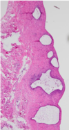
For hydrocele, provide description, epidemiology, pathophys, and diagnosis.

A
  • Hydrocele: painless scrotal enlargement
    • Epidemiology: newborns
    • Pathophysiology: collection of serious fluid between parietal and visceral layers of tunica vaginalis
    • Diagnosis: light to scrotum shows fluid
21
Q

For testicular torsion, provide description, pathophys, complications, and tx.

A
  • Testicular torsion: twisting of scrotal contents on spermatic cord
    • Pathophysiology: high attachment of tunica vaginalis on spermatic cord → testes free to swing and tangle on spermatic cord
    • Complications: Veins may become obstruction → hemorrhagic infarction
    • Tx: emergent surgical correction to preserve viability
22
Q

For Germ Cell Tumors of the testicles, provide epidemiology, risk factors, pathogenesis (genes), presentation, diagnosis, and types.

A
  • Germ cell tumors
    • Epidemiology: most common testicular tumor, ages 15-34, whites
      • Better prognosis in infants
    • Risk factors: cryptorchidism, Kleinfelter syndrome, testicular carcinoma in-situ, testicular dysgenesis syndrome, FHx of testicular GCT
    • Pathogenesis: KIT-activating mutations and i(12p)
      • i(12p)/CIS not associated with infant GCT
      • Retain expression of NANOG and OCT 3/4 pluripotent cells
    • Presentation: nodule, with dull aching pain
    • Diagnosis: scrotal US, staging CT, serum markers, orchiectomy (often found by patient or partners)
    • Types: seminoma and mixed GCT
23
Q

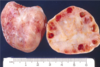
For seminomas of the testicles, provide epidemiology, description, diagnosis, prognosis, and pathology (gross and micro)

A
  • Epidemiology: most common testicular tumor, not common in infants
  • Description: remains localized within tunica albuginea
  • Diagnosis: radiosensitive (respond to radiotherapy), HCG production is rare
  • Prognosis: presents in clinical stage I (good prognosis); late metastasis
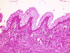
  • Pathology:
    • Gross: homogenous pale tan parenchyma (pic)
    • Micro: fibrovascular septae with lymphocytes
24
Q

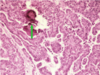
For mixed GCT: provide presentation, diagnosis, prognosis, gross pathology, and the different subtypes (in order from poor to good prognosis).

A

Mixed GCT (NSGCT):

  • Presentation: younger patients (compared to seminoma)
  • Diagnosis: not radiosensitive (surgery/chemo), AFP (alpha-fetoprotein) and/or HCG biomarkers
    • LDH correlates with tumor burden
  • Prognosis: poor, metastasizes earlier via heatogenous/lymphatic route
  • Gross pathology: hemorrhagic/necrotic appearances
  • Subtypes: from poor prognosis to better prognosis
    • Choriocarcinoma:
    • Embryonal carcinoma
    • Yolk sac tumor:
    • Teratoma
25
Q

What two GCT tumors are common in infants/children?

A

Yolk sac tumors and teratomas

26
Q

For spermatocystic seminoma: provide the epidemiology, and pathology. Is this a type of seminoma?

A
  • Spermatocytic seminoma
    • Epidemiology: old people, rare
    • Pathology: small cells (resembling spermatocytes), medium cells, and giant cells
    • Not associated with testicular CIS, i(12p), and not considered a subtype of seminoma
27
Q

For sex cord-stromal tumor: provide the two types.

For each type, provide a description, and complications/pathology if any.

A
  • Sex Cord-Stromal Tumor
    • Sertoli cell tumor
      • Description: benign tumor that resemble Sertoli cells lining seminiferous tubules
    • Leydig cell tumor
      • Description: benign estrogen/androgen producing tumor
      • Complications: precocious puberty, gynecomastia
      • Micropathology: cytoplasmic Reinke crystals
28
Q

For testicular lymphoma: provide epidemiology descriptions, complications, and prognosis.

A
  • Testicular Lymphoma:
    • Epidemiology: most common testicular in males > 60 y/o
    • Description: usually diffuse B cell lymphoma
    • Complications: disseminated/systemic disease found after testicular presentation
    • Prognosis: poor
29
Q

What is this?

A

Seminoma

  • Gross: homogenous pale tan parenchyma (pic)
30
Q

What is this?

A

Seminoma: fibrovascular septae with lymphocytes

31
Q

What is this? What do they produce?

A
  • Choriocarcinoma:
    • Micropathology: cells resembling placental cytotrophoblasts (mononucleated cells) and synctiotrophoblasts (polynucleated cells → produce HCG)
32
Q

What is this?

A
  • Embryonal carcinoma
    • Micropathology: poorly-formed papillae and glands (pic)
33
Q

What is this? What does this tumor produce?

A
  • Yolk sac tumor:
    • Epidemiology: most common in infants
    • Micropathology: Schiller-Duvall bodies (fibrovascular core surrounded by single layer of germ cells → produce alpha-fetoprotein), hyaline globules (red glossy) (pic)
34
Q

What is the micropathology of a teratoma?

A
  • Teratoma
    • Epidemiology: most common in infants
    • Micropathology: will appear as the tissue that it is derived from
35
Q

Generally for ovarian neoplasms

what is the epidemiolofy, presentation, risk factors

A
  • Epidemiology: mostly benign (young women), sometimes malignant (older women)
    • Less common than endometrial/cervical cancer, but more deadly
  • Presentation: advanced stage disease due to asymptomatic developing tumors
  • Risk Factors: nulliparity (can’t give birth), infertility, BRCA/p53/Her2 mutations, Turnery Syndrome (only one X chromosome)
36
Q

For tumors of epithelial origin

what is the epidemiology, presentation, grading, subtypes, tx

A
  • Epidemiology: majority of ovarian cancers, 20+ y/o
  • Presentation: the more solid a tumor is, the more malignant it is
  • Grading:
    • High grade serous tumors: in-situ carcinoma of the fallopian tube is a precursor lesion and is associated with p53/BRCA
  • Epithelial cell types involved: serous (fallopian tubes), mucinous (endocervix), endometroid
  • Tx: adjuvant chemo, surgery
37
Q

mucinous cystadenoma

is it benign or malignant?

what is the description, epidemiology, micropathology

A
  • benign
    • Description: single cyst often with KRAS proto-oncogene mutation (associated with pseudomyxoma peritonei)
  • Epidemiology: premenopausal women (30-40 y/o)
  • Micropathology: cysts lined with simple mucinous epithelium (pic)
38
Q

serous cystadenocarcinoma

benign or malignant?

what is the description, epidemiology, gross and micro pathology

A
  • malignant
    • Description: bilateral complex cysts (cysts within cysts)
  • Epidemiology: postmenopausal women
  • Pathology
    • Gross: papilla like projections (looks like cauliflower) (pic)
    • Micro: atypical nuclei with Psammoma bodies (calcifications) (pic)
39
Q

Boderline

is it benign or malignant ?

what is the description, criteria, tx

A
  • Description: unilateral lesions with features of both benign and malignant
  • Criteria: lack of invasion of stroma
  • Tx: conservative surgery
40
Q

generally, for germ cell tumors

what is the description, epidemiology, location

A
  • Description: tumors arising from primitive germ cell
  • Epidemiology: 15-20% of ovarian tumors, 0-25 y/o, mostly benign
  • Location: germ cell tumors can arise anywhere along the embryological migratory route
41
Q

subtypes for germ cell tumors

A

choriocarcinoma, teratoma, embtyonal carcinoma, yolk sac carcinoma, and dysgerminomas

42
Q

Choriocarcinoma

description, characterisitics

A
  • Description: malignant tumor composed of trophoblasts and synctiotrophoblasts; mimics placental tissue
  • Characteristics: beta-hCG+
43
Q

Teratoma

description, epidemiology and prognosis for mature versus immature cysts

A
44
Q

what is a Embryonal carcinoma

A

Description: aggressive malignant tumor composed of large primitive cells

45
Q

Yolk sac carcinoma (aka endodermal sinus tumor)

what is it? characterisitics?

A
  • Description: malignant tumor that mimics the yolk sac; common in children
  • Characteristics: AFP elevated, Schiller-Duval bodies (look like glomerulus)
46
Q

Dysgerminomas

description, epidemiology, pathology gross adn micro, tx

A
  • Description: uniform primordial germ cells (oocytes) –> analogous to seminoma
  • Epidemiology: 2% of ovarian tumors, ages 0-20
  • Pathology:
    • Gross: solid, yellow tan (pic)
    • Micro: uniform lymphocytes and germ cells; large cells with clear cytoplasm and central nuclei (pic)
  • Tx: highly radiosensitive, curable
47
Q

what is a sex cord tumor

A

Description: functioning ovarian tumors that are derived from granulosa/theca cells (10% of ovarian tumors)

48
Q

Granulosa-Theca cell tumor

epidemiology, characterisitics, grossa and micro pathology

A
  • Subtype: Granulosa-Theca cell tumor
    • Epidemiology: post-menopausal women
    • Characteristics: estrogen/steroid production in excess, associated with endometrial hyperplasia
    • Pathology:
      • Gross: unilateral solid yellow lesion
      • Micro: Call-Exner bodies between granulosa cells (pic)
49
Q
A

mucinous cystadenoma

50
Q
A

serous cystadenocarcinoma

51
Q
A

serous cystadenocarcinoma

52
Q
A

Borderline epithelial tumor of ovary

53
Q
A

Dysgerminomas

54
Q
A

Granulosa-Theca tumor

61
Q

normal histology of ovary

surface versus cortex

A
  • Surface: Coelomic epithelium (cuboidal)
  • Cortex:
    • Follicles – composed of granulosa cells and oocytes
    • Stroma – composed of theca cells and spindled cells
62
Q

Stages of ovarian development

what does it look like at different stage of life…

A
  • Infants: small developing ovaries (look linear)
  • Reproductive age: large pronounced oval shaped
    • Rupture during ovulation
  • Post-menopausal: fibrosis and shrunken
63
Q

Stages of follicle development

A
  • Primordial follicle (only the oocyte in stroma) → primary follicle (oocyte surrounded by granulosa [cuboidal] cells) → secondary follicle (contains an antrum) → Graafian follicle (antrum takes up most of the follicle) → corpus luteum (yellow body → white body [corpus albicans] if no fertilization)
64
Q

Histology of Granulosa cells (pic)

developed versus luteinized

A
  • Developing follicle – nucleus > cytoplasm
  • Luteinized follicles – nucleus < cytoplasm
65
Q

Ovarian infections

whhat is it? etiology, complications?

A
  • Description: Part of the pelvic inflammatory disease
  • Etiology: STI
  • Complications: infertility, sterility, pelvic mass/abscess
66
Q
A

normal ovary

67
Q

whats the difference

A

Developing follicle – nucleus > cytoplasm

Luteinized follicles – nucleus < cytoplasm

68
Q
A

Endometriosis

69
Q
A

Endometriosis

70
Q
A

Follicular cysts

71
Q
A

Polycystic ovarian disease

72
Q
A

Polycystic ovarian disease

73
Q

Endometriosis (super common)

what is it? location? presentation? pathophysiogy? tx?

A
  • Description: endometrial glands and stroma outside of endometrium
  • Location: ovary (most common), peritoneum, fallopian tubes
  • Presentation: pain, infertility
  • Pathophysiology: extra-uterine glands that respond to menstrual hormones → bleeding → fibrosis/adhesions
  • Tx: control bleeding/pain, correct infertility
74
Q

Endometriosis (super common)

gross and micro pathology

A
  • Gross: red-brown nodules (chocolate cysts), fibrous adhesions between organs/ligaments
  • Micro: glandular structures with hemosiderin due to bleeding, stroma (pic x2)
75
Q

non-functioning versus functiong cysts

A

Non-functioning – no hormones are made

Functioning cysts – hormones are secreted

76
Q

Surface inclusion cysts

description and pathology

A
  • Description: benign cysts due to the pinching off of surface epithelium after ovulation
  • Pathology
    • Micro: found in superficial ovarian cortex

non-functioning

77
Q

Follicular cysts

description and pathology

A
  • Follicular cysts
    • Description: common in newborn population, multiple simple cysts lined by granulosa cells that may secrete estrogen
    • Pathology:
      • Gross: cysts lined up linearly (pic)
78
Q

Polycystic ovarian disease aka Stein-Leventhal syndrome

description, presentation, pathophysiology, gross and micro pathology

A
  • Description: multiple ovarian follicular cysts due to hormone imbalance
  • Presentation: young women, oligomenorrhea (few menstruations), hirsutism (male-like hair growth), obesity/insulin resistant diabetes, virilism (male characteristics)
  • Pathophysiology: irregular hypothalamic control of pituitary → high LH levels/low FSH levels → excess androgen production
  • Pathology:
    • Gross: Numerous follicle cysts lining the periphery of the ovary, but no corpora lutea/corpora albicantia (so no signs of ovulation) (pic)
    • Micro: cysts – pearl necklace (pic)
86
Q

models of sexual response

which is linear?

A

Masters and Johnson (linear – assumes women have intrinsic desire)

Basson model (nonlinear)

87
Q

what is Basson model (nonlinear)

A
  • Assumes intrinsic desire for sex is not initially present
    • Desires is created with extrinsic factor including romantic environment, erotic stimuli, emotional intimacy, biologic/psychologic need
88
Q

what is involved with excitement

different physical changes?

NT/hormones invovled (excitatory and inhibitory)

clinical signs?

A
  • Changes: vasocongestion, lubrication, clitoral retraction
  • NTs and hormones involved
    • Pro-sexual: Norepi, dopamine, testosterone, oxytocin (nipple stimulation), serotonin, estrogen
    • Inhibitory: Prolactin, glutamate, vasopressin, GABA, serotonin
  • Clinical signs: Increase in BP, HR, RR
89
Q

excitment: vasocongestion

explain it?

A
  • Vasocongestion of the clitoris, vagina, vulva
    • Mechanism: parasympathetic release of NO, Ach, and VIP → smooth muscle relaxation →
      • Vascular: dilation of genital arteries → increased blood flow
      • Muscular: increase in vaginal length and diameter
90
Q

excitement: lubrication

explain

A
  • Introitus: Bartholin glands secrete mucous
  • Vaginal walls: increased blood flow → increased hydrostatic pressure → increased transudate leaking through
91
Q

excitement: Clitoral retraction

explain mech

A
  • Retraction due to decreased sympathetic tone (increased arterial flow/decreased venous return)
    • Perineal muscles contract via pudendal n.
92
Q

plateau

explain this

A
  • Plateau – brink of orgasm
    • Intensified excitement phase
93
Q

phases of sex

A

excitment, plateau, orgasm, resolution

94
Q

orgasm

A
  • Orgasm – involuntary muscle spasm
    • Mechanism: not well understood, S2-4 are involved (autonomic and somatic)
    • Signs: vaginal/uterine contractions, full body flushing, increasing BP, RR and HR
    • Cerebral event: decreased blood flow to the brain
95
Q

resolution

explain characteristics

A
  • Return to normal BP, RR, & HR
  • Decrease is blood flow & swelling of genitals
  • Fatigue
  • Refractory period (can have orgasm during this time in females)
96
Q

sexual dysfunction

epidemiology and risk factors

A
97
Q
  • Epidemiology: 40% women report issues
  • Risk factors: relationships, chronic illness, abuse, medications, stress, sleep
    • Chronic illness: diabetes, depression, arthritis, GYN surgery
    • Meds: SSRI/ SNRI (selective norepi reuptake inhibitor), oral contraceptives (eats up testosterone → reduces libido)
A
98
Q

DSM-V criteria for sexual dysfunction

A
  • Must be present for at least 6 months
  • Must result in clinically significant distress
  • Are not explained by a nonsexual mental condition, severe relationship distress, or other significant stressor
  • Are not secondary to use of a substance or medications or other medical condition
99
Q

what is Female orgasmic disorder ?

tx?

A
  • Female orgasmic disorder – marked delay/decrease in frequency or intensity of orgasm
    • Tx: Behavioral changes, CBT
100
Q

what is Female sexual interest/arousal disorder ?

tx (4)?

A
  • Female sexual interest/arousal disorder – lack of or reduced interest or arousal in 3 different areas
    • Tx:
      • Behavioral changes, cognitive behavioral therapy (CBT), couple therapy, mental/physical stimulation (vibrators or porn)
      • Flibanserin: centrally acting 5HT1a agonist → decreases serotonin and increases dopamine and norepi
        • SE: hypotension, syncope, avoid ETOH
      • Bupriopion: anti-depressant
      • Testosterone: severe cardiac SE
101
Q

what is Genitopelvic pain/penetration disorder ?

4 etiologys

A

Genitopelvic pain/penetration disorder – pain during intercourse or difficult penetration

  1. Pelvic floor dysfunction – tensing of pelvic floor muscles
  2. Vaginal atrophy – loss of rugae and lubrication in vaginal canal
  3. Vulvar vestibulitis – inflammation of vulva
  4. Endometriosis, chronic pelvic pain, anatomic causes
102
Q

tx for each of these

  1. Pelvic floor dysfunction – tensing of pelvic floor muscles
  2. Vaginal atrophy – loss of rugae and lubrication in vaginal canal
  3. Vulvar vestibulitis – inflammation of vulva
  4. Endometriosis, chronic pelvic pain, anatomic causes
A
  • Pelvic floor dysfunction – tensing of pelvic floor muscles
    • Tx: CBT, vaginal dilators, PT
  • Vaginal atrophy – loss of rugae and lubrication in vaginal canal
    • Epidemiology: post-menopausal women (lack of estrogen)
    • Tx: personal lubricant, vaginal moisturizer, topical estrogen
  • Vulvar vestibulitis – inflammation of vulva
    • Tx: treat infection, lidocaine, NSAIDS, PT, surgery (vestibulectomy)
  • Endometriosis, chronic pelvic pain, anatomic causes
    • Tx: Hormonal treatment, Pain medications, Surgery