Week 4 - Chapter 14,15,16 Flashcards

(94 cards)

1
Q

what is the skull ?

A

The skull is a rigid bony box that protects the brain and the special sense organs, it includes the bone of the cranium and the face

2
Q

How many cranial bones do we have ? what are they named ?

A

there are 4 cranial bones: frontal, occipital, parietal, temporal

3
Q

How do the cranial bones unite ?

A

They are meshed through immovable joints calleed sutures

4
Q

How do the sutures develope ?

A

During birth, the cranial bones are not firmly jointed together to allow for the mobility and chnage in shape that is needed for the birth process

The sutures gradually ossify during early childhood

5
Q

what are the different sutures present in the cranium ?

A
  1. Coronal Suture - crowns the head from ear to ear at the union of the frontal and parietal bones
  2. Saggittal suture - seperates the sides of the head lengthwise between the two parietal bones
  3. Lamboid Suture - seperates the parietal bones crosswise from the occipital bone
6
Q

How many facial bones are there ? which one does not articulate at the sutures ?

A

there are 14 facial bones - the Mandible does not articulate with the sutures

7
Q

what is the cranium supported by ? what is special about the C7 vertebrae ?

A

The cranium is supported by the cervical vertebrae (C1 atlas & c2 axis - all the way down to C7)

The C7 Vertebrae also has the name vertebrae prominens this is because it has an easily palpable long spinous process

8
Q

how many pairs of salivary glands are there ?

A
  1. Parotid Glands - found in the cheeks, mandible, anterior to and below the ear - they are the larges salivary glands
  2. Submandibular Glands - found beneath the manidble at the angle of the jaw
  3. Sublingual Glands - lie in the floor of glands
9
Q

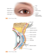
Where is the Temporal Artery located ?

A

Lies superior to the temporalis muscle and its pulsation is palpable anterior to the ear

10
Q

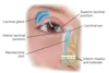
what are the blood vessels located in the neck ?

A
  1. Internal Carotid Artery - which branches off the carotid artery ad run inward and upward to supply the brain
  2. External Carotid Artery - supplies the face, salivary glands, superficial temporal area
  3. Internal Jugular Vein - lies beneath the sternomstoid muscle along with the Carotid Artery
  4. External Jugular Vein - runs diagnolly across the sternomastoid muscle
11
Q

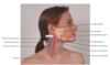
What are the neck muscles ?

A
  1. Sternomastoid - divides each side of the neck into two triangles (anterior triagle - base is along the lower mandible and apex is at the suprasternal notch ) - (posterior triangle - behind the sternomastoid muscle - base is the clavicle ) - innervated by Cranial Nerve XI - accomplishes head rotation and head flexion
  2. Trapzeuis muscle - forms trapezoid shape on upper back - arises from occipital bone & vertebrae and extends fanning out onto scapula & Clavicle - move shoulders and extend and turn the nek
  3. Omohyiod Muscle
12
Q

How many types of Trapezius muslces are there ? Where do they originate from? what is there function ?

A

There are two trapezius muscles - form trapezoid shape on the upper back - each of them arise from the occipital bone, the vertebrae

fans out from the scapula and clavicle

Trapzeius muscle moves the shoulders & extend & turn head

13
Q

How is the sternomastoid muscle divided ?

A

The sternomastoid is divided into the anterior & posterior triangle

the anterior triangle lies in the front between the sternomastoid and the midline of the body

The posterior triangle is behind the sternomastoid muscle

14
Q

What is the thyroid gland ?

A

The thyroid gland is an endocrie gland

straddles trachea in the middle of the neck

thyroid glad releases thyroxine (T4) Triiodothyronine(T3)

There are two thyroid lobes that are connected by the isthnus

15
Q

What is thyroid Cartilage ?

A

The thyroid cartilage is the adams apple

The highest structure i the neck is the Hyoid Bone

16
Q

how many lymph nodes are there ?

A
  1. Preauricular: in front of the ear
  2. Posterior Auricular (mastoid): superficial to mastoid process
  3. occipital: base of the skull
  4. submental: midline. behind the tip of the mandible
  5. submandibular: between the angle and the tip of mandible
  6. Tonsillar: under the angle of mandible
  7. Superficial Cervical: over;yin sternomastoid muscle
  8. Deep Cervical: deep uder sternomastoid muscle
  9. Posterior Cervical: in posterior triangle along the edge of the traps
  10. Supracavicular: just above and behind the clavicle
17
Q

What is the importance of the lymphatic system ?

A

The lymphatic system is important for the bodies immune system - its job is to detect and eliminate the foreign substances from the body - the nodes filter the lymph

18
Q

What are the four areas that are accessible for lymph examination?

A
  1. head & neck
  2. arms
  3. axillae
  4. ingiunal region
19
Q

What are the developement changes for infants and children ?

A

Neonatal skull is seperated by stutures called Fontanelles (spaces where the suture intersects) - sutures ossify during the first year

during the fetal period had growth is rapid - the childs head grows reaching 90% of its final size when the child is 6 years old

facial bones grow at varying rates

lymphoid tissues grow to adult size at age 6

Lymphatic issues grows rapidly beyong adult size at ages 10 and 11 before puberty but then slowlies atrophies

20
Q

What happens to women during pregnancy with regards to their thyroid gland ?

A

The thyroid gland enlarges slightly during pregnancy due to hyperplasia of the tissue and increased vascularity

21
Q

What are some changes that occur in adults ?

A

facial bones and orbits appear more prominent

facial skin sags - decreases elasticity, subcutaneous fat, moisture

22
Q

What are some questions that should be asked in regards to a headache ?

A
  1. any unsually frequent or severe headaches
  2. onset - when did the headache start
  3. gradual - over hours or a day or suddenly
  4. ever had this kind of headache before
  5. location of headache
  6. localized or on one side
  7. throbbing or aching pain
  8. mild, moderate, or severe
  9. course and duraion of headache
  10. how lond does it last
  11. what brings it on
  12. any other syptims associated with it
  13. any other illness
  14. medications
  15. what makes the pain worse
  16. pattern: any family history of headaches
  17. frequency of headaches
  18. what seems to help
23
Q

What are some questions in regards to head injuries ?

A
  1. Onset: when
  2. Setting: any hazardous conditions ? were you wearig a helmet or hard hat?
  3. before injury - were you dizzy or lightheaded
  4. did you have a blackout or a seizure
  5. did you loss conciousness or fall
  6. knocked unconcious or did you fall and lose conciousness
  7. any history of illness
  8. where did you hit your head
  9. how long were you unconcious
  10. any symptoms after head injury (vomiting, headache)
  11. any change in the level of conciousness
  12. any associated synpotoms: discharge from ears or nose
  13. pattern: have the symptoms become worse
  14. effort to reat
    15.
24
Q

Questions to ask in regards to Dizziness ?

A
  1. describe the dizziness
  2. onset: abrupt or gradual ? after change in position
  3. associated factors
25
Questions in regaards to neck pain and limitation of pain ?
1. onset 2. location - does it radiate 3. associted symptoms - any limitation of movement 4. percipitationg factors: what movements cause pain ?do you need to bend or lift at work 5. does stress trigger the pain 6. coping strategies
26
Questions in regards to Lumps or Swelling ?
1. any lumps or swelling in the neck 2. any recent infections ?any tenderness ? 3. how long have you had it ? has it changed it size ? 4. any hisory of prior irridation ? 5. difficulty swallowing 6. do you smoke ? for how long? how many packs ? 7. Last alcoholic drink ? how much alcohol do you drink in a day ? 8. ever had a thyroid problem ? how was it treated ? 9. history of head or neck surgery
27
What are some additional history questions for infacnts and children ?
1. did the mother use alcohol ot street drugs during pregnancy ? how often ? 2. how much used per episode 3. Delivery: was it vaginal or C section ? froceps used ? 4. Growth: what were you told about the babys growth ? was it on schedule ? did the head and the fontanelles grow on schedule
28
What does normocephalic mean ?
round symmetrical skull that is appropriately related to body size
29
What is temporal Arthitis ?
the temporal artery looks more tortuous and feels hardened and tender
30
What are some objective data to collect when examining the Head ?
1. Size and Shape - note deformaties like macro or microcephaly - note lumps, depressions or abnormal portrusions 2. Temporal Area - palpate the temporal artery in the zygomatic bone in between eye and top of ear
31
What are some objective data to collect when inspecting the face ?
1. Facial Structure - tense muscles - flat effect (depression) - edema in the face
32
What are some objective data to collect when inspecting the Neck ?
1. Symmetry 2. Range of motion - note any type of limited movement 3. Lymph Nodes: *Lymphadenopathy* is the enlargement of the lymph nodes 4. Trachea: note a tracheal shift when the trachea is shifted to the unaffected side or pulled to the affected side 5. Thyroid gland: you can asculate the thyroid fro *Bruit* you will hear whooshing sounds, blowing,soft, pulastile
33
what are some considerations when examining infants and children ?
1. measure infants head with measuring tape - infants head avergaes around 32 to 38cm - 2. observe infants head from all angles - note any bulges - *caput succedaneum* is edemtous swelling and ecchymosis of the presenting part of the head caused by birth trauma (no treatment is needed resolves within days) - *cephalhematoma* is a subperiosteal hemmorhage; result of borthing trauma; well ddeined over cranial bone and holds blood in pace; fluid is reabsorbed in the first few weeks 3. gently palpate skull and the fontanelles 4. note infants head posture and head control - inflants will show *tonic neck reflex* which is rotating the neck to one side causing the ipsilater extension of the estremeties 5. Facial features 6. Neck - a short neck or webbing may indicate congenital abnormalities
34
What is the *Palpebral Fissure ?* What is the *Limbus* ? What is the *Canthus* ? What is the *Carnucle* ?
is the elliptical open space between the eyelids the lower eyelid margin, border b/w sclera and cornea corner of the eye, angel where the eyelids meet small fleshy mass containing the sebaceous glands
35
What are *Tarsal Plates ?* What are *Meibomian glands ?* ​What is the *Conjuctiva ?* What is the *Lacrimal Apparatus ?* What is the *Puncta ?*
Tarsal Plates: are strips of the connective tissue that give it shape Meibomian Glands: the tarsal plates contain these, modified sebacious glands that secrete an oily lubricating material Conjuctiva: Transparent protective covering, thin mucous membrane folded like an envelope between the eyelids and the eyeball - conjuctiva merges with the cornea Lacrimal Appararus: provides constant irrigation to keep the conjuctiva and cornea moist and lubricated - lacrimal gland is located in the upper outer corner over the eye, sceretes tear Puncta: where the tears drain, visible on the upper and lower eyelids at the inner canthus
36
Where does the tears drain ?
The tears drain the nasolacrimal sac through the nasolacrimal duct and empties in inferior meatus inside the nose
37
how many muscles are attached to the eyeball in its orbit ?
![]()These muscles allow the both straight and rotary movements - humans can only view one image at a time and the eye movements are always parallel to each other 1) Superior Oblique 2) inferior Oblique 3) lateral Rectus 4) medial rectus 5) superior rectus 6) Inferior Rectus
38
What nerve stimulates the *extraocular muscles ?*
Three cranial nerves: 1) Cranial nerve VI innervates the lateral rectis muscle which moves it laterally towards the temple 2) Cranial Nerve IV (trochlear nerve) innervates the superior oblique muscle; moves it downwrad and inward toward the nose 3) Cranial Nerve III (occulmotor nerve) inneravtes at the other nerves ; superior, inferior, medial rectus and the inferior oblique muscles
39
how many layers are present in the eye ?
Three layers 1) Outer fibrous sclera 2) Middle vascular choroid 3) Inner nervous retina
40
Explain the aspects of the outer layer of the eye ?
The outer layer of the eye is called the sclera - it is a tough white protective covering - continuous anteriorly with smooth outer cornea which cover the iris and the pupil the cornea refracts the light so that it is focuseed on the retina within cornea is sensitive to touch; *corneal reflex* which causes blinking - the trimegnal nerve carries the afferent sensation into the brain and the facial nerve carries the efferent message that stimulates the blink
41
Explain the aspects of the middle layer of the eye ?
The *Choroid* has a dark pigmentatio to prevent light from reflecting internally and is hevaily vascularized to deliver blood to the retina anteriorly the choroid is continous with the ciliarly body and the iris - the muscles of the ciliary body controls the thickness of the lens the *iris* functions as a diaphragm, varying th eopening in the centre the *pupil*; this controls the amount of light that enters the retina In bright light the muscle fibres of the iris contract the pupil, and dilate when in dim light or far vision The *pupil* is round and regular; the size is determined by the parasympthatic and sympathtic nervous chains of the autonomic nervous system; the parasympathtic nervous branch through the cranial nerve III causes constricyion of the pupil while the sympathtic nervous system causes the dilation of the pupil and elevates the eyelid The *Lens* is a trasparent bioconvex disc located behind the pupil - serves as refracting medium - thickness controlled by cilliary body The *Anterior Chamber* is posteror to the cornea and anterior to the iris and lends while the *posterior chamber* lies behind the iris to the sides of the lens - continis flow of fluid delivers the nutrients to the surronding tissues
42
Explains the aspects of the inner layer ?
* Retina* is the visual receptive layer in which light waves are changed to nerve impulses * Optic Disk* is the area in which fibres from the retina converge to form the optic nerve * Retinal Vessels* include a paired artery and vein extending to each quadrant - the arteries are narrower and brighter than the veins - arteris also have a thin silver light * Macula* is located at temporal side of the fundus, it is a slightly darker pigmented regionsurronding the *Fovea centralis* this area has the sharpest and keenest vision; the macula recieves and trasduces light from the centre of the visual field
43
What is the *Pupillary light reflex ?*
It is the normal constriction of the pupils when bright light shines on the retina Cranial Nerve II ( optic nerve) carries it to the brain and Cranial Nerve III (oculomotor Nerve) carries it from the brain to the eye when light hits one eye, the one eye that recieves the light goes through DIRECT LIGHT REFLEX and the other goes through CONSENSUAL LIGHT REFLEX - this happens because the Optic nerve that is carrying the message to the brain synapses with both sides of the brain
44
What is *Fixation ?*
this is a reflex direction of the eye toward an object attracting a persons attention - this fixing ontains rapid ocular movements to put the target back on the fovea centralis these ocular movements are impaired by medicatio, alcohol, fatigue
45
What is *Accomadation ?*
is adapation of the eye for near vision which is accompolished by increasing the curvature of the lens through the movement of ciliary muscles You can measure the convergence that occurs with accomadation - convergence (motion toward) the eyeballs and pupillary constrictions
46
What are the developemental considerations for infants and children regarding the eyes ?
eye function is limited but matures during the early years the macula is abset at birth and begins developing at 4 months and finishes at 8 months eye movements are poorly coordinated at birth at 3 to 4 months the infats establishes binocularity and can fixate on a single image most neonates are born farsighted and decreases by ages 7 to 8 iris shows little pigmentation at birth & pupils are small
47
What are the developemental considerations for older adults regarding the eyes ?
Lacrimal glands have low tear production and feeling of dryness and burning pupil size decreases the lens loses elasticity, becomes hard and glasslike, which affects the lens ability for near vision called *Presbyopia* - onset is 40 years of age and by 70 senile cataracts occurs in which transparent fibres of the lens thicken and yellow visual acuity decreases at age 50 and even more at 70 decrease power of accomadation in the lens more light may be needed b/c of decreases adaptation to darkness
48
What are the most common causes of decreased visual functioning in older populations ?
1. Macular degenration: breakdown of cells in the macula; loss of central vision - the most common cause of blindness is Age related Macular degeneration - higher incidence in women 2. Cataract Formation or lens opacity: results from the clumpig of proteins in the lens 3. Glaucoma or increased interocular pressure - involves damage to optic nerve resulting in gradual loss of peripheral vision 4. Diabetic Retinopathy: highest in younger people below 65 - highest in diabetic patients
49
Describe the Snellen eye chart ?
used to measure visual acuity it has lines of letters in decresing in size place snellen chart in well lit spot at eye level - patient needs to be 20m from chart - ask them to cover the other eye with a opaque cover - if they are wearing contacts or glasses ask them to leave it on (only remove reading glasses) - start with line with the smallest letters - note how many letters the patient missed in each lie by writing minus 1 or minus 2 - the top number (20/20) in the fraction indates the distance the patient was at and the denominator indicates the distance in which a normal eye can read - of the patient cannot rad then bring the chart closer
50
How does one assess vision for people with near vision ?
test near vision with handheld vision screener with various sizes of print - instruct the patient to hold the card 14 in away from the eye which is equivalent to 20 ft - test each eye seperately with glasses - if no vision card ask patiet to read newspaper or maagzine
51
What is the Confrantation test and how is it conducted ?
this is ued to test the visual fields this is a gross measure of peripheral vision - patients peripheral vision is compared with your own position yourself at the same eye level as the patient abotu 60 cm away - direct the patient to cover one eye with an opaque card and look straight at you with the other eye - cover your own eye opposite to the patients covered one - you are testing the uncovered eye - hold you finger or pencil at the target midline between you and the patient, at the periphery of the vision and slowly advance it inward from the periphery in several directions - tell the patient to say "now" when it sees the target the location should match when you see the object
52
What is the Corneal light reflex (Hirschbergs test)?
asses the parallel aligment of the eye axes by shining a light toward the patients eyes - direct the patient to look straigh ahead and then shine a light 30cm away note the reflection of the light on the corneas - it should be on the same exact spot in each eye
53
What is the cover-uncover test ?
this test detects small degrees of deviated alignment by disrupting the fusion reflex that normally keeps the two eyes parallel ask the patient to stare straigh ahead at your nose - with a n opaque card cover one eye and examine the response of the uncovered eye - it should be a normal response to a steady fix gaze - then uncover the eye and observe it for moment and it should stare straight ahead - if it jumps to restablish fixation eye muscle weakness exists repeat test for both eye if there is muscle weakness the covered eye will drift into relaxed position -
54
What is the diagnostic Positions test ?
lead the eye throuh 6 different positions of gaze - this reveals any muscle weakness during movement ask th epatient to stand 30 cm away - ask them to follow your finger or light in 6 different positions - hold each position momentarily then back to centre then to the next position a normal response is parallel tracking with both eyes note any *Nystagmus* which is a fine oscillating movement seen around the iris - normal at lateral gaze but not normal at any other position note that the upper eyelid always overlaps the superior part of the iris even during downward movement - you should not see the white rim of sclera between the eyelid and iris this is called *lid lag*
55
What are the different ways to inspect the external ocular structures ?
1. General: examining there general movement throughout the room - how they move around - also note facial expressions 2. Eyebrows: 3. Eyelids and lashes: the upper eyelids cover the superior part of the iris 4. Eyeballs: no protrusion or sucken appearance 5. Conjuctive and Sclera: ask the patient to look up and drag the lower lids down to examine the conjuctive - normally it should be moist and glossy, numerous msall blood vessels should show - the sclera should be white and in people with dark skin it should be grey blue or muddy and sometimes in dark skinned people you will see macules on the sclera - yellowsih fatty deposits may be common below eyelids away from cornea in darker skinned people 6. Eversion of the upper lid 7. Lacriml Apparatus: ask the patient to look down and with your thumb slide the outer part of the upper eyelid up along the bony orbit to expose under the eyelid - inspect for redness and swelling - excessive tearing may indicate blockage of the nasokacrimal duct to check this press your index finger against the sac
56
what are the different ways to inspect the aterior eyeball structure ?
1. Cornea and lens: shine a light from the side across the patient cornea - it should be smooth and clear - note any cloudiness 2. Iris and pupil: note the size, shape, and equality of the pupils - a small number of pupils have two different sizes this is called *Anisocoria* - to test *pupillary light reflect* darken the room and ask the patient to gaze into the distance; advance a light in from the side and note the response; from this you should see a direct light reflex on the one sided pupil and a consensual light reflex in the other pupil \*\* in acute care setting, its important to compare the mm of the normal pupil size compared to after the light reflex\*\* \*\* to test for accomadation, ask the patient to focus on distant object this will cause the pupils to dilate then ask patient to shift gaze to closer object such as your finger 7 to 8 cm away from the nose - the normal response should be pupillary constriction and convergence of the axis of the eyes\*\* \*\*record PERRLA - pupils, equal, round, react to light, and accomadation\*\*
57
What are the different ways to inspect the occular fundus ?
Using the opthalomoscope, it enlarges the view of the eye so you can inspect the media ( anetrior chamber, lens, vitreous) and the ocular fundus ( internal surface of the retina) Hold instrument right up to the eye, darken the room , glasses need to be removed, lenses can stay - use larger aperture with white light if pupils are small use the smaller aperature - the light needs to be maximum brightness - instruct patient to look at marking on the wall start 25 cm away at a 15 degree lateral angle and note the *Red Reflex* which is a red glow that occurs caused by the reflection of the opthalmascope light off the inner retina by moving in on the 15 degree lateral angle you will view the optic disk, retinal vessels, general background and macula
58
What is the optic disk ?
it is located at the nasal side of the retina 1. Colour: creamy yellow orange to pink 2. Shape: round or oval 3. Margins: two varuations may occur around the margins; A *sclera crescent* is grey white new moon shape and a *pigment cresecent* is black due to accumulation of pigment in the crescent 4. cup disc ratio: physiological cup is brighter yellow-white compared to the rest of the disc
59
What is the retinal vessel and what aspects of it should be examined ?
1. Number 2. colour 3. artery vein ratio: ratio artey width to vein width 4. calibre 5. ateriovenous crossing 6. tortuosity: mild vessel twisting 7. Pulsations
60
what should be noted based on the general background of the fundus ?
colour varies from light red to dark brown clear, no lesions
61
What are some developemental considerations when examining the eyes of children and infants ?
Test light perception, by using blink reflex - by using a light - pupillary light reflex when the pupil restricts in response to light - noraml things to note: birth to 2 weeks: possible fixation on object; refusal to open eyes after light exposure; increase in alertness 2 to 4 weeks: fixatig on objecy 1 month: fixating and following 3 to 4 months: fixating anf following and reaching for object 6 to 10 months: fixating and following toy in all directions
62
What should be tested for children ages 3 and up ?
asses the visual field - note the time the childs eye deviate or head shifts to gaze at moving object Colour vision: uncommon in women b/c it is an xlinked trait - test only biys ages 4 to 8 years - use the *Ishihara's test* which a cards with pattern of dots printed against the a background - a patient with normal colour vision can see the pattern
63
What are somethings to note regarding children and infants regarding extraocular muscle function ?
test for *strabismus* (squint, cross eyed) it causes disconjugate vision because one eye deviates from the fxation point - to avoid diplopia or unclear images; the brain begins to suppress the data from teh weak eye - this then causes visual acuity in the other eye to deteroriate - must be tested before 6 years of age or after that it has a poor prognosis test this by misalignment by corneal light reflex (shining a light towrads the childs eye - the light should be on the same spot in the two corneas) and the cover uncover test
64
What are somethings to note regarding children and infants regarding external eye structures ?
infants eyes will open if you hold them supine or if you hold them at arms lengths and slowly turn the infant in one directions another thing to test for is vestibular function reflex - the babies eyes will look in the direction that it is being turned - when the tiurning stops the baby will look in the opposite direction
65
What are somethings to note regarding children and infants regarding eyelids and lashes ?
common in newborns is the *setting sun sign* the eyes deviate down and you can see the white rim of the sclera over the iris Many infants have *epicanthal fold* which is an excess skinfold extending over the inner corner of the eye, partly or totally overlapping the inner canthus - these folds give appearance of false misalignment this is called *psuedostrabismus*
66
what happens to the orbital structures of older adults ?
the eyes appear sunken due to the atrophy of orbital fat - the orbital fat may also herniate causing bulging at the lower eyelids and inner third of the eyelids eyes may look dry and lusterless due to the decrease in tear production of the lacrimal apparatus elevated yellowish nodules may appear on the sclera called *pingueculae* cornea may look cloudy with age called *arcus senilis* which is seen around the cornea which is grey-white arc or circle around the limbus *Xanthelasma* are soft raised yellow plaques pm the eyelids of the inner canthus
67
what are the six different positions in the diagnostic positions test ?
it tests the ocular muscles superior rectus (SR) later rectus (LR) Inferior Rectus (IR) Inferior oblique (IO) Medial rectus (MR) Superior Oblique (SO)
68
What is Periorbital Edema ?
Eyelids are swollen and puffy eyelid tissues are loosley connected so excess fluid is apparent sign of heart failure, renal failure, allergy or hypothyroidism
69
what is Ptosis ?
it is a positional defect that gives the patient a sleepy appearance and impairs vision caused by neuromuscular weakness sympathtic nerve damage or oculomotor cranial nerve III damamge
70
What is exopthalmos (protruding eyes) ?
eyeballs are displaced forward upper eyelid rests above the limbus and white sclera is visible
71
What Enopthalmos (sunken eyes) ?
appearance of the narrowed palpebral fissures eyeballs are recessed caused by loss of fats in orbits and occurs with dehydration
72
What upward palpebral slant ?
these slants combined with epicanthal folds, hypertolerism, and brushfields spots indicates down syndrome
73
What is ecotropion ?
lower eyelid is loose, rolls outward, and does not approximate the eyeball
74
what is Entropion ?
lower eyelid rolls inward
75
What is Belpharitis ?
inflammation of the eyelids red, scaly, greasy flakes, thickened crusted eyelid margins occurs with staphylococcus infection or sebrroheic dermatitis
76
What is Chalazion ?
A beady nodule portruding on the eyelid it is an infection or retention cyst of a meibomian gland
77
What are the three parts of the ear ?
External Middle Inner
78
What composes the external ear ?
the external ear is the *auricle or pinna* consists of moveable cartiladge and skin there is the external auditory canal that directs sound into the opening soundwaves than reach the eardrum or tympanic membrane - the tympanic membrane seperates the external ear and middle ear the small, slack, superior portion of the eardrum is called the *pars flacida* the remaining thicker and more taut part of the eardrum is *pars tensa* outer fibrous rims of the drum is *annulus*
79
what composes the middle ear ?
the middle ear is a tiny air filled cavity inside the temporal bone three auditory ossicles: Malleus, incus, stapes which are connected to the tympanic membrane middle ear has several opening - through the outer ear covered by the ear drum and opening to the inner ear via the oval window - another opening called the *eustachian tube* which connects the middle ear to the nasopharynx and allows of a passage of air *\*
80
what are the three functions of the middle ear ?
1. it conducts soud vibrations from the outer ear to the central hearing apparatus in the inner ear 2. protects inner ear by reducing the loud amplitude of loud sounds 3. th eeustachian tube allows the control of air pressure on each side of the ear drum so the membrane does not rupture
81
what are the three parts of the malleus ?
umbo manubrium short process
82
What composes the inner ear ?
the inner ear is embedded in bone it contains the *bony labyrinth* which holds the sensory organs for equillibrium and hearing *vestibule & semicircular canals* are contained with the bony labyrinth the semicircular canals are a part of the cochlea and vestibular apparatus functions of the inner ear cannot be assessed
83
What is the pathway of hearing ?
ear transmits sounds converts its vibration to electrical impulses the sound waves produce vibrations on your ear drums - vibrations are carried by middle ear ossicles to the oval window - the sound waves travel through the cochlea and along the way the basilar membrane vibrates at points specific to frequency the numerous fibres along the basilar membrane are celled the organ of corti (sensory organ of the ear) - as the hair cells bed they mediate the vibratiosn to electrical impulses which are conducted by the auditory portion of cranial nerve VII to the brain stem
84
What is Binaural Interaction ?
enables a person to locate the direction of sound in space The Cranial Nerve VII from each ear sends signals to both sides of the brain stem
85
What are the two pathways of hearing ?
air conduction (normal way) Bone conduction (bones of the skull vibrate)
86
What are the types of hearing loss ?
* Conductive Hearing Loss:* dysfunction of the external and middle ear - its is a partial loss b/c if the sound amplitude is high enough than the person can hear it - caused by foreign bodies, cerumen * Sensorineural hearing loss:* inner ear, cranial nerve VIII, or the auditory areas of the cerebral cortex - can be caused by gradual nerve degenration called *presbycusis* (aging or ototoxic medication) * Mixed Loss:* combination od conductive or sensorineural type
87
What are the developemental considerations when examining infants and children in regards to the ears?
the fifth week of gestation the inner ear develops infants eustachian tube is shorter and wider in its position and it is more horizontal than an adults - this means that it is easier for pathogens from teh nasopharynx to migrate to the middle ear infants are at greater risk for middle ear infections
88
What are some developemental considerations for adults in regards to ears ?
*osteosclerosis* common cause of conductive hearing loss in young aduts between 20 to 40 - it is caused by the gradual hardeining of the footplate of the stapes on the oval window which causes it to be more fixed which impedes sound transmission and causes progessiveness deafiness
89
What are some developemental considerations for older adults in regards to ears ?
cilia lining of the ear canal becomes coarse and stiff - impedes sound waves traveling down the ear canal causes cerumen to accumulate and oxidize which greatly reduces hearing people living inhigh noise polluted areas are at risk for hearing loss Presbycusis is a type of hearing loss that occurs with aging
90
How does one inspect and Palpate the external ear ?
1. size and shape - no swelling, no thickening 2. skin condition: is the skin intact ? no lumps or lesions - *darwin's Tubercle* a small painless nodule at the helix this is not sigificant 3. tenderness 4. the external auditory meatus: no swelling ,redness, discharge, cerumen should be present - colour should be grey yellow to light brown black
91
How does one inspect with an otoscope ?
1. hold the otoscope upisde down along your fingers 2. have the back of your hand along the patients cheek 3. pull ear up 4. insert slowly into the axis of the canal 5. put your eye on the otoscope When inspecting the external Canal ? 1. redness, swelling, lesions, foreign bodies, or discharge - if discharge note colour and odour When inspecting the Tympanic Membrane ? 1. colour and characteristic 2. position 3. Integrity of membrane
92
Different ways to test hearing acuity ?
1. should begin during the history - do you have trouble hearing ? 2. tones of random loudness are played for the patient while they are seated on the chair - when they hear the sound they raise there finger and when they no longer hear the sound they lower the finger 3. Whispered voice test: stand arms length from person and block the hearing of one of the ears with finger - ove your head to about half a meter from the persons ear - exhale fully and whisper a set of three numbers and letters - then the person needs to repeat it 4. Tuning fork tests: used to measure hearing by air conduction or bone conduction 5. The vestibular apparatus: Romberg test which is a test to see if the vestibular apparatus in the inner ear to help maintain standing balance
93
What are developmental considerations for infacts and children during hearing assessment ?
1. examining the exteral ear of an infant or young child is the same as an adult ( top of the pinna should be aligned with the corner of the eye) 2. Otoscopic examination: with assistance from paret or guardian - ask child to sit up and look to the side - pull the childs turgas down (if 3 years or younger) and inspect \*\* when examining a young child or infant - use apneumatic bulb attachment on the otoscope to direct a puff of hair toward the eardrum to check *vibratility \*\**
94
what are developement considerations when conduction hearing assessment of older adults ?
1. pendolous earlobes and wrinkling 2. eardrum may appear whiter, more opaque 3. high tone frequency loss 4. difficulty hearing in whispered test