1. Tibia & Fibula Flashcards

(59 cards)

1
Q

overall fxn of tibia

A

directly articulates w/ femur

weight is transferred thru condyles

2
Q

overall fxn of fibula

A

serves as attachment site for muscles

not much weight/if at all is transferred

3
Q

name the structure on the tibial plateau:

in green

what attaches here?

A

tibial tuberosity;

patellar ligament/tendon, & quadriceps tendon both attach here

4
Q

name the structure on the tibial plateau:

in BLUE

A

intercondylar eminence/ prominences

*aka “spines” or “processes”

5
Q
  1. name the structure(s) on the tibial plateau:

in yellow

  1. What attaches here?
A

anterior intercondylar area

ACL attaches here

6
Q
  1. name the structure(s) on the tibial plateau:

in purple

  1. What attaches here?
A

posterior intercondylar area

PCL attaches

7
Q

which color is the MEDIAL articular surface?

how do you know?

A

pink

•medial condyle is elongated, larger, less C-shaped,

whereas lateral is more C-shaped

8
Q

which bone is this?

identify the surfaces

A

tibia

  1. lateral surface - more concave
  2. medial surface - less concave
  3. posterior surface
9
Q

identify MEDIAL surface?

what attaches here?

A

2.

pes anserinas attaches here

10
Q

on what surface is the nutrient foramen located?

what is it a branch of?

A

posterior surface (3)

nutrient artery is a branch of the posterior tibial artery (near it’s origin)

11
Q

name the borders

A
  1. anterior border
  2. medial border
  3. lateral/interosseus border
12
Q

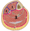
name the muscular compartments located on the tibia

which surface does it correspond with?

A

(1) anterior compartment muscles (on lateral surface)
(3) posterior compartment muscles (on posterior surface)

13
Q

describe the course of the anterior border of the tibia?

A

starts from TIBIAL TUBEROSITY proximally,

extends down anteriorly

forms anterior border of medial malleolus

14
Q

describe origin and course of soleal line

A

starts below articulation of tibia and fibula

descends obliquely towards medial side, joining medial border

15
Q

what inserts ABOVE soleal line?

what originates ON the soleal line?

A

popliteus inserts above soleal line (deep muscle of posterior compartment)

soleus originates ON the soleal line

16
Q

the vertical line of tibia separates which 2 muscle attachments?

A

Tibialis posterior (laterally) and

Flexor digitorum longus (medially)

17
Q

relationship of nutrient foramen and vertical line?

A

nutrient foramen is on posterior surface, LATERAL to the vertical line

18
Q

where does the fibula attach distally?

what type of joint is this?

A

at the fibular notch

forms a syndesmosis

19
Q

shape of articular facet on tibia for the talus?

A

comma shaped

20
Q

from the lateral view, which COLLICULUS extends further distally?

A

the anterior colliculus

21
Q

name the grooves, from medially –> laterally

on the posterior view of the tibia

A
  • medial malleolus
  • groove for TIBIALIS POSTERIOR
  • groove for FLEXOR HALLUCIS LONGUS
  • fibular notch
22
Q

name the inferior surface of the tibia, which articulates w/ dome of the talus

A

the PLAFOND

23
Q

of the plafond, which surface is longer?

(anterior/posterior)

A

Anterior is longer than posterior

24
Q

CC: Trimalleolar fx includes…

A

Posterior malleolus (can incl part of plafond), medial malleolus, and malleolus of talus

25
significance of colliculi of tibia?
• Important attachment sites for ligaments/muscles, like deltoid ligaments of the ankle
26
where is apex of fibula located, relative to tibia?
approx 2 cm inferior; lateral and posterior, on the tibial condyle
27
how to side a fibula?
take distal end; facet is inverted triangle, & malleolar fossa is where you place the thumb. You have the correct side if the thumb is in fossa and does NOT cross over the facet
28
identify bone: name the key landmarks.
fiibula: apex, head, neck, and medial flat portion to articulate w/ tibia
29
name key landmark on distal fibula
lateral malleolus
30
which muscles/ligs attach at: ## Footnote **head of fibula**
* biceps femoris muscle * lateral (fibular) collateral ligament
31
cc: clinical significance of neck of fibula
neck is weak area and is susceptible to fx
32
which nerve "Swoops" around the neck/superior aspect of fibula?
common fibular nerve
33
what are the 3 subcutaneous areas of the fibula?
1. head of fibula 2. triangular area immediately above lateral malleolus 3. lateral malleolus
34
identify/name the borders
1. top left: **anterior** 2. top right: **interosseus CREST** 3. bottomr right: **medial** 4. bottom left**: posterior**
35
identify/name the surfaces
1. top: **anterior/medial surface** 2. right: **posterior medial surface** 3. bottom: **posterior lateral surface** 4. left: **lateral surface**
36
name the compartments
1. anterior 2. posterior 3. lateral
37
identify the bones; name the compartments
right and larger is TIBIA; left and smaller is FIBULA 1. ANTERIOR 2. N/A 3. POSTERIOR 4. LATERAL
38
**COMPARTMENT 1**: identify * muscle category? * action? * pre or postaxial? * anterior or posterior division?
* anterior compartment; * **dorsiflexors/extensors** * post-axial * posterior division
39
**COMPARTMENT 2:** identify muscle category? action? pre or postaxial? anterior or posterior division of ventral rami?
* lateral compartment * evertors/plantar flexors * post-axial * posterior division
40
**COMPARTMENT 3**: identify * muscle category? * action? * pre or postaxial? * anterior or posterior division of ventral rami?
* posterior compartment * flexors/plantar flexors * preaxial * anterior division
41
name the 4 landmarks on the distal fibula
* purple: attachment of **interosseus tibiofibular ligament** * green: **triangular articular facet for talus** * blue: **apex of lateral malleolus** * yellow: **lateral malleolar fossa**
42
what is found behind the lateral malleolar fossa?
malleolar crest a combined tendon of the **fibularis longus and fibularis brevis** runs behind the lateral malleolus
43
**gluteus maximus and tensor fasciae latae via iliotibial tract:** 1. name region/compartment 2. list distal attachment/insertion
1. gluteal region 2. tubercle of IT band/Gerdy's tubercle
44
**quadriceps femoris:** 1. name region/compartment 2. list distal attachment/insertion
1. anterior comparmtent of thigh 2. tibial tuberosity
45
**sartorius:** 1. name region/compartment 2. list distal attachment/insertion
1. anterior compartment of thigh 2. superior part of medial surface of tibia (pes anserinus)
46
**gracilis:** 1. name region/compartment 2. list distal attachment/insertion
1. medial comparetment of thigh 2. superiror part of medial surface of tibia (pes anserinus)
47
**semitendinosus:** 1. name region/compartment 2. list distal attachment/insertion
1. posterior compartment of thigh 2. superior part of medial surface of tibia (pes anserinus)
48
**semimembranosus:** 1. name region/compartment 2. list distal attachment/insertion
1. posterior compartment of thigh 2. posterior part of medial condyle of tibia
49
**biceps femoris:** 1. name region/compartment 2. list distal attachment/insertion
1. posterior compartment of thigh 2. lateral side of head of fibula
50
**tibialis anterior** 1. name region/compartment 2. list distal attachment/insertion
1. anterior compartment of leg 2. lateral condyle and superior half of lateral surface of tibia & interosseus membrane
51
**extensor digitorum longus:** 1. name region/compartment 2. list distal attachment/insertion
1. anterior compartment of leg 2. lateral condyle of tibia, and superior 3/4 of medial surface of fibula and interosseus membrane
52
**extensor hallucis longus:** 1. name region/compartment 2. list distal attachment/insertion
1. anterior compartment of leg 2. middle part of medial surface of fibula & interosseus membrane
53
**fibularis tertius:** 1. name region/compartment 2. list distal attachment/insertion
1. anterior compartment of leg 2. inferior 1/3 of medial surface of fibula and interosseus membrane
54
**fibularis longus:** 1. name region/compartment 2. list distal attachment/insertion
1. lateral compartment of leg 2. head and superior 2/3 of lateral surface of fibula
55
**fibularis brevis:** 1. name region/compartment 2. list distal attachment/insertion
1. lateral compartment 2. inferior 2/3 of lateral surface of fibula
56
**soleus:** 1. name region/compartment 2. list distal attachment/insertion
1. SUPERFICIAL posterior compartment of leg 2. multiple attachments * posterior aspect of head & superior 1/4 of posterior surface of fibula * soleal line & middle 1/3 of medial border of tibia * tendinous arch extending between bone attachments
57
**popliteus:** 1. name region/compartment 2. list distal attachment/insertion
1. DEEP posterior compartment of leg 2. posterior surface of tibia; superior to soleal line
58
**flexor hallucis longus:** 1. name region/compartment 2. list PROXIMAL attachment/insertion
1. DEEP posterior compartment of leg 2. inferior 2/3 of posterior surface of fibula; inferior part of interosseus membrane
59
**tibialis posterior:** 1. name region/compartment 2. list PROXIMAL attachment/insertion
1. deep posterior compartment of leg 2. interosseus membrane; posterior surface of tibia inferior to soleal line; posterior surface of fibula