UW Flashcards
Blow flow per minute difference in pulmonary and systemic circulations
In order to maintain blood flow through the body, the blood flow (mL/min) in the pulmonary circulation must closely match the blood flow in the systemic circulation. This is true for conditions of both exercise and rest as the circulatory system is a continuous circuit. If the flow of blood through the pulmonary circulation is less than the flow of blood through the systemic circulation, the left ventricle would soon empty completely. Alternately, if the flow of blood is significantly greater in the pulmonary circulation than it is in the systemic circulation, the left ventricle would soon be overloaded.
The major exception to this is the bronchial circuit, which supplies oxygen and nutrients to the pulmonary parenchyma from the systemic circulation but drains mostly to the left atrium as opposed to the right atrium (creating a right to left shunt that acts as a partially independent circuit). However, this typically accounts for <5% of the systemic cardiac output.
Anthracyclines
daunorubicin, doxorubicin
Doxorubicin MOA
Binds with topoisomerase II to cleave DNA
Binds with iron to generate free radicals
Doxorubicin Toxicity
Dilated Cardiomyopathy
Bleomycin MOA
Indices free radical formation
Bleomycin Toxicity
Pulmonary fibrosis
Antracycline induced cardiomyopathy Tx
dexrazoxane, a chelating agent thought to both block the formation of iron-associated free radicals and inhibit the formation of anthracycline–topoisomerase II complexes in healthy cardiomyocytes.
Hypertrophic Cardiomyopathy mutation
Hypertrophic cardiomyopathy is an autosomal dominant disorder often caused by mutation of the beta-myosin heavy chain.
Autosomal recessive inheritance with 2 carrier parents

Pierre Robin sequence
In Pierre Robin sequence, hypoplasia of the mandibular prominence leads to micrognathia. Severe micrognathia causes posterior displacement of the tongue (glossoptosis), which blocks fusion of the palatine shelves, resulting in a cleft palate that is characteristically U-shaped. Difficulty breathing occurs because the tongue prolapses into the posterior oropharynx, blocking airflow. Breathing improves when the patient is in a prone position because gravity pulls the tongue anteriorly, opening the airway.

What is a sequence?
A sequence occurs when a single developmental defect causes a cascade of additional malformations.
VACTERL association
An association is a collection of malformations that are often seen together and do not have a known, common cause
V: vertebral anomaly
A: anorectal malformation
C: cardiovascular anomaly
T: tracheoesophageal fistula
E: esophageal atresia
R: renal &/or radial anomaly
L: limb defect
Disruption description
A disruption occurs when an external insult (rather than a genetic defect) interrupts and arrests normal fetal development (eg, when an amniotic band disrupts limb development)
Examples of Genomic imprinting and description
Genomic imprinting is a normal process that refers to selective activation of gene expression depending on the parent of origin. Aberrant imprinting occurs with uniparental disomy (ie, when a person receives 2 copies of a chromosome from the same parent and no copy from the other parent). Prader-Willi syndrome and Angelman syndrome (15q) are examples of conditions caused by dysfunctional imprinting.
What is a syndrome?
A syndrome is a collection of malformations that have a common cause but are not related anatomically. For example, upslanting palpebral fissures, atrioventricular canal defect, and single palmar crease are all due to trisomy 21.
Platelet plug formation mechanism
The formation of a platelet plug (primary hemostasis) is essential for preventing bleeding after damage to vascular endothelium; it occurs in 3 steps:
Platelet adhesion takes place via von Willebrand factor acting as a connector that binds platelets to underlying collagen
Platelets become activated and secrete multiple substances, including adenosine diphosphate, ionized calcium, and fibrinogen, from their alpha and delta (dense) granules.
Thromboxane A2 (Choice F) is released and acts as a vasoconstrictor and potent stimulator of platelet aggregation. Adenosine diphosphate also stimulates platelet aggregation.
Balance is required as excessive platelet plug formation can lead to a pathologic thrombus that restricts blood flow (eg, myocardial infarction). To oppose the functions of thromboxane A2, the endothelium secretes prostacyclin (prostaglandin I2), which is derived from arachidonic acid and synthesized from prostaglandin H2 by prostacyclin synthase. Once secreted, prostacyclin acts locally to inhibit platelet aggregation and adhesion to the vascular endothelium and to cause vasodilation. Nitric oxide aids in these functions as well. Atherosclerosis can impair the ability of endothelial cells to synthesize prostacyclin and nitric oxide, creating localized predisposition to excessive platelet thrombus formation.
A synthetic prostacyclin, epoprostenol, is used in the treatment of pulmonary hypertension, peripheral vascular disease, and Raynaud syndrome.

Epoprostenol MOA
Synthetic prostacyclin used to Tx Raynaud syndrome, Peripheral vascular disease and pulmonary hypertensions
NSAID induced acute kidney injury
NSAIDs inhibit COX and prevent the formation of prostaglandins (prostanoids) which reduce afferent arteriole vasodilation

Nonsteroidal anti-inflammatory drugs (NSAIDs) (eg, ibuprofen, aspirin, naproxen) exert their anti-inflammatory, analgesic, and antipyretic effects through the inhibition of the cyclooxygenase enzymes. These enzymes are the rate-limiting step in the formation of prostanoids (ie, prostaglandins, thromboxane), which are involved in mediating pain and inflammation.
Prostaglandins also help maintain renal perfusion by dilating the afferent arteriole, particularly in patients with intravascular volume depletion (eg, congestive heart failure, diarrhea, excessive diuresis) or chronic kidney disease. In such patients, increased prostaglandin synthesis is necessary to preserve renal blood flow and maintain glomerular filtration rate. In at-risk patients, inhibition of afferent dilation with NSAIDs results in reduced glomerular filtration and prerenal azotemia with elevations in creatinine and blood urea nitrogen (ratio >20:1).
NSAID-induced acute kidney injury is often diagnosed incidentally on laboratory tests performed for other reasons, and patients are generally asymptomatic. Urinalysis is typically bland without proteinuria, hematuria, or casts. Prolonged NSAID use can cause chronic kidney disease (analgesic nephropathy) due to papillary necrosis and chronic interstitial nephritis.
NSAIDs are a common cause of acute interstitial nephritis (AIN). However, urinalysis in AIN typically demonstrates white blood cells and white blood cell casts, and patients commonly develop fevers and rash.
Absent or deficient CD 18 causes
Leukocyte Adhesion Deficiency
Leukocyte Adhesion Deficiency pathophysiology
LAD is an autosomal recessive disorder characterized by the absence of CD18 antigens, which are necessary for the formation of integrins. Integrins are essential for leukocyte adhesion to endothelial surfaces and migration into peripheral tissues in response to infection or inflammation.
The failure of leukocyte chemotaxis results in characteristic LAD findings, including recurrent skin and mucosal infections (often due to Staphylococcus aureus or gram-negative rods) and periodontal disease. LAD-related infections are notable for lack of purulence because of the absence of leukocytes in peripheral tissues. Wound healing is also impaired, which can result in late umbilical cord separation (age >3 weeks).
Peripheral leukocytosis and neutrophilia are typical, particularly during active infection, because leukocytes cannot migrate out of the blood vessels
Leukocyte Adhesion Deficiency Clinical Features
Skin & mucosal infections (eg, cellulitis, periodontitis) without pus formation (non-purulent)
Impaired wound healing
Delayed umbilical cord separation (age >3 weeks)
Leukocyte Adhesion Deficiency Lab findings
Leukocytosis & neutrophilia because they cannot migrate out of the blood vessels to tissues
A 3-year-old boy is brought to the office due to abnormal motor development. He was born at 40 weeks gestation and had an unremarkable perinatal course. The boy developed normally during the first year of life. However, for the past 2 years, he has had progressive bilateral leg stiffness and abnormal involuntary movements. His cognitive and motor development is also delayed. There is no significant family history of neurological or muscular disorders. The patient’s height, weight, and head circumference are below the 3rd percentile. Examination shows bilateral spastic paresis of his lower extremities and frequent choreoathetoid movements. Comprehensive laboratory testing reveals significantly elevated arginine levels in plasma and cerebrospinal fluid. The deficient enzyme in this patient is normally involved in the production of which of the following?
Urea
Urea Cycle

Arginase Deficiency pathophysiology
features of arginase deficiency, including progressive development of spastic diplegia, abnormal movements, and growth delay in the setting of elevated arginine levels. Arginase is a urea cycle enzyme that produces urea and ornithine from arginine. Diagnosis is based on elevated arginine levels on plasma amino acid testing.
low-protein, arginine-restricted diet supplemented by essential amino acid Administration of a synthetic protein made of essential amino acids usually results in a dramatic decrease in plasma arginine concentration and an improvement in neurological abnormalities. Unlike other urea cycle disorders, patients with arginase deficiency have mild or no hyperammonemia.
Arginase Deficiency Tx
Treatment of arginase deficiency consists of a low-protein diet devoid of arginine.
low-protein, arginine-restricted diet supplemented by essential amino acid
Features of Nocardia
Gram-positive rod (beaded or branching)
Partially acid-fast
Aerobic

Mechanism of dietary iron absorption in gastrojejunostomy
Iron absorption occurs predominantly in the duodenum and proximal jejunum, and bypass of this segment of small bowel results in iron deficiency anemia. The post-surgical decrease in gastric acidity also diminishes iron absorption and may contribute to iron deficiency in these patients. Treatment is accomplished with pharmacologic iron supplementation, which allows for adequate iron absorption at secondary absorption sites in the distal small bowel.
Deficiency involving thiamine, folate, vitamin B12, fat-soluble vitamins (especially vitamin D), and calcium is also common following gastrojejunostomy.
Gastrojejunostomy implications
Partial gastrectomy with gastrojejunostomy is most often performed to treat complicated peptic ulcer disease (eg, perforation, malignancy, gastric outlet obstruction) or ulcers refractory to medical management. In a Billroth II gastrojejunostomy, the gastric antrum is removed to decrease gastrin production and for histopathologic evaluation. A side-to-side anastomosis is then made between the jejunum and the gastric body, bypassing the duodenum and proximal jejunum.
Iron absorption occurs predominantly in the duodenum and proximal jejunum, and bypass of this segment of small bowel results in iron deficiency anemia. The post-surgical decrease in gastric acidity also diminishes iron absorption and may contribute to iron deficiency in these patients. Treatment is accomplished with pharmacologic iron supplementation, which allows for adequate iron absorption at secondary absorption sites in the distal small bowel.
Deficiency involving thiamine, folate, vitamin B12, fat-soluble vitamins (especially vitamin D), and calcium is also common following gastrojejunostomy.

Carbon tetrachloride
Carbon tetrachloride (CCl4) causes free radical injury. Like many other toxic substances, CCl4 is oxidized by the P450 oxidase system in the liver. The result is the formation of the free radical CCl3, which reacts with structural lipids of cell membranes. The result is lipid degradation and hydrogen peroxide (H2O2) formation. This process is called lipid peroxidation. The peroxides go on to form new radicals, continuing the vicious circle of lipid degradation. Carbon tetrachloride cell injury develops rapidly and leads to swelling of the endoplasmic reticulum, destruction of mitochondria, and increased permeability of cell membranes. These processes culminate in hepatocyte necrosis.
Short acting Benzodiazepines
Triazolam, midazolam
Intermediate benzodiazepines
Oxazepam, alprazolam, lorazepam, clonazepam
Long-acting benzodiazepines
Diazepam, chlordiazepoxide, flurazepam
bile acid metabolism and reabsorption
terminal ileum
Bile acid implications of Crohn’s Disease
The terminal ileum is frequently involved in Crohn disease. Bile acids, which are necessary for the absorption of fat and other nutrients, are normally reabsorbed in the terminal ileum, recycled in the liver, and then reused in the absorptive process. When the terminal ileum is inflamed or resected, bile acids are lost with feces. Loss of bile acids causes fat malabsorption, which may lead to deficiencies in fat-soluble vitamins (A, D, E, K).
Vitamin K is a cofactor for several carboxylase enzymes that are necessary for coagulation factor II, VII, IX, and X activation. Coagulation disorders such as vitamin K deficiency typically result in easy bruising, large hematoma formation in deep tissues and joints (eg, hemarthrosis) after minor trauma, and prolonged bleeding after surgery.

Post MI ACE inhibition
This patient has a reduced left ventricular (LV) ejection fraction (<45%) due to acute myocardial infarction; echocardiography shows akinesia of the anterior and apical segments, findings consistent with infarction in those locations. In the months following infarction, the affected myocardium undergoes fibroblastic proliferation and fibrosis to repair the necrotic region (ie, initial remodeling phase); disproportionate thinning of the infarcted area can lead to ballooning of affected segments and marked LV cavity enlargement. Although LV cavity enlargement is initially beneficial as it acts as a compensatory mechanism for LV volume overload, progressive cavity enlargement (eg, eccentric hypertrophy) leads to overwhelming wall stress and further worsening of LV contractile dysfunction. With progressive dilation, the LV begins to assume a spherical, rather than its normal elliptical, shape.
Much of the deleterious remodeling that occurs following myocardial infarction is likely driven by neurohormonal signaling via angiotensin II. Accordingly, ACE inhibitors (eg, lisinopril) reduce the deleterious remodeling that takes place following myocardial infarction, minimizing LV dilation and helping preserve LV function.

What is this?

Neurocysticercosis

Neurocysticercosis histology
invaginated scolex, the anterior portion of a tapeworm. The scolex has refractile hooklets and suckers, which allow attachment to host tissue. These findings are consistent with neurocysticercosis (NCC).

Meningioma imaging aND histology
imaging reveals an extraaxial, dural-based mass that compresses brain tissue. In addition, histopathology often shows whorls of uniform, oval tumor cells with eosinophilic cytoplasm, as well as psammoma bodies.

Melanoma brain metastases
Brain metastases (eg, melanoma) often cause seizures due to focal brain edema. However, imaging usually reveals multiple masses with surrounding edema at the gray-white matter junction,

Toxoplasma brain imaging and histology
toxoplasmosis appears as ring-enhancing lesions on imaging, and histology shows spherical cysts and tachyzoites

Amebic encephalitis brain imaging and histology
Amoebic encephalitis typically appears as nonspecific brain edema on imaging, and histology shows trophozoites and cysts.

HMB-45
HMB-45 stain binds premelanosome protein
Melanoma

Tuberculosis histology
caseating granulomas with acid-fast bacilli.

Describe the Renin angiotensin aldosterone system and antihypertensives

Cervicitis
Acute cervicitis classically presents with purulent cervical discharge and a friable cervix that bleeds easily with contact (ie, postcoital bleeding). Microscopy showing inflammation (eg, neutrophils) without a visible pathogen is classic for cervicitis due to Chlamydia trachomatis or Neisseria gonorrhoeae, which are often identified by nucleic acid amplification testing.
Left untreated, cervicitis can compromise the endocervical barrier, allowing polymicrobial vaginal flora to ascend into the normally sterile uterus and fallopian tubes. In contrast to cervicitis, infection involving the upper genital tract (ie, pelvic inflammatory disease [PID]) can cause fever, abdominal pain, and cervical motion/uterine/adnexal tenderness due to increased bacterial load and spread.
When the infection extends to the fallopian tube, it creates an inflammatory exudate (ie, salpingitis) that causes the tubal walls to adhere. With prolonged infection, the exudate may be replaced by scar tissue, causing permanent tubal scarring and obstruction that impedes the fertilization and/or implantation of future pregnancies. Therefore, common complications of PID include tubal factor infertility and ectopic pregnancy.

HIV viral entry into cells mechanism
HIV attaches to host cells using the viral surface glycoprotein gp120. This glycoprotein binds to the CD4 molecule as the primary receptor and the chemokine receptor CCR5 (or CXCR4) as a coreceptor. Binding of the primary receptor and coreceptor induces a conformational change in gp120 that exposes the underlying transmembrane glycoprotein gp41, which mediates viral fusion to the host cell and release of the viral capsid into the cytoplasm.

Researchers develop a novel drug to treat HIV infection. In an in vitro experiment, wild-type viral isolates are cultured with human CD4 T lymphocytes in the presence of the drug. Microscopic evaluation of these cells reveals no cytopathic changes, and no intracellular viral particles can be detected. Further analysis reveals that in the presence of the drug the virus attaches to the cellular membrane, but fusion of the viral and cell membranes does not occur. Which of the following is the most likely target of this medication?
The drug described in this case does not interfere with viral attachment but does interfere with viral fusion. This implies that gp120 is still able to bind to the primary CD4 receptor but may be blocked from binding to the chemokine coreceptor, which would prevent the conformational change to gp120 required for viral fusion. Chemokine receptor antagonists (eg, maraviroc) block this step in the HIV replication cycle. The drug described in this case could also be a fusion inhibitor (eg, enfuvirtide), which blocks gp41 from fusing the viral and host plasma membranes.


ARR = control rate - treatment rate
25/1000 - 10/1000 = 15/1000
NNT = 1/ARR
1/(15/1000) = 66.67


PAthogenesis of left-sided heart failure -> pulmonary hypertension
This patient with dyspnea, orthopnea, and pulmonary crackles has left-sided heart failure (LHF) most likely due to long-standing, poorly controlled hypertension. Hypertensive heart disease typically manifests as heart failure due to concentric left ventricular (LV) hypertrophy and consequent LV diastolic dysfunction. LHF of any cause (eg, LV systolic dysfunction, valvular dysfunction) will result in higher diastolic filling pressures. This increase in pressure is transmitted backward to the left atrium and pulmonary veins, resulting in pulmonary venous congestion and consequent elevations in pulmonary capillary and pulmonary arterial pressure. The resulting pulmonary hypertension (PH) can lead to right-sided heart failure with jugular venous distension and peripheral edema.
Over time, remodeling of the pulmonary vasculature occurs with increased smooth muscle cell proliferation (medial hypertrophy) and collagen deposition (intimal thickening and fibrosis). The remodeling is less extensive than in (primary) pulmonary arterial hypertension; therefore, the PH is at least partially reversible with treatment of the LHF.

Question

Mucociliary clearance is responsible for removing the vast majority of inhaled particles that lodge within the bronchial tree. Ciliated mucosal epithelium lines the pulmonary airways from the trachea to the proximal portions of the respiratory bronchioles. Mucus and fluid secreted onto this epithelial surface act to trap particles suspended in the inspired air. The trapped particles are constantly swept upward from the bronchioles toward the pharynx by cilia that collectively beat in the direction of the pharynx. The mucus and debris are then swallowed or expectorated upon reaching the pharynx.
The terminal bronchioles are covered by ciliated cuboidal epithelium and club cells, which help with mucociliary clearance in this region.

Histology of bronchial mucosa

Cardiac Auscultatory locations

Aortic Area Ausculation location and findings
Right upper sternal border Aortic regurg and aortic stenosis

Pulmonic Area Ausculation location and findings
Left upper sternal border
pulmonic stenosis and ASD

Left mid sternal border Ausculation location and findings
Pulmonic regurg, aortic regurg, HCM

question

This pediatric patient with nephritic syndrome (eg, periorbital edema, hematuria, hypertension) following a recent skin infection most likely has poststreptococcal glomerulonephritis (PSGN). PSGN is the most common cause of nephritic syndrome in children and typically occurs 2-4 weeks after exposure to group A beta-hemolytic Streptococcus (eg, pharyngitis, skin infection). Antigens expressed on nephritogenic streptococcal species combine with antibodies to form immune complexes, which are deposited on the glomerular basement membrane (GBM) and induce complement activation and inflammation.
These immune complexes are visible on immunofluorescence microscopy as granular deposits of IgG, IgM, and C3 on the GBM and mesangium, producing a “starry sky” appearance. Electron microscopy can show the immune deposits as discrete, electron-dense, subepithelial humps on the GBM. The classic light microscopy finding in PSGN is enlarged, diffusely hypercellular glomeruli due to leukocyte infiltration (neutrophils and monocytes) and mesangial and endothelial cell proliferation.
Laboratory studies show decreased serum complement (eg, C3) due to consumption, and elevated titers of streptococcal antibodies (eg, anti-DNAse B, antihyaluronidase, antistreptolysin O [ASO, which is typically elevated with pharyngitis but often undetectable after skin infections]).

Immunofluoresence patterns in the glomerulus

question


question

B

This patient most likely has Broca (motor, nonfluent) aphasia. This condition classically results from damage to Broca area of the brain, the region responsible for all communicative motor planning. Individuals are able to communicate meaningfully, but their speech is slow and consists primarily of nouns and verbs. Speech may be punctuated by pauses after each word as the patient attempts to verbalize the next. Patients also often have difficulty writing and signing, and become frustrated as they have insight into their expressive language difficulties. They can understand spoken language and follow commands (intact speech comprehension) as Wernicke area is unaffected.
Broca area is in the caudal part of the inferior frontal gyrus of the dominant (usually left) hemisphere (Brodmann areas 44 and 45). Patients may have associated right hemiparesis involving the upper limb and face due to this region’s proximity to the primary motor cortex.
What is A
frontal eye fields

What is C

Lesions to the precentral gyrus (primary motor cortex) can cause slurred speech (dysarthria) due to paresis/paralysis of the skeletal muscles involved in movements of the mouth, tongue, and larynx.
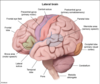
What is D?

This is the postcentral gyrus (primary somatosensory cortex). A lesion here would cause sensation loss in the corresponding area of the contralateral body.

What is E?

The caudal superior temporal gyrus (Brodmann area 22) is in the Wernicke area. A lesion here would cause a sensory (receptive) aphasia. Wernicke aphasia is also known as fluent aphasia in that speech flows readily but is meaningless (“word salad”). Patients typically lack insight into their problem

What is F?

A lesion to the visual cortex can cause cortical blindness or visual impairment that can make reading and writing difficult.

question

Water deprivation results in antidiuretic hormone (ADH) release from the posterior pituitary gland. This hormone stimulates V2 receptors on principal cells in the renal collecting ducts, causing translocation of aquaporin 2 channels into the apical cell membrane. Aquaporin 2 is a water channel that spans the luminal membrane, enhancing the water permeability of the principal cells. In the presence of high ADH, the tubular fluid osmolarity follows this pattern:
In the proximal tubule, water is reabsorbed along with electrolytes. The tubular fluid in this segment remains isotonic with plasma (300 mOsm/L) whether the final urine is concentrated or diluted (Choice A).
In the descending limb of the loop of Henle, free water is drawn out of the tubules into the renal interstitium and the tubular fluid becomes hypertonic (> 300 mOsm/L, typically reaching 1200 mOsm/L when ADH levels are high) (Choice B).
The thick and thin ascending limbs of the loop of Henle are the primary region of urine dilution. These regions are impermeable to water; electrolytes such as NaCl are passively reabsorbed in the thin ascending limb (Choice C) and actively reabsorbed in the thick ascending limb. The tubular fluid becomes increasing hypotonic (< 300 mOsm/L) within this region.
The distal convoluted tubule is relatively impermeable to water, so the tubular fluid remains hypotonic. Reabsorption of solutes continues to occur; thus, fluid in the distal tubules is the most dilute (lowest osmolarity, approaching 100 mOsm/L).
In the presence of ADH, the collecting duct is highly permeable to water. Water leaves the tubular fluid driven by the high osmolarity of the medullary interstitium, and hypertonic urine is formed (up to 1200 mOsm/L). The collecting duct system is the primary region of urine concentration through the mechanism of ADH-mediated water absorption.

Question


Stress-related mucosal Disease
Stress-related mucosal disease is characterized by acute gastric mucosal defects that develop in response to severe physiologic stress (eg, shock, extensive burns, sepsis, severe trauma, intracranial injury). Patients often have multiple, small (<1 cm), circular lesions in the stomach, ranging from superficial erosions to full-thickness ulcers. Ulcers may perforate or bleed, as in this patient.
The pathogenesis of these ulcers most often involves impaired mucosal protection due to local ischemia caused by systemic hypotension and splanchnic vasoconstriction. Ulcers arising in the proximal duodenum in association with severe trauma/burns are called Curling ulcers. Ulcers arising in the esophagus, stomach, or duodenum in patients with intracranial injury are particularly prone to perforation and are called Cushing ulcers. Cushing ulcers are a consequence of direct vagus nerve stimulation caused by elevated intracranial pressure, resulting in acetylcholine release and hypersecretion of gastric acid.

Question

This case is typical of hereditary nonpolyposis colon cancer (HNPCC), or Lynch syndrome, an autosomal dominant genetic predisposition to colon cancer. In patients with HNPCC, colon cancer occurs at a young age (age <50). Family history reveals a high incidence of colon cancer and, occasionally, extraintestinal (eg, endometrial) cancers in first-degree relatives.
With HNPCC, there is an inherited mutation in one of the genes responsible for DNA mismatch repair (eg, MSH2, MLH1). The products of these genes proofread DNA during replication. Patients with HNPCC inherit a mutation in an allele, and mutation of the second allele occurs during adult life. When 2 dysfunctional copies are present, malignancies will develop readily.

Question

This patient has symptoms consistent with an obstructed right brachiocephalic (innominate) vein. This may be the result of external compression by an apical lung tumor or thrombotic occlusion as can occur when a central catheter has been in place for an extended period. The right brachiocephalic vein is formed by the union of the right subclavian vein and the right internal jugular vein. The right external jugular vein drains into the right subclavian vein, so obstruction of the right brachiocephalic vein will also cause venous congestion of structures drained by the external jugular vein. It is important to note that the right brachiocephalic vein also drains the right lymphatic duct, which drains lymph from the right upper extremity, the right face and neck, the right hemithorax, and the right upper quadrant of the abdomen.

Right sided brachiocephalic vein obstruction
This patient has symptoms consistent with an obstructed right brachiocephalic (innominate) vein. This may be the result of external compression by an apical lung tumor or thrombotic occlusion as can occur when a central catheter has been in place for an extended period. The right brachiocephalic vein is formed by the union of the right subclavian vein and the right internal jugular vein. The right external jugular vein drains into the right subclavian vein, so obstruction of the right brachiocephalic vein will also cause venous congestion of structures drained by the external jugular vein. It is important to note that the right brachiocephalic vein also drains the right lymphatic duct, which drains lymph from the right upper extremity, the right face and neck, the right hemithorax, and the right upper quadrant of the abdomen.

question

his patient has inclusion cell (I-cell) disease, an autosomal recessive lysosomal storage disorder. I-cell disease occurs due to defects in protein targeting, a process by which proteins are transported to their appropriate intra- or extracellular location.
Normally, posttranslational modifications (eg, folding, glycosylation, phosphorylation) function as markers that help guide proteins to their final destination. For lysosome-bound proteins (ie, acid hydrolases), a Golgi body phosphotransferase enzyme catalyzes the phosphorylation of mannose residues on the proteins. Once tagged with mannose-6-phosphate, these proteins traverse the Golgi network and are ultimately transported to the lysosome, where they serve as catalysts for degradation of cellular components.
In I-cell disease, a defective phosphotransferase enzyme results in deficient phosphorylation (ie, incorrect targeting) of mannose residues on acid hydrolases, which are then inappropriately secreted to the extracellular space. This leads to lack of degradation of lysosomal cellular debris, which accumulates within the lysosome, forming the characteristic inclusion bodies. Patients with this disorder typically have failure to thrive, respiratory tract infections, and cognitive deficits in the first year of life, along with characteristic physical features (eg, coarse facies, corneal clouding, hepatosplenomegaly).

I-Cell Disease
this patient has inclusion cell (I-cell) disease, an autosomal recessive lysosomal storage disorder. I-cell disease occurs due to defects in protein targeting, a process by which proteins are transported to their appropriate intra- or extracellular location.
Normally, posttranslational modifications (eg, folding, glycosylation, phosphorylation) function as markers that help guide proteins to their final destination. For lysosome-bound proteins (ie, acid hydrolases), a Golgi body phosphotransferase enzyme catalyzes the phosphorylation of mannose residues on the proteins. Once tagged with mannose-6-phosphate, these proteins traverse the Golgi network and are ultimately transported to the lysosome, where they serve as catalysts for degradation of cellular components.
In I-cell disease, a defective phosphotransferase enzyme results in deficient phosphorylation (ie, incorrect targeting) of mannose residues on acid hydrolases, which are then inappropriately secreted to the extracellular space. This leads to lack of degradation of lysosomal cellular debris, which accumulates within the lysosome, forming the characteristic inclusion bodies. Patients with this disorder typically have failure to thrive, respiratory tract infections, and cognitive deficits in the first year of life, along with characteristic physical features (eg, coarse facies, corneal clouding, hepatosplenomegaly).

Question

This scenario describes a typical case-control study design. People with the disease of interest (ie, cases [women who have just delivered babies with neural tube defects]) and people without this disease (ie, controls [women who delivered apparently healthy babies]) are asked about previous exposure to the risk factor being studied (eg, acetaminophen use during the first 3 months of pregnancy). The main measure of association is the odds ratio (OR). The OR can be calculated as follows:
OR = (odds of exposure in cases) / (odds of exposure in controls)
