Neurological disorders Flashcards
Question

B. describing simularities between watch and ruler
The frontal lobes are involved in a wide array of functions, including personality, language, motor functions, and executive functions. Executive functions include abstraction, planning, impulse inhibition, attention, and working memory. Abstraction may be tested by asking a patient to describe the similarities between two objects (eg, a watch and ruler both measure things). Patients with frontal lobe damage may be unable to provide an answer or may provide concrete answers such as “they both have numbers.”
Because of its role in personality and impulse inhibition, processes that damage the frontal lobe (eg, traumatic brain injury, frontotemporal dementia) may lead to significant changes in behavior. Behavior change is more likely with bilateral, rather than unilateral, injury. Patients with frontal lobe injury may be disinhibited, socially inappropriate, irritable, and impulsive.
(Choice A) Impaired language comprehension (ie, Wernicke aphasia) results from lesions in the dominant superior temporal gyrus.
(Choices C and D) Patients with lesions in the dominant angular and supramarginal gyri (located in the parietal lobe) may have right-left disorientation and finger agnosia (ie, inability to recognize fingers). When these symptoms are accompanied by agraphia (ie, inability to write) and acalculia (ie, inability to perform arithmetic), it is known as Gerstmann syndrome.
(Choice E) The inability to recognize a familiar face (ie, prosopagnosia) may result from lesions in the fusiform gyrus (located in the temporal and occipital lobes).
Educational objective:
The frontal lobes are involved in personality, language, motor functions, and executive functions (eg, abstraction). Frontal lobe function may be diagnosed by testing abstraction ability (eg, asking about the similarities between two related objects) on the mental status examination.
Question

C. fluoxetine
Question

F. Tuberoinfundibular pathway

This patient likely has developed amenorrhea and galactorrhea as an adverse effect of dopamine-2 (D2) receptor blockade from treatment with antipsychotics. There are 4 major dopaminergic pathways in the brain (eg, mesolimbic, mesocortical, tuberoinfundibular, nigrostriatal); dopamine hyperactivity in the mesolimbic pathway is associated with positive psychotic symptoms (eg hallucinations, delusions).
The side effects of antipsychotic therapy are largely caused by D2 receptor blockade in other dopaminergic pathways. The tuberoinfundibular pathway connects the hypothalamus to the pituitary gland and is responsible for the tonic inhibition of prolactin secretion. Neurons in the arcuate nucleus of the hypothalamus secrete dopamine, which binds to D2 receptors on pituitary lactotrophs, resulting in decreased prolactin secretion from the anterior pituitary gland. Antipsychotics can interrupt the tuberoinfundibular pathway, causing increased blood prolactin levels (hyperprolactinemia), which may lead to galactorrhea (milky nipple discharge unrelated to pregnancy/breastfeeding) and menstrual irregularities (eg, amenorrhea).
(Choice A) The arcuate fasciculus is a neural pathway that connects the Broca and Wernicke areas, which are responsible for expressive and receptive language, respectively. Disruption of the arcuate fasciculus classically results in conduction aphasia, characterized by fluent speech, intact comprehension, and impaired repetition.
(Choice B) The hypothalamospinal tract projects from the hypothalamus to the ciliospinal center of the intermediolateral cell column (T1-L2), providing sympathetic innervation to the ipsilateral eye and face. Disruption of this tract typically results in ipsilateral Horner syndrome (eg, ptosis, miosis, anhidrosis).
(Choice C) The lateral medullary spinothalamic tract transmits pain and temperature signals from the contralateral body to the thalamus. Lateral medullary infarction (Wallenberg syndrome) presents with loss of pain and temperature sensation on the contralateral body and ipsilateral face as well as vertigo, hoarseness, dysphagia, and abnormal eye movements.
(Choice D) The therapeutic effect of antipsychotics arises from blockade D2 receptors in the mesolimbic pathway.
(Choice E) The nigrostriatal pathway projects from the substantia nigra to the caudate nucleus and putamen and primarily regulates the coordination of voluntary movements. D2 receptor blockade in this pathway results in extrapyramidal effects (eg, dystonia, akathisia, tardive dyskinesia) and drug-induced parkinsonism.
Educational objective:
Antipsychotic medications work by blocking dopamine-2 receptors in the mesolimbic dopamine pathway. Dopamine-2 receptor blockade in the tuberoinfundibular pathway can result in galactorrhea and amenorrhea.
Question

B. Increased stiffness of the LV wall
This patient has an S4 (low-frequency sound occurring just before S1), which is most likely secondary to concentric left ventricular hypertrophy (LVH) from longstanding hypertension (hypertensive heart disease). The calcifications revealed on chest x-ray likely represent degenerative mitral and aortic valve calcification, which is age related and typically accelerated by systemic hypertension.
An S4 is a sign of diastolic dysfunction. It occurs due to blood striking a stiffened ventricular wall during atrial contraction at the end of diastole. An S4 may be present in any condition that causes reduced ventricular compliance (eg, hypertensive heart disease, restrictive cardiomyopathy, acute myocardial infarction). A left-sided S4 is best heard with the bell of the stethoscope over the cardiac apex with the patient in the left lateral decubitus position; it will intensify during expiration due to increased blood flow from the lungs to the left atrium.
(Choices A and D) Age-related aortic valve calcification can cause restricted motion of the aortic valve cusps and lead to aortic stenosis. There may be a high-frequency ejection click heard just after S1, followed by the crescendo-decrescendo systolic murmur of aortic stenosis created by increased flow velocity through the aortic valve (and best heard at the right upper sternal border). Aortic stenosis is also a potential cause of concentric LVH and an S4, but it is a less common cause than chronic hypertension.
(Choice C) An S3 is caused by blood filling an enlarged (or overfull) ventricular cavity during early passive diastolic filling. It occurs shortly after S2 and is often heard in patients with heart failure with reduced ejection fraction or other causes of ventricular volume overload (eg, mitral or aortic regurgitation).
(Choice E) Systolic anterior motion of the mitral valve can exacerbate left ventricular outflow tract obstruction in patients with hypertrophic cardiomyopathy (HCM). HCM is expected to cause a systolic ejection murmur best heard at the left sternal border. An S4 is also sometimes heard with HCM.
Educational objective:
An S4 is a low-frequency, late diastolic sound that immediately precedes S1. It is caused by blood striking a stiffened ventricular wall during atrial contraction; concentric left ventricular hypertrophy due to chronic hypertension (or less commonly due to aortic stenosis) is a common cause of an S4.
Question

E. Tangle of large blood vessels with thickened walls
This patient with recurrent headaches and seizures has multiple flow voids in the left temporoparietal lobe demonstrated by MRI, consistent with a cerebral arteriovenous malformation (AVM).
AVMs are vascular malformations in which blood courses directly from arteries to veins, without passing through an intervening capillary bed. These tangled vessels with turbulent flow can lead to aneurysm development, bleeding, and damage to the surrounding brain. Microscopy of AVMs shows abnormal vessels, including irregular arteries and large veins with thickened walls, and gliotic brain tissue.
AVMs frequently present in children and young adults with headaches and seizures as well as intracranial hemorrhage (eg, intraparenchymal, subarachnoid hemorrhage). Some patients are initially asymptomatic and diagnosed incidentally by imaging. A nest of abnormal vessels with “bag of worms” appearance may be seen on angiography. MRI may reveal prior hemorrhage and multiple dark flow voids, indicative of swift blood flow.
(Choice A) Dense perivascular aggregates of atypical lymphoid cells are a histologic feature of primary CNS lymphoma, which is typically a diffuse large B-cell lymphoma. Although clinical symptoms may include seizures, it usually presents in older adults or immunocompromised patients. MRI would likely reveal an enhancing mass.
(Choice B) Lipid-laden macrophages with phagocytized myelin debris are seen in the demyelinating plaques of multiple sclerosis. Symptoms are variable but commonly include unilateral visual disturbance (eg, optic neuritis) and focal sensory and motor deficits. MRI typically reveals periventricular white matter lesions.
(Choice C) Histologic findings in cerebral Toxoplasma gondii infection include necrotic debris with surrounding inflammation, tachyzoites, and pseudocysts of bradyzoites. Although seizures and headaches are a frequent manifestation, CNS toxoplasmosis usually occurs in immunocompromised patients. Imaging would likely reveal ring-enhancing lesions.
(Choice D) Necrosis surrounded by malignant cells (pseudopalisading necrosis) and microvascular proliferation are features of glioblastoma, a high-grade infiltrating astrocytoma. It commonly presents with seizures, headache, and neurologic deficits in middle-aged or older adults (rather than children). MRI would likely reveal a mass with central hemorrhagic necrosis, vasogenic edema, and possible extension to the contralateral hemisphere.
Educational objective:
Cerebral arteriovenous malformations (AVMs) are vascular malformations histologically characterized by a tangle of abnormal vessels, including large veins with thickened walls and irregular arteries. AVMs may present with intracranial hemorrhage, headache, and/or seizures. Radiologic findings include abnormal vasculature with a “bag of worms” appearance and multiple dark flow voids.

Whats this?

multiple flow voids in the left temporoparietal lobe demonstrated by MRI, consistent with a cerebral arteriovenous malformation (AVM).
AVMs are vascular malformations in which blood courses directly from arteries to veins, without passing through an intervening capillary bed. These tangled vessels with turbulent flow can lead to aneurysm development, bleeding, and damage to the surrounding brain. Microscopy of AVMs shows abnormal vessels, including irregular arteries and large veins with thickened walls, and gliotic brain tissue.
AVMs frequently present in children and young adults with headaches and seizures as well as intracranial hemorrhage (eg, intraparenchymal, subarachnoid hemorrhage). Some patients are initially asymptomatic and diagnosed incidentally by imaging. A nest of abnormal vessels with “bag of worms” appearance may be seen on angiography. MRI may reveal prior hemorrhage and multiple dark flow voids, indicative of swift blood flow.

AVM histology

Cerebral toxoplasmosis histology

Glioblastoma histology

Glioblastoma radiology

Question

C. Delta Waves
This girl’s nighttime ambulation associated with incomplete arousal and amnesia for the event is consistent with sleepwalking, a non-REM (NREM) parasomnia. Sleepwalking is more common in children, peaks at age 8-12, and often remits spontaneously. NREM-related parasomnias such as sleepwalking and sleep terrors occur during deep, slow-wave sleep. Deep, slow-wave sleep is more prominent during the first half of the night and, as a result, NREM-related parasomnias occur more commonly during this time.
Sleep is divided into NREM and REM sleep. Most dreams occur during REM sleep. NREM sleep is further divided into 3 substages based on depth of sleep (N1, N2, and N3). Brain waves of different frequencies are associated with different stages of sleep. Delta waves (frequency up to 3.99 Hz) are characteristic of deep, slow-wave sleep (stage N3). Alpha and beta waves are characteristic of wakefulness and REM sleep (Choices A and B). K complexes and Sleep spindles are characteristic of stage N2 (light, stable sleep) (Choices D and E).
Educational objective:
Sleepwalking, a common non-REM parasomnia of childhood, occurs during slow-wave sleep (stage N3), which is characterized by delta waves on EEG. Sleepwalking typically occurs during the first half of the night, when slow-wave sleep is most prominent.
Sleep stages
This girl’s nighttime ambulation associated with incomplete arousal and amnesia for the event is consistent with sleepwalking, a non-REM (NREM) parasomnia. Sleepwalking is more common in children, peaks at age 8-12, and often remits spontaneously. NREM-related parasomnias such as sleepwalking and sleep terrors occur during deep, slow-wave sleep. Deep, slow-wave sleep is more prominent during the first half of the night and, as a result, NREM-related parasomnias occur more commonly during this time.
Sleep is divided into NREM and REM sleep. Most dreams occur during REM sleep. NREM sleep is further divided into 3 substages based on depth of sleep (N1, N2, and N3). Brain waves of different frequencies are associated with different stages of sleep. Delta waves (frequency up to 3.99 Hz) are characteristic of deep, slow-wave sleep (stage N3). Alpha and beta waves are characteristic of wakefulness and REM sleep (Choices A and B). K complexes and Sleep spindles are characteristic of stage N2 (light, stable sleep) (Choices D and E).
Educational objective:
Sleepwalking, a common non-REM parasomnia of childhood, occurs during slow-wave sleep (stage N3), which is characterized by delta waves on EEG. Sleepwalking typically occurs during the first half of the night, when slow-wave sleep is most prominent.

Non-REM Stage 1 EEG pattern and characteristics

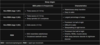
Non-REM Stage 2 EEG pattern and characteristics


Non-REM Stage 3 EEG pattern and characteristics


REM EEG pattern and characteristics


Question

A. Alpha synuclein
This patient’s repeated nocturnal episodes of complex motor behaviors that reflect dream enactment are consistent with REM sleep behavior disorder (RBD). RBD is a parasomnia characterized by dream enactment that occurs because the muscle atonia usually accompanying REM sleep is absent or incomplete. When awakened, patients may be transiently confused but very quickly become fully alert. They may not recall their movements during sleep but can frequently remember their dreams.
RBD is more likely to occur in men age >50 and is strongly associated with alpha-synuclein neurodegenerative disorders. Alpha-synuclein is a synaptic protein that accumulates in neurodegenerative conditions such as Parkinson disease, dementia with Lewy bodies, and multiple system atrophy. Spontaneous (ie, not associated with medications) RBD is considered a prodromal syndrome of alpha-synuclein neurodegeneration because up to 90% of patients with idiopathic RBD eventually develop one of these conditions.
(Choices B and D) Extracellular accumulation of beta-amyloid and/or tau proteins occurs in Alzheimer dementia, which typically presents with early and prominent memory impairment with language deficits and spatial disorientation. Although sleep disturbances commonly occur in patients with Alzheimer dementia, they usually impact sleep initiation and continuity and occur later in the disease process.
(Choice C) Excessive accumulation of misfolded, infectious proteins (ie, prions) causes Creutzfeldt-Jakob disease, a fatal disease that most commonly presents with rapid mental deterioration, behavioral abnormalities, and myoclonus.
(Choice E) Accumulation of abnormally ubiquitinated TAR DNA-binding protein-43 (TDP-43), which normally functions as a transcription inhibitor and DNA repair protein, is associated with both amyotrophic lateral sclerosis and frontotemporal dementia. Patients with amyotrophic lateral sclerosis frequently have sleep difficulty, which is usually related to an inability to change position due to muscle weakness, pain due to muscle cramps, and comorbid anxiety.
Educational objective:
REM sleep behavior disorder (RBD) is a parasomnia characterized by dream-enactment behaviors due to a loss of atonia during REM sleep. Most patients with idiopathic RBD eventually develop a disorder of alpha-synuclein neurodegeneration, most commonly Parkinson disease.
Question

C. Arachnoid Granulations
Cerebrospinal fluid (CSF) is secreted by the choroid plexus of the lateral and fourth ventricles. The path of CSF flow is as follows: lateral ventricles → interventricular foramen of Monro → the third ventricle → cerebral aqueduct → the fourth ventricle → the foramina of Luschka and Magendie → subarachnoid space. In the subarachnoid space, CSF is absorbed by arachnoid granulations and then enters the venous sinuses. The dynamic balance between production and absorption of CSF allows stable intracranial volume (120-150 ml) and pressure (50-180 mm H2O).
If the normal flow of CSF from the ventriculi to the subarachnoid space is disrupted, non-communicating hydrocephalus occurs. Ventriculi above the obstruction are enlarged, while those below the obstruction are normal. Congenital anomalies, such as aqueductal stenosis, Arnold-Chiari or Dandy-Walker malformations, cause non-communicating hydrocephalus. In all cases, the obstacle to CSF flow exists within the ventriculi.
In communicating hydrocephalus, there is no obstruction to CSF flow from the ventriculi to the subarachnoid space. Communicating hydrocephalus usually occurs secondary to dysfunction or obliteration of subarachnoid villi. This dysfunction is usually a sequelae of meningeal infections (including tuberculosis meningitis, as in this patient) or subarachnoid/intraventricular hemorrhage. In communicating hydrocephalus, all ventriculi are symmetrically enlarged, as in the image above.
(Choice A) The emissary veins pass through apertures between the intracranial sinuses and veins outside the cranial vault. They do not participate in absorption of CSF.
(Choice B) Arachnoid trabeculae pass from the arachnoid through the subarachnoid space to the pia mater. They are not involved in the metabolism of CSF.
(Choice D) The choroid plexus is the site of CSF secretion; in communicating hydrocephalus, CSF absorption, not secretion, is impaired. If present, a choroid plexus papilloma might cause a symmetric ventricular enlargement that appears virtually identical to communicating hydrocephalus. Communicating hydrocephalus is far more common than choroid plexus papilloma, however, making Choice D a plausible answer, but not the most likely one.
(Choice E and F) Neither dural septae nor communicating arteries are implicated in the pathogenesis of communicating hydrocephalus.
Educational Objective:
Symmetrical enlargement of the ventriculi is characteristic of communicating hydrocephalus. Communicating hydrocephalus usually occurs secondary to dysfunction or obliteration of subarachnoid villi. This dysfunction is usually a sequelae of meningeal infection (including tuberculosis meningitis) or subarachnoid/intraventricular hemorrhage.
Path of CSF
Cerebrospinal fluid (CSF) is secreted by the choroid plexus of the lateral and fourth ventricles.
The path of CSF flow is as follows: lateral ventricles → interventricular foramen of Monro → the third ventricle → cerebral aqueduct → the fourth ventricle → the foramina of Luschka and Magendie → subarachnoid space.
In the subarachnoid space, CSF is absorbed by arachnoid granulations and then enters the venous sinuses. The dynamic balance between production and absorption of CSF allows stable intracranial volume (120-150 ml) and pressure (50-180 mm H2O).

Foramen of Lushka goes to
cerebellopontine cistern

Foramen of Magendie flows to
cisterna magna
