UW2 Flashcards
question

Angiotensin-converting enzyme (ACE) inhibitors (typically named “-pril”) are one of the most important agents in treating hypertension, heart failure, and renal failure with or without proteinuria. They work by preventing the conversion of angiotensin I to angiotensin II. This prevents the efferent arteriole from constricting more than the afferent arteriole, thus decreasing the glomerular pressure and glomerular filtration rate (GFR). It is expected for the GFR to decrease in all patients initially. Most clinicians are generally not concerned by this unless the creatinine increases by greater than 30% because the long-term benefits of ACE inhibitors are well studied. Other common side-effects of ACE inhibitors include hyperkalemia and cough.

Myasthenia Gravis pathophysiology
MG is most commonly caused by autoantibodies against postsynaptic nicotinic acetylcholine receptors. Binding of antibody to these receptors results in blockade of the receptor’s active site, receptor internalization and degradation, and damage to the motor end plate due to complement fixation. Overall, this leads to decreased numbers of functional acetylcholine receptors at the neuromuscular junction. The decrease in the number of available cation channels reduces the end plate potential following acetylcholine release. Because the threshold potential is not reached, the muscle cells do not depolarize. Synaptic concentrations of acetylcholine are unaffected, unlike in botulism or Lambert-Eaton syndrome.
Pilocarpine MOA
Pilocarpine is a nonselective muscarinic receptor agonist.
Glycopyrrolate MOA
Selective muscarinic antagonists (eg, glycopyrrolate, hyoscyamine, propantheline) can be used to reduce the side effects of cholinesterase inhibitors in sites where acetylcholine action is mediated by muscarinic receptors (ie, gastrointestinal tract). Because of their selectivity, these drugs improve side effects without affecting the action of cholinesterase inhibitors on skeletal muscle, which uses nicotinic receptors.
MEchanism of Collagen synthesis

Ehlers-Danlos syndrome
Ehlers-Danlos syndrome (EDS) is a group of hereditary disorders involving a defect in collagen synthesis. EDS usually manifests clinically as hypermobilejoints, overelasticskin, and fragile tissue susceptible to bruising, wounds, and hemarthrosis. Common mutations leading to EDS phenotypes include deficiencies of the lysyl hydroxylase and procollagen peptidase enzymes responsible for collagen synthesis.
Actions of aldosterone in the collecting ducts

question


Failure of the urachus to obliterate
Around 3 weeks gestation, the yolk sac forms a protrusion (allantois) that extends into the urogenital sinus. The upper part of the urogenital sinus gives rise to the bladder. The allantois, which originally connected the urogenital sinus with the yolk sac, becomes the urachus, a duct between the bladder and the yolk sac. Failure of the urachus to obliterate before birth leads to several abnormalities:
Complete failure of obliteration of the urachus results in a patent urachus that connects the umbilicus and bladder. Patients present with straw-colored urine discharge from the umbilicus, which is exacerbated by crying, straining, or prone position. Local skin irritation can cause erythema.
Failure to close the distal part of the urachus (adjacent to the umbilicus) results in a urachal sinus. This presents with periumbilical tenderness and purulent umbilical discharge due to persistent and recurrent infection.
Failure of the central portion of the urachus to obliterate leads to a urachal cyst.

Question

C. Potassium Channels
This patient’s clinical presentation is suggestive of Jervell and Lange-Nielsen syndrome, an autosomal recessive disorder characterized by profound bilateral sensorineural hearing loss and congenital long QT syndrome, which predisposes individuals to syncope and sudden cardiac death.
This syndrome occurs secondary to mutations in genes (eg, KCNQ1, KCNE1) that encode the alpha and beta subunits of voltage-gated potassium channels. These subunits contribute to the slow-acting component of the outward-rectifying potassium current, which is responsible for ventricular repolarization during phase 3 of the cardiac action potential. Mutations in the potassium channel lead to a decrease in potassium current with prolongation of action potential duration and the QT interval. QT interval prolongation predisposes to the development of life-threatening ventricular arrhythmias, such as torsades de pointes and ventricular fibrillation.

ECG to Ventricular action potential

question

D. Increased Intraabdominal pressure

Several physiologic changes in pregnancy contribute to SUI. The gravid uterus applies pressure on the bladder and stretches the connective tissue and muscles that normally support the pelvic organs. In addition, increased progesterone levels relax the muscles responsible for maintaining urinary continence: the external urethral sphincter and pelvic floor muscles (levator ani muscle complex). Normally, the external urethral sphincter compresses the urethra and creates a high urethral closing pressure. The pelvic floor muscles usually stabilize the urethra against the anterior vaginal wall and contract to decrease the urethrovesical angle, thereby kinking the urethra closed.
Because of decreased urethral sphincter tone and pelvic floor muscle laxity, the compression and position/angle of the urethra are compromised such that sudden increases in intraabdominal pressure (eg, coughing, sneezing) can cause the pressure within the bladder to exceed the urethral closing pressure. This leads to intermittent leakage of urine.
Question

Congenital hypothyroidism is one of the most common causes of preventable intellectual disability. Most cases are due to thyroid dysgenesis (agenesis, hypoplasia, or ectopy), and iodine deficiency is a common cause in areas endemic for iodine deficiency (eg, Europe).
Neonates initially have no significant symptoms due to the presence of maternal thyroxine (T4) from transplacental transfer. T4 is responsible for the stimulation of protein synthesis as well as carbohydrate and lipid catabolism in many cells; as maternal T4 wanes, metabolism is impaired and marked by a slowing of physical and mental activity (lethargy, poor feeding, constipation, hypotonia). Accumulation of matrix substances cutaneously and internally results in nonpitting edema (eg, “puffy” face), umbilical hernia, protruding tongue, and a large anterior fontanelle. In addition, T4 is essential for normal brain development and myelination during early life, and infants are at risk of severe and irreversible intellectual disability.
Treatment with levothyroxine by age 2 weeks can normalize cognitive and physical development.
Therefore, universal newborn screening for congenital hypothyroidism is performed in the United States but not in all other countries.

Standard deviations of a normal distribution

% of data within +/- 1 standard deviation
68%

% of data within +/- 2 standard deviation

% of data within +/- 3 standard deviation
99.7%

Congenital adrenal hyperplasias

17 alpha-hydroxylase deficiency
This patient is genetically male (46,XY) with features suggestive of 17 alpha-hydroxylase deficiency, a rare cause of congenital adrenal hyperplasia (CAH). The enzyme 17 alpha-hydroxylase is active in the adrenal glands and gonads and is responsible for converting pregnenolone to 17-hydroxypregnenolone and progesterone to 17-hydroxyprogesterone. Deficiency of this enzyme impairs synthesis of sex hormones (eg, testosterone, estradiol) and cortisol but does not inhibit mineralocorticoid production.
Due to the absence of androgens in utero, genetic males (46,XY) with 17 alpha-hydroxylase deficiency have undervirilized, ambiguous genitalia. In severe cases, they appear phenotypically female but lack internal female genitalia, as in this case. In contrast, genetic females (46,XX) develop normal internal and external genitalia. At puberty, impaired synthesis of sex hormones in the gonads leads to absent secondary sexual characteristics in both sexes and primary amenorrhea in girls.
Excess mineralocorticoid production results in hypertension and hypokalemia that are usually detected around the expected time of puberty. Low cortisol provides positive feedback to increase ACTH production, further stimulating the mineralocorticoid pathway. This allows for the excess production of weak glucocorticoids (ie, corticosterone), preventing the detrimental effects of low cortisol.
11β-Hydroxylase deficiency
*Low aldosterone but excessive, weak mineralocorticoid (11-deoxycorticosterone).
High mineralocorticoids and androgens and low glucocorticoids
ambiguous genitalia in girls

17α-Hydroxylase
**Low cortisol but excessive, weak glucocorticoid (corticosterone)
Low androgens but high mineralocorticoids and glucocorticoids
ambiguous genitalia in boys

21 hydroxylase deficiency
Low mineralocorticoids and glucocorticoids but high androgens
Ambiguous genitalia in girls

A 57-year-old man comes to the hospital due to nausea, vomiting, and severe crampy pain in the right flank. He has had no fever or chills. Several days ago, the patient had similar, but less severe, pain that resolved spontaneously. Medical history is significant for type 2 diabetes mellitus, obesity, hyperlipidemia, hypertension, and gout. Temperature is 37 C (98.6 F), blood pressure is 160/100 mm Hg, and pulse is 98/min. Physical examination shows right flank tenderness. Blood urea nitrogen and serum creatinine are normal. Abdominal ultrasound reveals right-sided hydronephrosis and proximal ureteral dilation. Urinalysis in this patient would most likely reveal which of the following?
This patient has acute, recurrent flank pain associated with ureteral dilation; this presentation is typical for acute ureterolithiasis. Although ultrasound is relatively sensitive for ureteral and calyceal dilation due to an obstructing stone (hydronephrosis), small stones themselves may not be visible.
Kidney stones usually cause disruption of the ureteral epithelium with resulting gross or microscopic hematuria due to the presence of free red blood cells (RBCs) in the urine. When bleeding into the renal collecting system or lower urinary tract occurs, RBC morphology is normal. In contrast, glomerular bleeding (eg, glomerulonephritis) causes formation of RBC casts due to trapping of RBCs by precipitating Tamm-Horsfall protein (Choice C); the cells are typically dysmorphic due to mechanical and osmotic trauma as they pass through the nephron.
Inspection of urine sediment in patients with acute ureterolithiasis may identify crystals corresponding to the type of stone. This patient has risk factors for uric acid stones, including gout and metabolic syndrome (obesity, diabetes mellitus, hyperlipidemia), and urinalysis may show polygonal uric acid crystals (which are morphologically distinct from the needle-shaped monosodium urate crystals seen in synovial fluid in acute gout).
question


Thiamine deficiency
Beriberi (peripheral neuropathy, heart failure)
Wernicke-Korsakoff syndrome
Riboflavin deficiency
Cheilosis, stomatitis, glossitis
Normocytic anemia
Niacin Deficiency
Pellagra (dermatitis, dementia, diarrhea)
Pantothenic acid deficiency
Distal paresthesia (rare)
Pyridoxine deficiency
Peripheral neuropathy
Cheilosis, stomatitis, glossitis
Biotin deficiency
Dermatitis, conjunctivitis, alopecia, neurologic changes
Folate deficiency
Megaloblastic anemia
Neural tube defects (fetus)
Cobalamin Deficiency
Megaloblastic anemia
Neurologic deficits
Ascorbic Acid Deficiency
Scurvy (perifollicular hemorrhage, gingivitis, muscle pain)
Glucocorticoid MOA

question


Question

C.

This patient with acute-onset chest heaviness and shortness of breath and ECG showing ST-segment elevation has a myocardial infarction (MI). Localization to the lateral leads is consistent with involvement of the left ventricle. Hypotension and cold and clammy extremities (evidence of poor tissue perfusion) suggest cardiogenic shock due to MI-induced acute left ventricular (LV) failure.
A large MI can cause profoundly decreased cardiac contractility. The resulting LV systolic failure leads to markedly reduced cardiac output and hypotension. In response to the hypotension, the aortic and carotid baroreceptors stimulate peripheral vasoconstriction to increase systemic vascular resistance in an effort to maintain blood pressure. Heart rate is also increased (ie, tachycardia) to help increase cardiac output.
Failure to eject blood from the left ventricle also increases LV end-diastolic pressure, which is transmitted backward to increase pulmonary capillary wedge pressure (an estimate of left atrial pressure) and cause pulmonary edema. This increase in pulmonary venous pressure also necessitates higher pulmonary arterial systolic pressure to maintain forward blood movement, which may precipitate acute right ventricular failure.
Question

D.
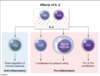
Interleukin-2 (IL-2) is a cytokine produced endogenously by activated CD4 cells, CD8 cells, and natural killer (NK) cells. High-dose IL-2 infusions can also be administered to patients with advanced renal cell carcinoma or metastatic melanoma in order to increase antitumor lymphocyte activity, as follows:
CD4 cells: IL-2 converts activated CD4 cells into type 1 T-helper cells, which then secrete inflammatory cytokines (eg, IFN-gamma, TNF-alpha, IL-2) that drive an antitumor response.
CD8 cells: IL-2 expands the pool of activated CD8 cells and increases their cytotoxic killing with granzymes and perforins.
NK cells: IL-2 triggers proliferation of NK cells and dramatically increases their cytotoxic activity; most of the antitumor effect of IL-2 therapy comes from increased NK cell activity.
Although patients with advanced renal cell carcinoma who undergo IL-2 therapy may enter long-lasting remission, high-dose IL-2 treatment is nonspecific and causes significant adverse effects due to excessive cytokine production (eg, capillary leak syndrome). Therefore, most patients are now treated with newer immunotherapy agents such as CTLA-4 inhibitors (eg, ipilimumab) or PD-1 protein inhibitors (eg, nivolumab) that deliver more targeted therapy and are better tolerated.
Question

E.

The graph compares the respiration curve for a normal, healthy individual (black line) and this patient (blue line), who has chronic obstructive pulmonary disease (COPD); the tracings show resting breaths along with a maximal air intake and expulsion effort. In a resting breath, a single tidal volume (TV) is exchanged. With maximal inhalation, the curve includes both the TV and the inspiratory reserve volume (IRV), which together equal the inspiratory capacity. With maximal exhalation, the curve reflects the expulsion of the IRV and the TV, as well as the amount of additional air that can be expired, which is the expiratory reserve volume (ERV). The air remaining in the lungs following full exhalation is the residual volume (RV).
COPD causes air trapping and hyperinflation of the lungs, so these patients breathe at higher baseline lung volumes (higher FRC). The absolute volume of air in the lungs that is not respired, the RV, is substantially increased. The total lung capacity (TLC) also increases but to a lesser extent than RV. Therefore, the fraction of air in the lungs that is not involved in respiration, the RV/TLC ratio, is also increased.
Lung volumes

Transesophageal Echocardiography

Question

Etoposide is a semisynthetic derivative of the plant alkaloid podophyllotoxin, which targets topoisomerase II.
The topoisomerases are enzymes that relieve the DNA supercoiling that occurs during DNA replication as a result of separation and unwinding of the double helix. Topoisomerase I makes single-stranded nicks to relieve negative supercoiling, while topoisomerase II induces transient breaks in both DNA strands simultaneously to relieve both positive and negative supercoiling. Etoposide and podophyllin specifically inhibit topoisomerase II’s ability to seal the strand breaks it induces, causing chromosomal breaks to accumulate and eventual cell death. Two major uses of etoposide are in testicular cancer and small cell lung cancer. Podophyllin is used topically to treat genital warts.

Question

C.

Tamoxifen and raloxifene are selective estrogen receptor modulators (SERMs). They interact with the estrogen receptor and have agonist or antagonist activity depending on the tissue. In breast tissue, tamoxifen has an anti-estrogenic effect, and it is used for adjuvant treatment of estrogen receptor-positive breast cancer. Tamoxifen reduces the risk of recurrent cancer as well as estrogen-dependent benign breast lesions (eg, fibroadenoma, cystic changes) (Choice D).
In endometrial tissue, however, tamoxifen has a stimulatory effect and can lead to development of endometrial hyperplasia and endometrial cancer. This risk is not seen with raloxifene.
Question

B.
The PR interval
question

A. Acute Hep B
This patient has features of acute hepatitis B (HBV), including systemic, skin, and joint symptoms; hepatomegaly; and elevated transaminase levels. Hepatitis B is a DNA virus with an incubation period of 30-180 days. Transmission can occur sexually, parenterally, or vertically. Onset is dominated by nonspecific symptoms, although patients may develop a serum sickness-like syndrome with joint pain, lymphadenopathy, and a pruritic urticarial vasculitis rash. Right upper quadrant pain can also be present.
Acute HBV is characterized by significant elevations in aspartate aminotransferase and alanine aminotransferase, often >10 times the upper limit of normal. Most patients will have nonicteric hepatitis, but icteric hepatitis with jaundice and elevated bilirubin is also common. Impaired hepatic synthetic function, as indicated by a prolonged prothrombin time, confers a poor prognosis. The most important early marker of acute infection is hepatitis B surface antigen (HBsAg), which may be detectable prior to symptoms or changes in transaminase levels. IgM anti-hepatitis B core (anti-HBc) may also be positive. Hepatitis B e antigen (HBeAg) and HBV DNA counts correlate with infectivity.
Question

D. Impaired neutrophilic respiratory burst

This patient has a severe respiratory infection with Aspergillus fumigatus, an opportunistic, catalase-positive fungus usually seen in immunocompromised individuals. He also has a history of recurrent infections caused by Staphylococcus aureus, another catalase-positive organism, as well as normal immunoglobulin levels and an appropriately elevated leukocyte count in response to infection (ie, no leukopenia). These findings are suggestive of chronic granulomatous disease (CGD).
CGD is most commonly due to an X-linked recessive mutation affecting NADPH oxidase. This enzyme is the first step in the respiratory burst pathway, initiating the formation of reactive oxygen species (ROS). In addition to directly damaging phagocytized pathogens, ROS also activate granule proteases that aid in the destruction of organisms within the phagolysosome.
Without NADPH oxidase, patients with CGD have impaired respiratory burst. Although protease degranulation occurs normally (Choice A), these enzymes are not activated due to the absence of ROS, resulting in defective phagocytic intracellular killing. Catalase-positive bacteria and fungi are most pathogenic in CGD because they can degrade their own metabolically produced hydrogen peroxide (ie, ROS), further limiting microbicidal protease activation. Patients commonly develop S aureus abscesses (eg, lymphadenitis) and severe lung infections, as seen in this patient.
Question

E.
2
Question

D. Lipoprotein Lipase

This patient has acute pancreatitis presenting with abdominal pain, epigastric tenderness, and an elevated serum amylase level. In addition, he has severe hypertriglyceridemia, which is the likely cause of his pancreatitis. Triglyceride-induced pancreatitis (TIP) is usually seen in patients with triglyceride levels >1,000 mg/dL. The risk is elevated in patients with diabetes mellitus, obesity, or excessive alcohol use.
TIP is initiated by impaired blood flow in pancreatic capillaries due to excessive concentrations of chylomicrons and VLDL particles. The resulting tissue ischemia releases pancreatic lipases into the plasma, hydrolyzing triglycerides to yield very high local concentrations of inflammatory free fatty acids.
Management of TIP requires rapid lowering of triglyceride levels, often with an insulin infusion. Insulin upregulates lipoprotein lipase, which hydrolyzes triglycerides in chylomicrons and VLDL; the liberated fatty acids are largely taken up and esterified in adipose tissue, and plasma triglyceride levels are lowered. In addition, insulin reduces the activity of hormone-sensitive lipase in adipose tissue (Choice B), suppressing the release of fatty acids.
Question

D. IL 6
This patient with headaches, visual and muscular symptoms, an enlarged temporal artery, and elevated erythrocyte sedimentation rate has typical features of giant cell arteritis ([GCA] temporal arteritis) with associated polymyalgia rheumatica. GCA is a common systemic vasculitis and occurs almost exclusively in patients age >50.
Although both humoral and cellular immune mechanisms have been implicated in the pathogenesis of GCA, cell-mediated processes are of primary importance. The inflammatory infiltrate in affected vessels is composed of lymphocytes (predominantly CD4-positive T cells) and macrophages and frequently contains multinucleated giant cells. The production of cytokines, in particular interleukin-6 (IL-6), appears to closely correlate with the severity of the disease; a monoclonal antibody against IL-6 (tocilizumab) is effective in treating GCA.

Giant Cell arteritis histology

Sympathetic pathway of pupillary dilation

A 65-year-old man with a history of hypertension is brought to the hospital due to sudden-onset vertigo and dizziness that started an hour ago. On physical examination, there is ptosis and pupillary constriction in the left eye. Extraocular movements are normal in both eyes. Pain and temperature sensations are decreased over the left side of the face, and the direct corneal reflex is absent on the left. Pain and temperature are also decreased over the right side of the body. Muscle strength is 5/5 in all 4 extremities with normal deep tendon reflexes. Which of the following is the most likely cause of this patient’s asymmetrical pupils?
This patient with acute vertigo, sensory loss, and abnormal eye findings most likely suffered a stroke affecting the brainstem. The presence of unilateral ptosis and miosis indicates impaired sympathetic activity (ie, Horner syndrome). Sympathetic innervation of the eye follows a complex pathway:
First order axons originate in the hypothalamus and descend in the lateral brainstem (next to the spinothalamic tract)
They exit the spinal cord at T1 and T2 to reach the paravertebral sympathetic chain
Second order axons ascend to the superior cervical ganglion
Third order axons then continue to ascend along the carotid artery via the carotid plexus
They then traverse the ciliary and oculomotor nerves to innervate the:
Pupillary dilator muscle (dilates pupil)
Superior tarsal muscle (elevates upper lid)
This patient also has a unilateral loss of the direct (afferent) corneal reflex, which is mediated by the ophthalmic nerve (CN V1). In combination with the loss of pain and temperature sensation on the same side of the face, these findings suggest damage to the ipsilateral spinal trigeminal nucleus in the lateral medulla. The patient’s vertigo (vestibular nucleus) and contralateral loss of pain and temperature sensation of the body (spinothalamic tract) are further consistent with damage to the lateral medulla (ie, lateral medullary syndrome).

Lateral medullary syndrome

Question

E.
This patient has herpes zoster ophthalmicus, a condition caused by reactivation of latent varicella-zoster virus (VZV) in the ophthalmic division of the trigeminal nerve (CN V1).
Following primary VZV infection (ie, chickenpox), the virus is transported retrograde along sensory nerves to cranial nerve and dorsal spinal ganglia, where it lies dormant for years. Reactivation of the virus most commonly occurs in elderly or immunosuppressed patients and typically causes sensory disturbances (eg, burning or shooting pains) and eruption of a painful rash (eg, scattered vesicles) in the associated dermatome (eg, unilateral forehead, side of the nose, periorbital area). A prodrome of systemic symptoms (eg, headache, malaise, fever) can also occur.
Because CN V1 also conveys sensory innervation of the cornea (sensory limb of the corneal/blink reflex), involvement can cause acute keratitis (ie, corneal inflammation) and corneal ulceration (eg, linear, branching ulcerations on fluorescein stain). Involvement of the side or tip of the nose (ie, Hutchinson sign) correlates highly with ocular involvement because the nasociliary branch of CN V1 innervates both the eye and the side/tip of the nose.
Question

F.
This patient who was recently hospitalized for mitral valve replacement now has fevers and bacteremia with catalase-positive, coagulase-negative, gram-positive cocci in clusters. This is consistent with infection due to coagulase-negative staphylococci (CoNS). These organisms produce a polysaccharide slime facilitating prosthetic device adherence. As a result, CoNS are a major cause of infection in patients with indwelling catheters or implanted foreign bodies (eg, peritoneal dialysis catheters, vascular grafts) and a common cause of prosthetic valve endocarditis (PVE). For example, Staphylococcus epidermidis (a type of CoNS) can cause an indolent endocarditis following valve replacement; if left untreated, it can lead to intracardiac abscess, prosthetic valve dehiscence, and septic embolization. As CoNS are a component of normal skin flora, it may be difficult initially to distinguish blood culture contamination from true infection; however, recovery of the same organism from several blood culture bottles indicates infection.
Most (>80%) CoNS strains are methicillin-resistant. Therefore, particularly with nosocomial infection (as is likely in this patient), CoNS should be assumed to be methicillin-resistant until proven otherwise. Vancomycin should be a component of initial therapy. Additional antimicrobial agents (eg, gentamicin and/or rifampin, which kills bacteria on foreign material) are administered in some cases of deep-seated methicillin-resistant CoNS infection or staphylococcal PVE. If susceptibility results later demonstrate a methicillin-susceptible isolate, vancomycin can be switched to a semi-synthetic β-lactamase-resistant penicillin such as nafcillin or oxacillin. Native valve endocarditis due to methicillin-sensitive staphylococci is best treated with nafcillin or oxacillin
Question

E. Release of Norepinephrine
Norepinephrine-releasing agents (eg, phentermine) are sympathomimetic weight-loss drugs indicated for short-term (≤12 weeks) treatment of obesity. They work by stimulating release and inhibiting reuptake of norepinephrine and, to a lesser extent, serotonin and dopamine. Subjective effects include an increased sensation of satiety and subsequently reduced caloric intake.
Because of the increased sympathetic activity associated with these drugs, they can raise blood pressure and should be avoided in patients with comorbid hypertension or heart disease. Norepinephrine-releasing agents can induce brisk initial weight loss but are notable for poor long-term benefit, and most patients regain the lost weight following discontinuation.
Question

Peptic ulcers (defects in the stomach or duodenal mucosa) are most commonly due to Helicobacter pylori infection or nonsteroidal anti-inflammatory drug (NSAID) use. The majority of duodenal ulcers are associated with H pylori infection, but the prevalence is decreasing in the United States due to improved hygienic practices. H pylori has been adequately ruled out in this patient; therefore, he most likely has an NSAID-related duodenal ulcer.
Nonselective NSAIDs (eg, ibuprofen, naproxen, aspirin) exert analgesic, anti-inflammatory, and antipyretic effects through the inhibition of both forms of the cyclo-oxygenase (COX) enzyme. In the gastrointestinal tract, the COX-1 subtype is responsible for the synthesis of prostaglandins, which are critical for the maintenance of intestinal mucosa. Inhibition of COX-1 leads to increased gastric acid formation, decreased mucosal bicarbonate production, and decreased mucosal blood flow, which can increase the risk for gastritis and peptic ulcer formation. Patients who are age >65 or those on long-term or high-dose NSAID therapy are at the highest risk.

Question

A.
Upper motor neuron lesion in the Cortical spinal tract so would have hypereflexia
Question

D. Superior displacement of the Larynx
Normal swallowing involves a complex sequence of voluntary and reflexive processes that move food from the mouth to the esophagus while preventing it from entering the airway (ie, aspiration). Three main airway-protective movements occur during normal swallowing:
- Displacement of the larynx superiorly and anteriorly under the base of tongue, which allows food to be directed into the more posteriorly located esophagus
- Tilting of the epiglottis to block the airway
- Closing of the glottis by adduction of the vocal folds
Because of the complexity of swallowing, patients who sustain strokes often have persistent dysphagia and/or aspiration. If a neurologic deficit cannot be corrected, behavioral modifications can sometimes improve the safety of swallowing. A chin-tuck maneuver (ie, flexion of the head and neck during swallowing) sometimes helps. The maneuver seems to decrease the distance from the hyoid bone to the larynx (simulating elevation of the larynx), as well as to narrow the distance of the laryngeal entrance, leading to decreased aspiration.