Ear Flashcards
Vessels closely associated with the horizontal ear canal?
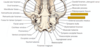
caudal auricular superficial temporal retroarticular (retroglenoid) v.


S-shaped incision in the concave skin followed by staggered parallel rows of MATRESS sutures placed parallel to the blood vessels. Tied on CONVEX surface
Hapharzzard in cats to allow blood supply
Other option CO2 lazer tx
Bandage important

Aspiration of hematoma fluid with IV 2 mg/kg of dexamethasone = success 88.9%
Daily aspiration with irrigation with 0.2 to 0.4 mg of dexamethasone in saline along with 0.5 mg/kg of dexamethasone orally was successful in 92.9% of cases
Drainage with injection of 0.5 to 1.0 mL of methylprednisolone into the cavity with or without the use of systemic corticosteroids = 90% to 98%
A window of skin overlying the vertical canal is prepared. B, The skin strip is elevated, exposing the lateral surface of the auricular cartilage. C, The vertical canal is cut to the level of the auricular–annular junction. D, The lateral wall is reflected ventrally, creating a “drainage board.” E, The deepest sutures are placed first at the “hinge” to minimize tension on the appositional repair.
Lateral wall resection

A T-shaped skin incision is made over the vertical canal. B, The tissues overlying the auricular cartilage are dissected free. C, The vertical canal is separated from the surrounding tissues circumferentially and reflected ventrally. D, The ear canal is amputated distal to the affected tissue. E, The lateral and medial walls of the remaining ear canal are spatulated. F, The dorsal and ventral cartilage flaps are sutured to the skin, and the T-shaped incision is closed routinely.
Vertical EAR canal ablation

A ventrally based skin flap is advanced across the defect of the external auditory meatu


euter Bobbin vent tube for tympanostomy drainage

Ventral bulla osteotomy in a cat. A, Location of skin incision. The upper finger indicates the larynx; the lower finger indicates the angle of the mandible. B, Retraction of external maxillary and linguofacial veins with mandibular salivary gland. C, Exposure of the ventral aspect of the osseous bulla. D, Removal of mucus from the hypotympanic chamber. E, Osteotomy of the roof of the hypotympanic chamber to permit access to the mesotympanic chamber.
triangular area bounded by the mandibular symphysis, the caudal border of the mandible, and the larynx
dissect through platysma and sphincter colli muscles
digastricus and mylohyoid muscles are separated by blunt dissection, and the underlying hyoglossus and styloglossus muscles are retained
avoid hypoglossal n. near lingual a.

Lateral view of the canine skull. 1, Orbital ligament (inset); 2, infraorbital foramen; 3, orbit; 4, pterygopalatine fossa; 5, optic canal, orbital fissure, and rostral alar foramen; 6, retroarticular process; 7, retroarticular foramen; 8, external acoustic meatus; 9, tympanic bulla; 10, stylomastoid foramen; 11, paracondylar process; 12, occipital condyle; 13, nuchal surface; 14, mastoid process; 15, zygomatic arch; 16, temporal fossa; 17, nuchal crest


- Maxillary Nerve
- Mandibular Nerve
- Optic Nerve
- Optic Canal
- Oculomotor Nerve
- Orbital Fissure
- Trochlear Nerve
- Oval Foramen
- Rostral Alar Foramen
- Abducens Nerve
- Stylomastoid Foramen
- hypoglossal nerve
- Caudal Alar Foramen
- hypoglossal canal
- Ophthalmic Nerve
- vagus nerve
- Facial Nerve
- Ethmoidal Foramina34.5%
- accessory nerve34%
- Tympano-occipital fissure33%
- glossopharyngeal nerve33%


Lateral canthotomy. A, The lateral canthus is incised full thickness. B, The appearance at closure.

Posterior aspect of the medial commissure of the eyelids.

Action and innervation of the eyelid muscles. C, Contracts the palpebral fissure; E and D, enlarge the fissure

Hotz-Celsus procedure for entropion correction.

Arrowhead resection for lateral canthal entropion seen most commonly in large-breed dogs with broad skull conformation. Lateral canthal ligament transection, if performed, should be performed first.

Kuhnt-Szymanowski procedure for correction of entropion caused by macroblepharon. A, The lower eyelid is stretched tautly while the first incision is made. B, The lower eyelid is relaxed, and the skin and muscle flap is undermined after the second laterally located incision is made. C, A close-up view of the most medial aspect of the wedge taken from the tarso-conjunctival flap. D, The completed surgery after a wedge was removed from the lateral aspect of the skin muscle flap and the sutures are placed.

V-Y blepharoplasty for the correction of cicatricial ectropion.

“Pocket” medial canthoplasty.

Electroepilation for the treatment of distichiasis. A Bishop-Harmon forceps can be used to stabilize the eyelid if positioning of the needle is hindered by a chalazion clamp.

Two-layer closure used to appose the eyelid margin. A, Eyelid defect. B, Mattress suture to appose tarsus and orbicular is muscle layer. C, Figure of eight suture. The numbers represent the order in which the bites are taken to ensure that the knot is away from the margin. Bites should be small (1 to 2 mm), and bites on one side should mirror those on the opposite side of the cut margin. D, Final appearance

Sliding pedicle advancement flap. This technique is typically used to reconstruct central lower eyelid defects but can be used anywhere.

Semicircular flap technique for eyelid reconstruction of upper or lower eyelid defects.

Myocutaneous pedicle graft as described by Dziezyc and Millichamp27 for the repair of eyelid agenesis. The conjunctiva can be harvested from the anterior surface of the third eyelid as depicted or the graft can be lined with mucosa from a different site, buccal mucosa, or not lined at all.

Lip-to-lid mucocutaneous subdermal plexus flap

roper suture placement in a temporary tarsorrhaphy. Stents help distribute the forces over the skin so the sutures do not migrate into the skin and allow the tarsorrhaphy to loosen, exposing the sutures to the cornea.

Cutaway drawing of the surgical field used for a parotid duct transposition. The facial muscles have been omitted so that the essential features can be seen. 1, Dorsal buccal nerve; 2, anastomosis of dorsal buccal and ventral buccal nerves; 3, ventral buccal nerve; 4, parotid salivary gland; 5, parotid duct; 6, papilla of parotid duct; 7, facial vein; 8, upper carnassial tooth.
