Basic bio Flashcards
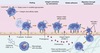
Neutrophil extravasation. Leukocyte extravasation is a multistep process orchestrated by both hemostatic and cell–cell interactions. Margination and rolling of leukocytes along the vascular endothelium are mediated through interactions between endothelial selectins with their corresponding leukocyte ligands. Chemokines stimulate increased expression and enhanced binding affinity of leukocyte integrins, leading to firm adherence to endothelial cell integrins (e.g., intracellular adhesion molecule [ICAM]-1). Leukocyte diapedesis is facilitated by the adhesion molecule, platelet–endothelial cell adhesion molecule (PECAM)-1, and leukocytes follow chemokine gradients to the site of injury. IL-1, Interleukin-1; TNF, tumor necrosis factor
Proinflammatory response to alarm signals. In response to pathogen-associated molecular patterns (PAMPS) or danger-associated molecular patterns (DAMPS), toll-like receptors (TLRs) on the surface of resident macrophages induce various molecular signaling pathways. Many of these pathways lead to the translocation of nuclear factor kappa B (NFκB) into the cell nucleus, where it acts as a transcription factor, regulating the production of proinflammatory cytokines. The cytokines act directly and indirectly on a number of cell types. Interleukin (IL)-6 induces hepatic production of acute phase proteins, which in turn influence a number of inflammatory systems. Chemokines induce recruitment of inflammatory cells, which produce additional mediators. If the process is not properly balanced by antiinflammatory responses, tissue damage and systemic inflammation may result in serious consequences. CRP, C-reactive protein; NO, nitric oxide; ROS, reactive oxygen species.

The arachidonic acid pathway. Arachidonic acid is metabolized by the cyclooxygenase or lipoxygenase pathway to produce prostaglandins or leukotrienes and proresolution lipoxins, respectively. The inhibitory effects of several drugs on specific enzymes are denoted by a red X. COX, Cyclooxygenase; HETE, hydroxyeicosatetraenoic acid; HPETE, hydroperoxyeicosatetraenoic acid.

Cellular Origins and Functions of Prostaglandins

Functions of nitric oxide. Endothelial-derived nitric oxide synthase (eNOS) functions to maintain normal vascular tone via the vasodilatory effects of nitric oxide on vascular smooth muscle. In addition, nitric oxide modulates the interactions of platelets and leukocytes with the vascular endothelium. At increased levels, inducible nitric oxide synthase (iNOS) facilitates nitric oxide–derived free radical production and removal of target pathogens by macrophages. NO, Nitric oxide.

Complement pathway activation and effector functions. The complement cascade is activated via three different pathways, all of which culminate in cleavage of C3 into C3b and C3a. Complement proteins and breakdown products facilitate several aspects of inflammatory responses as well as pathogen removal via phagocytosis and membrane attack complex (MAC) production

The central dogma of molecular biology. Genomic DNA (gDNA) is transcribed to mRNA, starting at the first exon (E1), after the initiation of transcription. The whole gene sequence, not including the promoter region (P), is transcribed before splicing removes the introns (I). Translation of the mature mRNA sequence produces the protein.

The polymerase chain reaction (PCR). RNA or DNA (gDNA, genomic DNA) can be evaluated, but RNA is usually reverse transcribed into complementary DNA (cDNA) before the PCR occurs. First, the sample is heated to separate the DNA into single strands (denatured). The sample is then cooled to allow the primers to bind to their target sequence (annealing). Finally, the mixture temperature is increased to the optimum for DNA polymerase use. The DNA polymerase then synthesizes a new DNA template (extension or elongation). After each PCR cycle, the number of templates is doubled

The polymerase chain reaction (PCR). RNA or DNA (gDNA, genomic DNA) can be evaluated, but RNA is usually reverse transcribed into complementary DNA (cDNA) before the PCR occurs. First, the sample is heated to separate the DNA into single strands (denatured). The sample is then cooled to allow the primers to bind to their target sequence (annealing). Finally, the mixture temperature is increased to the optimum for DNA polymerase use. The DNA polymerase then synthesizes a new DNA template (extension or elongation). After each PCR cycle, the number of templates is doubled

Figure 5-1 Total body water (TBW) fluid compartments.

Intracelluar and extracellular compartment % of total body water?
Intracellular = 66% total body water and 40% total body weight
Extrecellular 33%, 20% total body weight = plasma 25%, interstital 75%
What is the osmolarity of body fluid compartments?
290-310mOsm/L
What precursors are used in fluids as buffer?
Lactate - liver to bicarb
Acetate - muscle
Gluconate - cells
What isotonic fluid is “unbalanced” and what happens when this is given as a bolus?
0.9% NaCL
mild increase Na, Marked increase Cl, moderated decrease bicarb and K (acidfying effect)
What percent of isotonic fluids (extracellular expanding fluids) are redistributed to the intersitial space?
75%
only 25% remain in intravascular space
What are hypotonic fluids?
- 45% saline
- 5% dextrose with 0.45% saline
2.5% dextrose with half strength LRS
Normosol M
Plasmalyte 56
D5W
large volumes can rapidly decrease osmolarity and cause cerebral edema
What are the side effects related to synthetic colloids?
decrese factor VIII and vW factor
impairment platelet function
interference in stability of fibrin clots
dose hypoproteinemia = 0.5-2ml/kg/day
shock dose 5-10ml/kg
What are the standard doses for blood products?
pRBC and FFP = 10-15ml/kg
whole blood = 20-25ml/kg
What is the formula for blood adminstration based on target PCV?
V rbc = blood V x (target PCV- current PCV/ donor PCV)
blood V = 90ml/kg dog and 50ml/kg cat
what is the PCV of packed rbc?
80%
lifespan 20-35 d at 4 C
How do you calculate fluid replacement?

What is the formula for daily water requirement?
FOR CATS: BW(KG)^75 × 80 = ML/DAY
FOR DOGS: BW(KG)^75 × 132 = ML/DAY
Isotonic Crystalloid Compositions?

Albumin accounts for what percent % of plasma oncotic pressure?
80%
Causes of Hyponatremia

Causes of Hypokalemia

Causes of Hyperkalemia

Causes of Hypocalcemia

Box • 5-7 Causes of Hypomagnesemia

What is Wipple’s triad?
A low blood glucose concentration with concurrent clinical signs of hypoglycemia, as well as resolution of clinical signs when the blood glucose level is normalized, constitutes a clinical definition of hypoglycemia known as Whipple’s triad
Box • 5-8 Causes of Hypermagnesemia
A serum concentration of magnesium greater than 2.51 mg/dL in dogs and greater than 2.99 mg/dL in cats is consistent with hypermagnesemia, but clinical signs are generally seen at levels exceeding 4 mg/dL. Electrocardiographic changes that may occur in patients with hypermagnesemia include prolongation of the PR interval, widening of the QRS complex, and, at higher concentrations, heart block and asystole. Hypermagnesemia may cause hypotension and can interfere with normal clotting and coagulation. Hypermagnesemia also has muscular effects similar to hypercalcemia, including weakness and decreased tendon reflexes.77,78 Treatment of hypermagnesemia includes fluid therapy and administration of loop diuretics (furosemide). Severe cases can be treated with calcium gluconate, as calcium is an antagonist of magnesium at the neuromuscular junction. Anticholinesterases such as physostigmine may also help reduce the actions of magnesium at the neuromuscular junction
Causes of Hypophosphatemia

Causes of Hyperphosphatemia

Causes of Corrected Hypochloremia

Causes of Corrected Hyperchloremia

Causes of Hypoglycemia

Causes of Hyperglycemia

Reabsorption of filtered [HCO3−] by H+ secretion in the proximal tubule.

Where does the majority of bicarb resorbtion occur?
Ninety to 95% of filtered HCO3− is reabsorbed in the proximal tubule, and the rest is reabsorbed in the loop of Henle, distal tubule, and collecting duct.
Table • 5-8 Normal Blood Gas Values in the Dog and Cat (mean ± SD)

Serum Anion and Cation Concentrations (mEq/L) in the Dog and Cat

Compensatory Responses to Simple Acid-Base Disturbances in Dogs

Septic chapter equations

Box • 6-3 Defects in Oxygen Uptake
Diffusional shunting: This is caused by slow blood velocity (prolonged transit time), which favors diffusional oxygen transport over convective oxygen transport (i.e., the movement of oxygen following a concentration gradient prevails over transport of oxygen within the bloodstream). It may occur in the intestine during shock states. The intestine relies on countercurrent blood flow to deliver blood to the villi. If the rate of flow is so slow that oxygen from the arterial blood can diffuse to the venous blood before it is delivered to the tip of the villus, the intestine will be hypoxic, but venous saturation will actually be increased.
Diffusional resistance: For cells to utilize oxygen, O2 must diffuse from the vascular space through the tissues, into the cell, and finally to the mitochondria. Oxygen is poorly soluble in aqueous solutions; therefore tissue edema will increase diffusional distance and limit oxygen availability to the tissues. Hypoxia may increase edema by increasing vascular permeability, thus leading to a “feed-forward” cycle of hypoxia and edema and more hypoxia. Impaired diffusion will result in a smaller gradient, less driving pressure for the oxygen to leave the hemoglobin, and thus higher venous saturation, despite tissue hypoxia. •
Arteriovenous shunting: One of the most obvious reasons for failure of oxygen uptake by the cells is the loss of normal capillary flow. In sepsis, trauma, and diseases associated with systemic activation of the inflammatory response, capillary obstruction can result from neutrophil-endothelial interaction, platelet aggregation, thrombi, or loss of normal vasodilatory function. As a result, arterial blood bypasses the hypoxic bed, leading to an increase in venous oxygen saturation.
Perfusion/metabolism mismatch: A cause of inadequate tissue oxygenation is perfusion/metabolism mismatch. Despite the normal excess (approximately four-fold) of oxygen delivery in relation to tissue demand, in some tissues and in some disease states the increased metabolic demand combined with reduced blood flow can lead to tissue hypoxia. A classic example is sustained tachycardia, when c_oronary perfusion is reduced because of shortened diastole, yet the metabolic demand of the myocytes is increased_.
Cytopathic hypoxia (mitochondrial dysfunction): Cytopathic hypoxia is classically associated with sepsis. Experimental studies of sepsis have demonstrated that despite direct measurement of adequate cellular oxygen, mitochondrial respiration is impaired. Proposed mechanisms to explain this “cytopathic hypoxia” include the following: •Inhibition of pyruvate dehydrogenase and subsequent lactate accumulation •Nitric oxide–mediated inhibition of mitochondrial electron transport •Poly-(ADP-ribosyl)-polymerase activation by reactive oxygen species and subsequent depletion of NAD+/NADH for oxidative phosphorylation

Vasoactive and Inotropic Agents—Intravenous Constant Rate Infusion

SIRS Criteria for Dogs, Cats, and People

Diagnostic Criteria for Veterinary Acute Respiratory Distress Syndrome (ARDS)

Differential Diagnoses of Prolonged Coagulation Times: Prothrombin Time and Activated Partial Thromboplastin Time

Differentials thrombocytopenia
Diffenertials thrombopathia

Disorders secondary hemostatsis

Plasma Component Transfusions

Platelet Transfusions

Antithrombotic Drugs: Preoperative Discontinuation and Therapeutic Recommendations in Cases of Uncontrolled Bleeding

Recommended Safe Storage Times for Sterile Packs Using Various Wrapping Materials

The “anatomy” of an articulating instrument (a needle holder)

The four blades that fit a #3 scalpel handle (i.e., 10, 11, 12, and 15) and four blades that fit a #4 scalpel handle (i.e., 20, 21, 22, and 23)

A Beaver surgical knife handle and selected beaver blades (#64 and #67

Three commonly used scissors: A, Curved Mayo; B, curved Metzenbaum; and C, operating (utility) scissors

A pair of Westcott scissors

Wire-cutting scissors, including A, the “notch” and B, serrated cutting edges

Four different rongeurs: two (A, Stille-Luer and B, Ruskin) double-action type and two (C, Lempert and D, Kerrison) single-action type.

Four periosteal elevators: A, Freer, B, Sayre, C, ASIF (Synthes/AO), and D, Langenbeck

This figure shows each of the following: A, Bone chisel. B, Osteotome. C, Gouge. D, Gigli saw. E, Bone-cutting forceps, and F, a trephine

Mayo-Hegar and B, Olsen-Hegar needle holders.

Castroviejo needle holder.

Right-angle; B, Babcock; C, Allis; and D, Ochsner-Kocher forceps.

Noncrushing forceps: A, Doyen intestinal; B, DeBakey; and C, Satinsky forceps

Patterns for thoracic and vascular DeBakey (A) and Cooley (B) forceps

Three commonly used hemostatic forceps: A, Halsted mosquito; B, Kelly; and C, Crile forceps

Brown-Adson; B, DeBakey, C, dressing; and D, Adson forceps

Backhaus (penetrating) clamps; and B, Lorna (nonpenetrating) clamps.

Figure shows five bone-holding forceps: A, Kern; B, Lane; C, Vebrugge; D, reduction forceps with a speed lock; and E, reduction forceps with points and a ratchet

malleable; B, Hohmann retractors; and C, spay (Snook) hooks.

Lister bandage scissors; and B, Spencer stitch scissors.

Jewelers’ forceps; B, vessel dilators; C, dissecting forceps; and D, tying forceps.

Nonlocking microsurgical needle holders with three different types of tips (straight, curved, and extra fine).

A microsurgical vessel clamp

An Alm microsurgical retractor

Comparison of the breaking strength of different tissues across time

