Musculoskeletal Flashcards
What are the fibrillar collagens?
Major 1,2,3
Minior 5,11,24,27
major component of macromolecular collagen fibrils
What are fibril-associated collagens with interrupted tuple helices (FACIT)?
9,12,16,19,20,21,22
do not assume fibrillar structure
assocted with fibrilla
important roles in 3D organization and interaction fo fibrillar collagens
Basmentmembrane collagens?
4, 7, 15, 18
Filamentous collagen
6
What is the structure of collagen?
triple helix made of 3 separate polypeptide molecules (alpha chains)
homotypic (same 3 chains) = 2, 3
heteroypic = 5
Describe collagen biosynthesis
transcription of gene and translation mRNA = pre-proalpha chains (pre-pro-collagen)
proline hydroxylation (rate limiting step)
O-linked glycoslation reactions
triple helicies stablized by disulfide bonds
folding = molecular chaperones, cis-trans isomerization (prolylpeptidyl isomerase) = triple helical procollagen
globular teloprptide somains cleaved metalloproteinases = trocollagen
fibrillogenesis (trocollagen +ECM) = fibrils = fascicles = macroscopic fibers
Descrine the structure of a proteoglycan.
Glucosaminoglycan = heparin sulfate, keratan sulfate, D-glucosamine
Galatosaminoglycans = chondroitin sulfate, dermatuan sulfate, D-galactosamine
Hyaluronic acid = nonsulfonated glucosaminoglycan, repeating unitsD-glucuronic acid and D-glucosamine
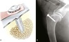
Diagram of the basic structure of an aggregating proteoglycan. Proteoglycan monomers consist of a core protein with numerous covalently linked glycosaminoglycans. Keratan sulfate and chondroitin sulfate, the most common glycosaminoglycans in articular cartilage, are depicted. In a typical aggregating proteoglycan, hundreds of proteoglycan monomers may associate with a single hyaluronic acid backbone. This association is noncovalent and is stabilized by link proteins. The positions of the globular domains (G1, G2, and G3) of the core protein relative to the glycosaminoglycan binding region are shown. G1 contains a hyaluronan binding domain, and G3 contains domains that bind a variety of extracellular matrix components. The function of G2 is unknown

What are matrix metalloproteinases
Zinc dependent endoprptidases that cleave ECM protieins (collagenaes, gelatinases, stromolysins)
What is elastin?
protein biopolymer = monomeric component is protein tropoelastin (65-70kDa)
formation requires fibrillin microfibrils and fibronectin as scaffold for tropoelastin
elastic deformation or strain of approx 70% restin length, maximum extension of 220% before loss of strength
How does the modulus of elasticity compare between type 1 collagen and elastin?
300-600 kPa elastin compated with 10^6 kPa for type collagen
Sturcture of bone - cortical.
Haversian units
Volkman’s canal

What makes up periosteal ECM?
Type 1 collagen, proteoglycans and elastin
What orchestrates osteoclastic recruitment?
monocyte colony stimulating factor (M-CSF) and receptor activator of NFkB ligand (RANKL)
expressed by osteoblasts, decreases expression of osteoprotegerin (blocks RANKL and prevents differentiation of osteclast precusors)
What is sclerostin?
Released from osteocytes - exerts inhibitory effects on proliferation and biosynthetic activity of osetoblasts
What is another name for a resorption pit?
Howship’s lacuna
What is the mineral composition of bone?
70% mineral, resists compressive force
calcium hydrocyl-apatite
What is the organic matrix of bone composed of?
90% collagen (primarily type 1, also 5 and 3)
What law describes bones ability to remodel in response to mechanical load?
Wolff’s Law 1892
What is the make up of articular cartilage?
70% water
Dry weight = 50% collagen (85-90% type 2- provides tensile stiffnes and strength)
- 35% proteoglycan
- 10% gylcoprotien
minerals and lipis
- 2-10% chondrocytes
Other components: firoenectins, type 11 (stablizes lateral gowth fibrils), Aggrecan (240kDa, major proteglycgan, 90% carbohydrate), leuine rich proteoglycans (chondroitin/dermatan sulfate- decorin, biglycan and keratin sulfate - fibromodulin) modulate fibrillogensis
What are the zones of cartilage?
1: superfical/tangential zone - fibrils tangential (tension)
2: transitional zone - fibrils parallel and branching (shear and compression)
3. radiate zone - fibrils perpendicular (compression)
tidemark
- calcified cartilage zone
conc proteoglycan increase with depth from surface
collagen fibrils more concentrated at surface

How much does osmotic pressure contribute to compresive stiffness of cartilage?
50%
GAGs account for 75% of the osmotic pressure of PGs
What kind of collagen makes up fibrocartilage?
Type 1
Small amounts of proteoglycans
makes up annulus fibrosus, menisci and parapartellar isetions of quadriceps mm.
What are types of tendons?
Aponeuroses
Positional tendons - DDFT
Energy storing tendongs = calcaneal tendon - greater elastic fiber component
Stress-strain relationship of tendons.
intial pahse - straighten fibrils
linear elastic region
rapid loaging = fibrillar elongation and stress relaxation and creep = interfibrillar shear
yeild point
larger diameter = greater stiffness
small diameter - greater surface area and viscoelastic properites
healed collagen fibrils remain smaller and more uniform = inferior properties

Name parts of muscle
Z-band
A-band = conjoined myosin fibers
I-band = centerd on z disc and actin fibers

Describe muscle contraction
Action potential → acetylcholine released → binds sarcolemma and depolarizes causing Ca release from sarcoplasmic reticulum
→ confrom change in tropmyosin exposes myosin site on actin → globular head myosin filament engages with ATP → shorten sarcomere
What is the ECM of muscles?
Epimysium: fasicles assembed to mm. bellies
Perimysium: Fibers into fasicles
Endomysium: myofibers into fibers (satellite cells = myoblastic progenitor cells)
basement membrane basis of ECM in each layer: Composed of nerves, vessels, lymphatics, Type 1 collagen, proteglycans, fibroblasts
ECM <10% skeletal m
ECM provides structure for force transfer and elastic recoil
What are the 2 types of muscle fibers?
Type 1 = slow twitch = mitochondria rich, sustained low velocity, low force (oxidative metabolism) = improved aerobic capcity, stimulated by prolonged low levels force
Type 2 = fast twitch = few mitochondria, rich in myofibrils, transient high velocity and force = high intesity conditioning = hypertrophy type 2, synthesis myofibril proteins, enlarment and increase contractile capabilites
Define viscoelastic and anisotropic.
Visoelastic = strength depends on the rate the bone loaded (bone stronger when rapidly loaded)
Anisotropic = mechanical properties depend on the direction of loading
Define stress and strain
Stress = N/m^2 = newton/ meter squared
Stain = local deformation = L1-L2 / L1
Illustration of stress (A) and strain (B). A, Force (F) is applied to a block, producing stress (σ). When the surface area is decreased by one half, the stress is doubled (2σ) for the same amount of force. B, The mathematical definition of strain is the change in length divided by the original length

Define strength and stiffness
Strength = load material can withstand before failure
Stiffness = rate at which material deforms with load
Describe stress strain curve.
Illustration of a stress/strain curve produced when a material is loaded. The slope of the ascending linear portion of the curve is the stiffness of the material. Point Y is the yield point of the material. Point U is the ultimate strength of the material. Toughness is defined as the area under the curve (red shading)

How does cortical bone compare to cancellous bone in porosity and stres/strain curves?
Porosity (volume open space to bone): cortical 5-10%, cancellous 75-95%
Cortical bone short plastic phase, cancellous bone long plastic phase
Cortical bone higher stiffness and strength
Total energy absorbed can be greater for cancellous due to prolonged plastic phase
Note the prolonged plateau in cancellous loading representing the collapse of trabeculae.

Describe bending forces:
moment
pure bending
cantileve ending
3 point bending
4 point bending
Cantilever = break at fixation point
3-point = break at middle force (where bending point present)
4 point = constant loads between middle points
moment = tendency of a force to twist or rotate an object, expressed in units of torque
Illustration of bending moment diagrams for various loading conditions. A, Pure bending when opposite torques are applied to each end of a beam. B, Cantilever bending with an applied load at the end of a beam. C, Three-point bending with two equal loads applied at each end and a third load applied between them. D, Four-point bending with two equal loads applied at each end and two additional loads applied between them

Describe the fracture associated with the following forces.
Compressive = oblique fracture
tensile and bending = transverse
bending with compression = butterfly fragment
torsional = spiral
Primary gap healing
Primary gap healing. A, The gap is initially filled with transversely oriented lamellar bone. B, A cutting cone moves across the lamellar bone, producing osteoid that is mineralized to form longitudinally oriented lamellar bone. This process is known as longitudinal Haversian remodeling
Occurs when gap <1mm, Strain <2%
- Lamellar bone forms in gap early = Deposited within days, No cartilage intermediary, Transverse orientation, Little stability
- 3rd week of healing = Longitudinal remodeling = Osteons (with osteoprogenitor cells) cross gap, Longitudinally oriented osteons placed (still weak)
- By 8 weeks = secondary remodelling = Similar to contact healing cascade

Primary healing - no gap
A, Because of direct contact with bone ends, lamellar bone is not formed transversely. Cutting cones cross the fracture line directly and form new longitudinally oriented lamellar bone. B, Mature osteons cross the original fracture site, connecting the segments and providing strength to completely repaired bone

Secondary bone healing

Plt-rich hematoma formation
- Release of TNF-α, IL-1, IL-6
- MSC release from soft tissues, periosteum, bone marrow
- BMPs induce proliferation and differentiation of MSCs
- Granulation tissue replaces hematoma
- Angiogenesis (VEGF, angiopoietin)
- Hard callus (woven bone) forms by intramembranous ossification from cambium layer of periosteum
- Fracture gap bridges by soft callus
- Endochondral ossification replaces soft callus with bone
- Remodelling: hard callus replaces with lamellar bone

What are types of bone healing?
Endrochondral ossification (cartilagenous precusor)
Intramembranous (direct differentiation of mesenchymal cells to ostoblasts) - how flat bones formed, compaction of trabeculae to cortical bone
Direct (no evidence of callus)
Indirect aka secondary bone healing
List the strains associted with bone healing for various tissues
Granulation / hematoma up to 100% - 40 degrees
Fibrous tissue 20%
Cartilage 15%
Fibrocartilage 10% - 5 degrees
Woven bone < 10%
lammelar Bone <2% - 0.5 degrees
How does distraction osteogensis work?
regnerate bone via intramembranous ossification
- fibrous interzone (central radiolucent zone) = once distraction stops the zone mineralizes
During distraction osteogenesis, osteoid is laid down in parallel columns that extend from osteotomy surfaces centrally. Lamellar bone develops within these columns if the fracture is sufficiently stable

What is the user agreement for open fractures? And what is the Gustil-Anderson scheme?
60% agreement
Type 1: Open fracture with wound <1cm, soft tissues mild/mod contused, usually inside out
Type 2: wound >1cm w/o extensive soft tissue damage, flaps or alvusions, usually from outside
Type 3: extensive soft tissue damage
3a = adequate soft tissue coverage despite extensive trauma
3b = extensive soft tissue loss, periosteal stripping, bone expsoure, usually massive contamination
3c = associated with arterial injury requiring repair
What is the orthopedic trauma associated scheme for open fractures?
Each given subscore of 1, 2, or 3 for mild, mod or severe
S-skin defect
M- muscle injury
A- arterial injury
B - bone injury
C- contamination
SMABC = M-CABS
What are risk factors for infection for open fractures?
Lack abx
resistent organisms
too much time from injurgy to starting abx
extensive soft tissue trauma
positive port-debridement/irrigatation culture
What is the infection rate for open fractures and what common organisms?
human: within 3 hours = 4.7% vs 7.4% for +4 hours
Type 1 and 2 fractures: ~6% with fluoroquinolone or cephalsporin/gentamycin = rec. 1st/2nd gen cephalporin
Type 3: 7.7% with ceph/genta vs. 31% with cipro = rec. ceph + fluoro
Common = staph, strep, kleb, pseudomonas, clostridium, enterobacter, e. coli
abx beads = decreased from 12% to 3.7%
Does early surgical debridment effect sucess?
Human studies no difference, old dogma = debride within 6 hr
What are the advantages of negative pressure wound healing?
Decreased interstitial edema
Increased blood flow
Accelerate formaiton of granulation tissue
increased bacterial clearence
promotes wound contraction
secure skin graft
Which bone graft is safe for an open wound?
Cancellous is safe
Cortical = risk for infection/sequestum formation
What are the delayed/non-union rates for open fractures?
Type 1 = 0-5%
Type 2 = 1-14%
Type 3 = 2-37%
Physeal cartilage healing. A, Salter-Harris Type I physeal fracture occurs through the hypertrophied zone of cartilage. B, If reduced accurately, these physeal fractures heal by continued formation of cartilage. C, If the fracture involves the reserve zone or if the germinal cells are damaged, healing occurs by endochondral ossification

What is the tensile strength of wire?
related to cross-sectional area
π x r^2
How do single, double and twist orthopedic wire conformations compare?
Peak load = twist knot superior
Tension lower twist (70N vs 165N single loop) but similar load to losening (268N vs 259N loop)
double loop more tension (391N) and higher load to losening (661N)
Resting tension drops <30N after collapse of only 1%
What are the principles of circlage wire?
at least 2 wires
space 1/2 bone diameter apart
shaft completely rebuilt
What sizes do K wire come in?
0.035mm 0.045mm 0.062mm
How do you determine stiffness and strength of a pin?
Area moment of inertia = r ^4
Stiffness also influenced by the length of the pin (shorter = stiffer)
How much of the medullary diameter should be filled by a Steinmann pin?
70% when pin is used as only intramedullary device
What is the advantage of scre-cone pegs in interlocking nails?
self locking and self centering
What are advantages of the traditional interlocking nail?
Placed neutral axis of bone = direct axial compression
Large AMI = more resistence to bending
- nail AMI = r^4 vs plate AMI = thickness ^3 (aka 8mm nail AMI 6.8x 3.5mm DCP plate)
Locking mechanism = stability in torsion and compression
What are flaws of the traditional interlocking nail?
Weakest when bending parallel to the long axis of the screw
- screw holes concentrate stress, small hole = stronger nail (3.5mm to 2.7mm in 6mm nail = 52x increase in nail fatigue life)
Plate rod may be biomechanically superior (ILN more slack in bending and torsion - upto 33 degrees)
Rotational instability = nonhealing rate of 14% for 2nd gen ILN
How can you increase stability of a ILN?
Bolts not scews
Placement of IM pin with ILN
Excessive angling of screws
Use of bolts attached to ESF
How does Angle stable ILN compare to traditional ILN?
ASLIN elminates all slack in bending (both planes) and torsion
- ASLIN smaller maximal deformation, 50% deformation in ILN due to slack
— ASLIN stable directly after sx
- ASLIN decreases interfragmentary motion
Titanium ILN increased stiffness, strength in torsion, compression and bending
Lower lameness scores at 8w
Clinical union at 8-10w vs. 12-18w (stiffer/stronger under torsion = calus matured faster)
What are the 2 modes the ILN can be placed in?
Dynamic mode = need axial loading bc only secured to 1 fragment
- minmal resistence to rotation, locking prevents migration, associated with prolonged healing and the need for removal of locking device but potential for increased micromotion and healing
Stable mode - can be destabilized at 6-10w
Non-Reaming of the canal associated with less infection, fat metabolism and histological more rapid healing.
Angle stable vs normal ILN appearence
Examples of interlocking nail systems. Regular interlocking nail attached to extension piece and aiming device; drill bit is placed through the sleeves into the most distal nail hole. The guide sleeve closest to the top of the attachment guide contains a trocar, which is used to create a path through soft tissues and to score the bone, so that the drill bit purchases the bone at the appropriate site (A). Interlocking screw and bolt (B). Screw-cone peg (C). Angle-stable interlocking nail with screw-cone pegs (D–E)

How do you pick a nail for an ILN?
Should not exceed 70-90% of isthmus
3.5-4.7mm nails for cats/sm. dogs
6mm 15-30Kg
8-10mm for large or giant breeds
What is the general technique for ILN?
Blunt tips
Normograde placement - entry hole, extension piece to nail
Controlled impacted with mallet (more secure than twisting)
Etch marks assess depth
2 Locking devices in prox and distal = 1-2 bone diameters from fracture and in metaphyseal region
Locking device not sucessfully placed through distal hole in 28%
How should the ILN be placed in the humerus?
Normograde: lateral junct. crest of greater tubercle and the greater tubercle
Seated proximal to supratrochlear foramen (or retrograde)
Prox locking device should be distal to level of greater tubercle
What is the sucess and complication rate fo the ILN?
83-96% success, 95% heal w good function by 3m (median 6w vs 8w)
Complication = 4-23%
- Pulmonary embolism (2 dogs)
- bending/breaking drill bit, nail, screw or bolt
- fracture of bone through hole, fx prox or distal to nail
- osteomylitis
- nerve damage (radial or sciatic)
_ quads contracture, psuedoartho]\rosis, granuloma at tip, windshield wiper effect, pain, infection, seroma, instbility, delayed/non-union
Screws with different thread type and arrangement and design have different uses and indications. All screws in this figure are 3.5 mm in outer diameter. The cortical screw (A) has a thread pitch and depth designed for dense, hard bone. Cancellous screws (B–C) have greater thread pitch and depth to optimize purchase in trabecular bone. These screws are available in a fully or partially threaded design. The partial thread creates a lag effect when the screw is placed. The shaft screw (D) is a partially threaded screw; the diameter of the shaft is the same as the diameter of the threads. This design modification results in a stronger screw and improves contact between the screw and the bone in the near cortex. The thread design is cortical in nature. The cannulated screw (E) has a hole through its length. It is often used after closed reduction or when exposure of the fragments is difficult. A Kirschner wire is used to maintain reduction. The screw is placed over the Kirschner wire, thus ensuring accurate placement. The self-tapping screw (F) has a cutting flute in the tip that allows insertion without a tap to cut the thread pattern into the bone. It must be advanced so that the cutting flute passes completely through the far cortex to achieve holding power similar to a tapped screw

When is it difficult to place a lag screw?
if fx line is <1.5x the diameter of the bone
Configuration of screws, demonstrating differences between cortical and cancellous screws. Root diameter is also commonly referred to as core diameter
Pull out strength determined by outer diameter and strength of material it is placed in
Bending strength = core diameter

Types of material plates are made out of?
316L stainless steel
steel (reconstruction plates may be softer)
Titanium plates: theoretical advantage with respect to failure, not as stiff or strong as stainless steel
What are the advantages of LC-DCP vs. DCP?
- Even stress distribution - AMI less between screws, can bend over the whole plate
- Periosteal damage less
- Holes allow screw angulation more (80 degress longituindally, 14 degrees side to side
- Bidirectional compression
- Strength compared to DCP – similar, maybe sl. weaker, no significant difference
Comparison of the inner (core or root) and outer diameters of cannulated, shaft, cortical, and cancellous, screws


Examples of eight-hole dynamic compression plates from the Association for the Study of Internal Fixation system: 2.0 (A); 2.7 (B); 3.5 standard (C); 3.5 broad (D) (this plate has the same dimensions as the 4.5 narrow, but the screw holes are smaller); 4.5 narrow (E); 4.5 broad (F). The screw holes are staggered to improve the holding strength and to distribute the holes within the bone farther from each other.

How do reconstruction plates compare to DCP?
Softer steel
Weaker than DCP
V notched = contour in 3 directions
Sizes: 2.0,2.7,3.5 (no 4.5)
Examples of specialized plates. The cuttable plate (A) is pictured in the 2.0 size; a size suitable for 1.5 mm screws is also available. The semitubular plate (B) is thinner than the dynamic compression plate and is rarely used on load-bearing bones. The lengthening plate (C) has a central solid section that can be used to span a defect. The reconstruction plate (D) is pictured in the 3.5 size; a size suitable for 2.7 mm screws is also available. Reconstruction plates are V notched and are made from softer metal to enable contouring to irregularly shaped bone

Pull-out of standard screws and locking head screws. A, Fixation with cortex screws. B, With conventional plating, if axial load exceeds the frictional force between the plate and the bone, plate loosening occurs, and screws are subject to an axial pull-out force and to sequential screw loosening. C, Fixation with locking head screws. D–E, Failure of the locking system requires concurrent axial pull-out of all implants or compressive failure of the bone surrounding the screws. The force or load required to cause failure of the bone or of all screws greatly exceeds that required to cause failure in a sequential fashion

What is plate-span ratio and what is plate screw density?
Importance of the plate-span ratio and plate-screw density in bridge plating technique. The schematic drawing shows mechanically sound fixation of a multifragmentary diaphyseal fracture in the lower leg. The ratio between the length of the plate and the length of the fracture is known as the plate-span ratio. In this case, the ratio is high enough, that is, approximately 3, indicating that the plate is three times longer than the overall fracture area. The plate-screw density is shown for all three bone segments. The proximal main fragment has a plate-screw density of 0.5 (three out of six holes occupied); the segment over the fracture has a density of 0 (none of the four holes occupied); and the distal main fragment has a density of 0.75 (three out of four holes occupied). The higher plate-screw density in the distal main fragment has to be accepted, because for anatomic reasons, there is no way of reducing it. The overall plate-screw density for the construct in this example is 0.43 (six screws in a 14-hole plate).

What are indications for use of a locking plate?
Poor bone quality
Comminuted metaphyseal or diaphyseal fx
Periprothetic fx
Complex periarticular fx
What are common locking systems?
LCP (synthes, locking compression plate)
String of pearls (standard cortical screw)
Advanced locking plate system (ALPS, Kyon)
Fixin (traumavet Italy)
What factors are associated with implant failure and how is biological osteosynthesis statistically better than ORIF?
extensive soft tissue dissection, disruption of the fracture hematoma, multifocal periosteal necrosis secondary to plate compression, and iatrogenic trauma associated with interfragmentary implants such as lag screws and cerclage wires. In this study (human), the best predictor of success was the use of longer bridging plates with fewer plate and interfragmentary screws.
Biological osteosynthesis, time to union decreased from 20 to 13 weeks, nonunion rates decreased from ≈10% to ≈4%, and revision surgery rates decreased from 43% to 13%. Overall, the success rate increased from 62% to 87%, despite a drastic decrease in the use of bone grafts, from 30% to 4%.
What is elastic plate osteosynthesis?
Immature animals <5-6m
Due to weak bone, failure of bone-screw interface with pullout complication of stiffer implants
long thin plate that span entire bone with few screws, increased working distance
Clinical union 2w, union by 4w in all cases
Immediate and 4-week postoperative radiographs of a comminuted diaphyseal fracture treated using a locking compression bone plate applied in bridging mode. Note that in the absence of anatomic reconstruction, no load sharing occurs between the bone and the plate, which now sustains all forces at the fracture gap. The plate is secured onto the bone using two to three screws at each end. This results in increased compliance of the construct and decreases the risk of fatigue failure. Indirect reduction and percutaneous plate fixation were performed to preserve the fracture site and optimize bone healing potential. These principles serve as the basis of biological osteosynthesis and are applied to promote early formation of callus and rapid clinical union as observed by 4 weeks postoperatively in this case

In a plate rod construct, how much of the medulary cavity should be filled and how does stiffness change as filling increased?
Recommend 35-45%
For each 10% increase, strain reduced by 20%
Overal stiffness increased 6% for 30%, 40% for 40% and 78% for 50% (too ridgid)
How does brdiging differ from butress plating?
anatomic location (metaphysis, trans-cortical defects for bridging)
relative compliance (increased bridging?)
limited reliance on plate screws near the fx site
elimination of interfragmentary implants
limited use of bone grafts
MIPO
longer plates (high plate bridigng ratio), fewer screws (low plate screw density), low plate span ratio (plate to fracture length ratio)
Examples of commercially available clamps designed for use with linear external skeletal fixation systems. A, Kirschner-Ehmer clamp; single and double clamps (IMEX Veterinary Inc., Longview, TX). B, SK clamp; single and double (IMEX Veterinary Inc.). C, Securos external skeletal fixator clamp (Securos, Sturbridge, MA). D, Titan external skeletal clamp (Securos). Black arrow, position for transfixation pin; white arrow, position for connecting bar

How does adding a unilateral plate agumentation steel connecting plate or 2nd steel connecting bar add to the stiffness of a type 1a fixator?
Steel plate = 4.5x increase axial stiffnes and med/lat bending, 2x increased cr/cd bending and stiffness
Double steel connecting bar = 80% stiffer in axial and 170% stiffer in med/lat beneing compared to external plate.
= stiffness of IIb or 50% IIa
Which is more important, bar diameter or pin number?
larger diameter bar to increase stiffness negated when 2 or more full pins used
What factos improve stiffness type 1 fixators?
stonger frame
hybrid design
Tie-ins
threaded pins
At what point will pin number not increase stiffness?
>4 per segment
What size pins should be used and at what spacing for ESF?
no greater than 20-30% bone diameter = stress riser
Evenly spaced, no closer than 3x pin diameter or 1/2 bone diameter from joint/fracture
What can be done to decrease bone resorption around the pins?
reduce pin-bone interface stress
smooth pins at 70 degrees to long axis
position bar closer to the bone = stiffer construct
Pin stiffness porportinal to the pin length^3 (shorter is stiffer)
No chuck (wobbles), power drill <150rpm
60 degree pin offset = 4-5x stiffness in 1a (acylics)
What are the advantages of circular ESF?
1- increased stiffness in bending/shear and decreased stiffness in axial compression
- small fragmenrs 1-1.5cm
- adjustable after placement
What are the 15 principles of ESF application?
- Aseptic technique
- Proper locaiton for pin insertion
- Select most suitable ESF
- Auxillary fixation when indicated
- Maintain stabilization and reduction when applying frame
- Insert pins without damaging soft tissues
- proper pin insertion technique
- Engage both corticies
- insert smooth pins and neg. profile pins at 70 degree angle
- Insert all pins in same plane when using bar
- Even distribution - optimize mechanical stability
- 3-4 pins in each fragment
- Optimal size implants
- Optimal distance between clamps and skin
- Cancellous graft in sig cortical defects
How does the use of ESF effect surgery time and healing time for comminuted tibia fractures?
Decreased surgery time 45%
Decreased healing time 27%
What is the incidence of mal/non-union in small breed dogs treated with exernal coaptation for radial fractures?
83%
What is this?

Robinson sling
Other wierd slings = schroeder thomas
What are the guidelines for external coaptation?
Reduction = at least 50% contact of cotical fracture fragments
Proper alignment
Neural standing angle
immobilize joints above and below
What are grades of sprains?
Grade 1 = overstretch ligament
Grade 2 = partial tear
Grade 3 = complete tear
What are types of orthoses?
Non-ridgid, semi-ridgid, Rigid
Static vs. Dynamic
Stifle braces: prophylactic, rehabilitative, funcitonal
Contracture/assist type braces
What are the types of non-unions?
Viable = mechanical issue (motion, fracture gap), biologically OK
- Hypertrophic
- Moderately hypertrophic
- Oligotrophic - some biologic failure as well (loose implant at fracture- prevent bone proliferation and vascularization.
Non-viable = biologically inactive
- Dystrophic - compromised vasculature/non-viable bone 1 or both sides
- Necrotic - infected dead bone = sequestrum
- Defect - gap too large and filled with non-bone (fibrous or muscle)
- Atrophic- result of above types, host removes bone but doesn’t replace
What are less conventional methods to deal with delayed/non-unions?
Extracorporeal shock wave - hypertrophic but not atrophic
Pulse electromagnetic field
Low intensity pulsed US
What is the mean value for the mLDHA?
mechanical lateral distal humeral angle = 83.7 +/- 2.9 degress

What is the definition of an elbow straight rad?
1) No appearance of medial or lateral surfaces of the anconeal process
2) The distance from the medial epicondyle to the med cortex olecranon is 45% of the transcondylar distance
What are the normal angles of the proximal and distal radius?
aMPRA = 83
aLDRA= 86
aCdPRA = 85
aCdDRA = 22
Procurvatum = 27
How do you calculate procurvatum?
procurvatum = (90-aCdPRA) + (90-aCdDRA) + theta

What are the landmarks used to measure joint angles and antomic axis for the radius?
proximal radial joint orientation line (frontal plane) = proximolateral edge of the radial head and the medial portion of the coronoid process OR distal aspect of the humeral condyle
distal radial joint orientation line = lateral-most aspect of the articular surface and the medial aspect of the articular face, ignoring the styloid process.
anatomic axis is drawn by connecting three points with a best-fit line that bisects the radius at levels within the metaphyses and mid-diaphysis. (aMPRA) and (aLDRA)

How do you measure anatomic and mechanical axis of the femur?
distal joint orientation line = distal-most aspect of the lateral and medial femoral condyles
proximal joint orientation line = center of the femoral head to the dorsal-most aspect of the greater trochanter of the femur.
anatomic axis = line that connects points selected 33% and 50% below the proximal aspect of the femoral neck in the middle of the femur (normal distal femoral varus) (aLDFA), (aLPFA)
mechanical axis = line that runs from the center of the femoral head to the center of the distal femoral joint orientation line. (mLDFA), (mLPFA) i

How do you define angle of inclinaiton?
Femoral angle of inclincation = angle formed by the proximal anatomic axis and a line that bisects the femoral head/neck
coxa vera = decreased AOI
coxa valga = increased AOI
Normal angle of inclination = 134
What is angle of anteversion?
anteversion angle of femoral head and neck = angle between the neck and frontal plane of the caudal aspect of the condyles
Normal = 27-31
In 4 studies, axial range = 16-30.8, oblique planer analysis = 31.3, CT 19.6

What are the normal anatomic and mechanical axis of the femur?
Labs, Golden, GSD, Rotties
aLDFA = 97, 97, 94, 98
aLPFA = 103, 98, 101, 96
mLDFA = 100, 100, 97, 100
MLPFA = 100, 95, 97, 93
How do you measure the anatomic axis of the tibia?
the frontal plane, prox orientation line = distal points of the subchondral bone concavities of the medial and lateral tibial condyles
distal point angle = most proximal points of the subchondral bone of the two arciform grooves of the cochlear tibiae
mechanical axis = point in the center of the proximal-most aspect of the intercondylar fossa of the femur and at the most distal point of the subchondral bone of the distal intermediate tibial ridge (mMPTA, mMDTA)
Saggital proximal tibial joint orientation line = cr. and cd. aspects medial tibial condyle
Distal tibial joint orientation line = distal aspect of the distal intermediate ridge of the tibia cranially, and the caudodistal aspect of the cochlea tibia caudally.
mechanical axis = midpoint between the apices of the two tibial intercondylar eminences and the center of the circle created by the talus. (mCdPTA, and mCrDTA)

What are normal mechanical tibial angles?
mMPTA = 93
mMDTA = 96
mCdPTA=64
mCrDTA = 82
Important pointes when determining Center of Rotation Angulation (CORA)
Torsion > 15 degrees = >5 degree miscalculation in frontal deformation
Each CORA had a location, plane and magnitude
What are opening and closing CORA?
CORA on convex = opening
CORA on concave = closing
A distal antebrachial deformity with resulting intersection of anatomic axes (red). Bisection of the mediolateral angles formed by the intersecting axes results in determination of the transverse bisecting line (tBL), which is an infinite line of centers of rotation of angulation (CORAs); the CORAs are named opening (light blue) if on the convex side of the neutral CORA, and closing (dark blue) if on the concave side. Basing the angulation correction axis on an opening or a closing CORA results in an opening or closing wedge ostectomy, respectively

How to measure CORA
Canine antebrachium with distal uniapical deformity as determined by the intersection of proximal and distal anatomic axes (red) as determined from joint orientation lines (green). The magnitude of the center of rotation of angulation (CORA) is calculated as the angular difference between the axes (α). The location of the CORA is measured from a nearby anatomic landmark, such as the radiocarpal joint.
If angulation is apparent in both orthogonal planes, then an oblique plane deformity is present. It is necessary to determine the plane of the deformity. This can be done by using a graphical interpretation of oblique plane analysis, as discussed in the next section.

What is Paley’s nomenclature for angular limb deformities?
of CORAs = Uniapical, biapical multiapical
If biapical = partially compensate (valgus and varas) or noncompensated (2 of the same)
Translation deformitiy
Oblique plane deformities
hondrodystrophic dog might be classified as having a biapical, partially compensated radial deformity with a proximal varus and distal valgus and concurrent procurvatum and external torsion. Similarly, a giant-breed dog with a grade IV medial patellar luxation may have a uniapical distal femoral varus deformity with external torsion.
What are Paley’s rules of osteotomies?
Paley’s rules of osteotomies. A, Rule 1: If the osteotomy (black line) and the angulation correction axis (ACA) (yellow circle) pass through any of the centers of rotation of angulation (CORAs) along the transverse bisecting line (tBL) (dotted line), then the bone is appropriately realigned through angulation. B, Rule 2: If the osteotomy is completed at a level different from the CORA, but the ACA is based on a CORA, appropriate realignment of the bone occurs through angulation and translation. C, Rule 3: If the osteotomy and the ACA occur at a level different from the CORA, then co-linearity of the segments does not occur, and iatrogenic translation results.

Correcting a uniapical oblique deformity with a hinged circular external skeletal fixator. Schematic shows the relationship between the motor and the angulation correction axis (ACA) and the center of rotation of angulation (CORA) plane (blue arrow), as determined by the graphical method of oblique plane determination. Note that the ACA is perpendicular to the CORA plane.

What are the advantages and disadvantages of a radial osteotomy for ALD?
Advt = maintatins bone length, only 1 cut, maintains apposition, resists shearing loads
Dis = can only be performed on uniplaner deformities
What are the advantages of the dome blade forALD?
advantages of a dome osteotomy include all of those associated with cylindrical osteotomies along with the versatility to correct deformities in three planes. Thus, torsion angulation deformities can potentially be corrected with the completion of a single cut. Limitations arise, however, with application to bones that are nonuniform in cross-section, such as the canine radius, which is ovoid and thus possesses diameters that differ in the orthogonal planes. This occurs because the dome osteotomy blade must be size-matched with the bone in the widest dimension (in the case of the radius in the frontal plane), which results in size-mismatching in the sagittal plane and large decreases in postcorrectional apposition and correctional accuracy.10 Still, the potential usefulness of the saw blade in correcting oblique plane torsional angulation deformities in bones with circular cross-sections, such as the canine femur, is intriguing.
What is reactive hyperemia?
Area of bone surrounding ischemic bone that has increased osteoclastic activity
What is thought to be the location and general pathogensis of hematogenous osteomylitis?
Metaphyseal region
imcomplete basement membraneof capillaries in the region
downregulation fo Tcell immunity and cytokines
Most common bacteria in osteomyelitis?
Staph 60%, mostly intermedius
What are the 4 stages of biofilm formation?
- reverisble attachement
- irreversible attachment
- growth and differentiaton
- Disseminaiton
What are the 3 main components of biofilm?
Microbe
microbe induced glycocalyx
host-bacteria surface
What are 3 possible mechanisms for biofilm antimicrobial resistence?
- Molecular filter
- Slow growth/reproduction of bacteria
- Changing the microenviroment - loweing pH, increasing PCO2, decreasing P)2, and hydration levels neg. affect antimicrobials
What are growth factors potentially involved with bone growth
BMP 2,4,7
TFG beta
Insulin GF/GH
platelet dervied GF
fibroblast GF
What are the 4 stratagies of bone graft in enhancing healing?
Osteogensis - supply and support bone forming cells: autogenous cancellous bone graft, bone marrow
Osteoconduction - scaffhold
Osteoinduction - induces bone formation where no bone would form: chemoattraction/migration, induce proliferation of stem cells
- demineralized bone matrix: decalcifcaiton without inactivation cytokines (BMP, TNF, etc.) and organic matrix
Osteopromotion - enhances regenerating bone - platelet rich plasma
Why is autogenous cancellous bone graft gold standard?
Osteoblasts (osteogensis)
cytokines/GF (osteoinduction)
scaffold (osteoconductoin)
blood clot - IGF, PDGF, TGFbeta (osteopromotion)
What are locations for autogenous bone graft?
Proximal humerus - large amount
Wing of illium - no prob if fx
Proxmed tibia
Subtrochanteric portion femur
Fermoral condyles
Cdventral Mandibule/rib
Biofilm containing bacteria on a metal implant. The biofilm or (slime) is made up of the implant passivation layer, host extracellular macromolecules (fibrinogen, fibronectin, collagen), and bacterial extracellular glycocalyx (polysaccharide). Staphylococci are bonded on the implant, and this inhibits phagocytosis. Failure of antibiotics to cure prosthesis-related infection is due to the diminished antimicrobial effect on bacteria in the biofilm environment

Normal osseous circulation to a growing tubular bone. Nutrient arteries (1) pierce the diaphyseal cortex and divide into descending and ascending (2) branches. These latter vessels continue to divide, becoming fine channels (3) as they approach the end of the bone. They are joined by metaphyseal vessels (4) and, in the region of the physis, form a series of end-arterial loops (5). The venous sinuses extend from the metaphyseal region toward the diaphysis, uniting with other venous structures (6) and eventually piercing the cortex as a large venous channel (7). At the ends of the bone, nutrient arteries of the epiphysis (8) branch into finer structures, passing into the subchondral region. At this site, arterial loops (9) are again evident, some of which pierce the subchondral bone plate before turning to enter the venous sinusoid and venous channels of the epiphysis (10). At the bony surface, cortical capillaries (11) form connections with overlying periosteal plexuses (12). Note that in the growing child, distinct epiphyseal and metaphyseal arteries can be distinguished on either side of the cartilaginous growth plate. Anastomoses between these vessels either do not occur or are infrequent.

Hematogenous osteomyelitis occurs most commonly in the metaphyseal region. In this illustration, increased pressure in the medullary cavity eventually results in extension of inflammatory exudate through the Haversian systems of the cortex and beneath the periosteum. The elevated periosteum will lay down a sleeve of new bone (the involucrum) around the infected bone segment. This reaction is likely to be prominent in young animals and tends to be less prominent in mature animals

What size are morselized cortical bone used for allografting?
125-1180 microns
Describe DBM (Demineralized bone matrix)
20-35% calcium reduced to 3% - favorably influences bone healing
Osetoinductive - acid resistnet BMPs and other GFs
Can be mixed with other sustances including bone chips (=osteoconduction)
Time to destablization of arthrodesis was same for dogs treated with DBM, autograft or both
Where are mesenchymal stem cells most abundant?
periosteum
bone marrow
fat
Difference in cell behavior depening on source
What are strategies to get mesynchaml stem cellls?
1) Culture epanded autogenous - isolated from BM by density gradient centrifugation
2) Culture expanded allgenic - potentially can cause T-cell mediated cell rejection, but not seen in the clinical setting and healing similar to autogenous MSC
3) Selective MSC retention - large bone marrow aspirate passed over demineralized cortical fibers and mineralized cancellous bone chips as an allomatrix
- femoral defect - 33% allomatrix alone, 50% BM, 100% selective retention
What makes a bio-ceramic good for stem cell ingrowth?
chemistry/sintering = different avilable levels of stiffness, hardness and brittleness
Interconnective porosity = allows vasular ingrowth = if low O2, cells become fibroblastic, chondroblastic or adiopoblastic
pores 300-500 microns
What are various types of synthetic materials used for bone graft substitues?
Ceramics
Calcium phosphate ceramics - hydroxapatite, others
Coralline bone graft - Ca carbonate→Ca hydroxapatite, 200-500 microns
Tricalcium phosphate - Ca:Phos 1:5, hydroapatite 1.67:1= more soluable, granules do not provide support
Biphasic calcium phosphate = combo of Tricalcium and hydroxyapatite
Nanocrystalline calcium phosphate - hardens with endothermic rx
calcium sulfate aka plaster of paris - rapid absorption, not suitable for sturctural support
Which BMP cause MSC → osteoprogenitor cells and which cause osteoprogenitor → osteoblastic cells?
MSCs → osteoprogenitor = BMP 2,6,9
osteoprogentior to osteoblastic = BMP 2,4,7,9
Only 2 and 7 commerically available
What percent of scapular fx have concurrent injuries?
56-70%
What are 2 classification schemes that have been described for scapular fx?
Classified as I (fx body/spine/acromion), II neck, III glenod fx
OR
Stable extra-articular, unstable extra-articular, intra-articular (sx rec for last 2)
What recommendations have been made regarding plating of the scapular body?
Recommend inverted tubular plates as “best fit”
Recommend plating dorsal fractures caudally and ventral fractures cranially - do to thickness of bone
Locking plates may be benefical but not evaluated
Screw angles at 45 degress to spine
double vs single plate = double stronger but not stiffer, equally properties with cyclic loading
Scapular anatomy

Proper positioning for a craniocaudal radiographic view of the scapula. The patient is rotated 30 degrees away from midline

Photograph of positioning a patient for a distoproximal (axial) radiographic view of the scapula. The spine of the scapula is perpendicular to the table. B, Radiographic image made from the positioning in A

How much scapula can be removed with good function?
60% = excellent outcome
total - fair outcome in 1 case
Transverse computed tomography (CT) section of the midbody of the scapula. The right arrow indicates the recommended angle of screw placement at the base of the scapular spine (see text for comment regarding placement of the plate along the cranial or caudal aspect of the scapular spine). Note also the thicker bone available at the caudal aspect of the scapular body (left arrow)

Caudocranial radiograph of an overriding scapular body fracture. Surgery is indicated because of the impact on joint function. Interfragmentary wire (B) and scapular body plate fixation (C) for scapular body fracture. Note that the location of the interfragmentary wires requires greater overall exposure than plate fixation. The wires are placed in the thicker cranial and caudal aspects of the scapular fossae. The plate is placed along the thicker bone at the base of the scapular spine

Fracture or osteotomy of the acromion is stabilized using Kirschner wires with a figure-of-eight tension band (A), interfragmentary wires (B), or single interfragmentary wire in small dogs and cats (C)

Which direction is the neck displaced with a scapular neck fx and how can you tell the suprascapular n is damaged?
Distal neck displaces medially
Atrophy of the supra and infraspinatus
What are methods to repair a scapular neck fracture?
Cross pin fixation (visualize supraglenoid tubercle and caudal aspect of glenoid
Divergent pin fixation (through supraglenoid)
Plate (T or L plate)
Place in Velpeau post op

What are additional methods to gain exposure to the neck of the scapula?
osteotomy of the greater tubercle of the humerus
tenotomy of the infraspinatus and/or teres minor muscles.
A recent study described a muscle separation approach to the scapular neck, which avoids the need for acromial osteotomy. After dissection through the deep brachial fascia and cranial retraction of the omotransversarius muscle, the fascial plane between the supraspinatus muscle, the acromial head of the deltoideus muscle, and the infraspinatus muscle is dissected. The supraspinatus muscle is retracted cranially, while the infraspinatus muscle and the acromial head of the deltoideus muscle are retracted caudally
What is the percentage of articular fractures and what are the 2 most common types?
28% are articular
58% cranial glenoid
23% T or Y fx
How is an avlusion fracture of the supraglenoid tubercle treated?
Tubercle = accessory center of ossifcation, fuses at 5m
Osteotomy of greater tubercle or myotomy of supraspinatus m.
Lag screw +k wire OR tension band OR excise fragment
If excision = tenodesis of biceps brachii

How do you perform a supraspinatus myotomy?
Approach in which a longitudinal myotomy of the supraspinatus muscle precludes the need for an osteotomy. The myotomy begins at the level of the midbelly of the supraspinatus muscle and continues distally to the level of its humeral insertion.
How to repair glenoid fx?
In picture should have been a lag screw rather than a pin

approach may include osteotomies of the greater tubercle or acromion, as well as tenotomy of the tendon of insertion of the infraspinatus or teres minor muscles, or any combination of osteotomy and tenotomy. With T or Y fractures, the articular surface should be reduced anatomically first, using a screw in lag fashion (Figure 50-9). The screw is typically directed in a craniocaudal direction. The neck is then reduced and stabilized to the scapular body as described earlier. Fractures of the medial or lateral labrum of the glenoid are less common, and in small breeds, inadequate bone may be available for adequate implant purchase (Figure 50-10). In larger dogs, if adequate bone is available, one or two lateral-to-medial directed screws may be placed in lag fashion

What are options for highly comminuted articular fx?
Excision of the glenoid
OR
scapulohumeral arthrodesis
What is this?

Ununited accessory ossificaiton center of the caudal glenoid
Often bilateral
Treat with arthroscopic removal if causing pain
How do you fix this?

Scapular luxation is typically stabilized using 20 or 22 gauge cerclage wire that is passed around the fifth, sixth, or seventh rib and through holes drilled near the caudodorsal border of the scapula in the area of the origin of the teres major muscle.13 Additionally, the insertion of the serratus ventralis muscle on the scapula may be reconstructed through drill holes at the craniodorsal angle of the scapula
Movement of the upper arm
2/3 glenohumeral joint
1/3 scapulothoracic synarcosis
When do the glenoid and humeral physes fuse?
glenohumeral = 6m
proximal humeral = 1yr
What composes the rotator cuff?
Medially = subscapuaris, coracobrachialis
Laterally = supraspinatous, infraspinatous and teres minor
What is the normal flexion/extension for the shoulder joint in the dog/cat?
dogs 57-165 degrees
cats 32-164 degrees
What are the stabilizers of the shoulder joint?
Passive = limited joint volume, adhesion/cohesion mechanism, concavity compression, capsuloligamentous restraints
Active: supraspinatous, infraspination, teres minor, subscapularis, biceps brachii, long head triceps, deltodieous and teres major
Describe how the shoulder is a moderately congruent joint
The glenoid provides relatively little coverage of the humeral head, and the joint, based on topographic distribution of cartilage thickness, is classified as being moderately congruent. The cartilaginous glenoid lip (labrum glenoidale or labrum) surrounds the glenoid on all sides, is wider on the lateral side, and extends the surface area of the glenoid by 25% to 30%. The labrum is triangular in cross-section and is highly vascularized, except along the free margin.75 Both articular surfaces are covered in hyaline cartilage of moderate thickness (approximately 1 millimeter thick in 20 to 25 kg dogs), as is typical of moderately congruent joints
Describe the ligaments of the shoulder
Type 1, 2, and 3 mechanoreceptors present in the collateral ligaments of the shoulder allow the ligaments to serve not only as passive restraints but as sensory structures that actively contribute to joint stability.51 Type 1 (Ruffini) receptors are most common and are more densely concentrated at the cranial aspect of the scapular side of the ligament

What is the most common locaiton of OCD in the shoulder?
Caudal central or caudal medial aspect of humeral head
27-68% bilateral
Usually large breed dogs
What occurs in 10% of OCD shoulder cases?
nonmineralized cartilage flaps trapped within the biceps tendon shealth - why US or MR or contrast may be useful
What are the 2 appraoches to the shoulder joint that can be used to treat OCD?
craniolateral (Hohn)
Caudal (Gahring)
The caudal approach requires a surgical assistant to provide tissue retraction for adequate viewing of the osteochondritis dissecans flap but results in less loss of shoulder range of motion and increased weight bearing (as evidenced by increased peak vertical forces when compared with the lateral approach), at least within the first month after surgery. The lateral approach offers increased exposure of the caudal humeral head.
What is this?

Glenoid dysplasia with med luxation
Treat with excision arthroplasty (glenoid +/- humeral head) or arthrodesis
How is a glenoid excision arthroplasty performed?
A lateral approach111 to the shoulder is recommended, and the glenoid is excised by making a distolateral-to-proximomedial osteotomy of the scapular neck with an osteotome or, preferably, a sagittal saw (Figure 51-4). The suprascapular nerve should be identified and protected while the osteotomy is performed
No evidence to support humeral head excision

How do you perform a shoulder arthrodesis?
In small dogs, arthrodesis can be performed by placing a large transarticular screw or diverging Kirschner wires (with or without tension band wire); this method is not recommended in medium- or large-breed dogs, and clinical outcomes are generally better when plates and screws are used for all sizes of dogs and cats. Arthrodesis of the shoulder using bone plate and screws is performed through a craniolateral approach. The insertion of the trapezius muscle and the origin of the omotransversarius muscle are elevated from the cranial edge of the scapular spine as needed. The incision is continued distally along the cranial border of the acromial head of the deltoideus muscle. If greater exposure is required, an osteotomy of the acromion can be performed and the acromial head of the deltoideus muscle retracted caudally. The omobrachial vein (and cephalic vein, if necessary) is divided and the incision follows the lateral aspect of the brachiocephalicus muscle to its insertion. The insertion of the superficial pectoral muscle is incised and the muscle elevated and retracted cranially. An osteotomy of the greater tubercle allows elevation and retraction of the supraspinatus, or, alternatively, the insertion of the supraspinatus muscle on the greater tubercle is incised and elevated as needed to allow placement of a bone plate and screws along the cranial aspect of the humerus. The elevation of the supraspinatus muscle is continued proximally through the entire supraspinous fossa until the muscle can be retracted cranially. The suprascapular nerve should be identified, carefully retracted, and protected at all times. If necessary, the insertional tendon of the infraspinatus muscle is transected and the muscle retracted caudally. This approach exposes the entire craniolateral aspect of the scapula and the cranial aspect of the humerus. The lateral collateral ligament is transected and the joint capsule incised to allow luxation of the humeral head from the glenoid fossa. A motorized burr is used to remove the articular cartilage from the surfaces of the humeral head and the glenoid fossa.

What is this?

Mediolateral radiograph of the shoulder of a dog affected by multiple epiphyseal dysplasia. Observe the severely misshapen and irregular humeral head
Radiographically evident by 8w, usually severe lameness by 5-8 months
euthanasia
bone changes similar to congential hypothyroidism
What is the joint angle for arthrodesis of the shoulder?
105-110 degrees
What is chondrocalcinosis?
Disease in plateu humeral head greyhounds and femoral head GSD
White spots on cartilage = hydrosyapatite (pseudogout)
Incidental finding??
What are the 3 palpation tests for Biceps tendinopathy?
Biceps tendon test
Drawer test
Biceps retraction test
What are radiographic and other modalities to test for biceps tendonopathy?
Radiographs = flexed crainodistal-cranioproximal skyline veiw, allows surpraspinatus vs. biceps
Contrast arthrography - may be more sensitve than US
US: Sonolucent line around the tendon; an enlarged, hypoechoic tendon with fiber pattern disruption; irregular or proliferative synovium; and, in chronic cases, irregularities in the surface of the bicipital groove - not all cases have US changes
MRI: in one study all biceps tendopathy had concurrent joint problems
Arthroscopy
What is this?

Skyline radiographic view of the cranial aspect of a dog shoulder demonstrating a small mineralization within the tendon of the supraspinatus muscle (arrow). The tendon of origin of the biceps brachii muscle lies medial to the greater tubercle, within the intertubercular groove.
What are methods to Tx biceps tendopathy?
1-2 injections of methylpred (10-40mg) or trimacinolone (5mg) intra-articluar with 4-6 weeks cage rest
Tenodesis - open or arthroscopy, attach to prox. humerus
Tenotomy - open or arthroscopy
Drawing of a biceps tenodesis performed with the use of a cannulated screw and washer. B, Mediolateral radiograph of a biceps tenodesis performed with the use of a cannulated screw and washer in a dog
What other disease that effect the biceps tendon besides biceps tenopathy?
medial displacement with rupture transverse humeral ligament: Varying degrees of atrophy of the deltoideus, infraspinatus, and supraspinatus muscles, pain with range of motion, and a “popping” sound with range of motion. variable duration and severity of lameness. Varying degrees of atrophy of the deltoideus, infraspinatus, and supraspinatus muscles, pain with range of motion, and a “popping” sound with range of motion have been reported
rupture of the tendon
rupture of the tendon sheath
dystrophic mineralization
Supraspinatus tendiopathy
with or without calcification
Rotties, labs
Supraspinatus contraction: trauma or VonWillenbrand or end-stage supraspinatus tendinopathy
Pain>biceps tendopathy
Tx: rest and NSAID, extracorporeal shock therapy
Sx excision of calcified tissue within tendon/muscle
Axial T2-weighted magnetic resonance image of the proximal portion of the right humerus of a dog. Observe the partial medial displacement of the tendon of origin of the biceps brachii muscle (solid arrow) from the intertubercular groove by the increased mass of the tendon of insertion of the supraspinatus muscle
The enlarged tendon of the supraspinatus muscle may also impinge on the adjacent tendon of the biceps brachii muscle, and excision of diseased tissue may decompress the tendon of origin of the biceps brachii muscle.47 Impingement on the tendon of origin of the biceps brachii muscle may be positional and may be best evaluated when the joint is extended

What are possible causes of shoulder joint instability?
- loss of concavity compression - abnormally small or flat glenoid, torn or avulsed glenohumeral ligaments, fracture of the lesser tubercle, or a decrease in the depth of the concavity as a result of injury or chronic repetitive wear
- disruption of glenohumeral balance (uncentered net joint reaction force)- dynamic muscle imbalance, abnormal angulation of the glenoid as it interdigitates with the humeral head, and disruption of the capsuloligamentous restraints
How do you dx shoulder joint instability?
Most commonly middle aged, large breed aiwth chronci lameness
Positive biceps and shoulder drawer
Abduction angle (normal 30, abnormal 50) - measured btwn scapula spine and humerus, must stabilize scapular spine
Rads (varus and valgus stressed) - DJD in absence of OCD strongly inficative of instability
MRI
Arthroscopy - cartilage erosion caudal region of head aand medial ridge of glenoid
US NOT useful for medial
What are treatments for subluxation of the shoulder?
Transposition of the biceps tendon or supraspinatus - both result in abnormal biomech and OA
Augmentation of the exsisting medial collateral - anchor large monofilament suture at the origins and insertions of the cranial and caudal bands of the medial glenohumeral ligament in a V-shaped manner
inbrication of tendon of the subscapularis - craniomedial approach and was imbricated with two to five horizontal mattress sutures of polydioxanone suture (PDS)
Radiofrequency induced thermal modificaiton (RITM) - Only arthroscopic tx- thermal energy is applied to the joint capsule, shrinking the collagen bundles
Excision arthroplasty or arthrodesis
Bilateral rotator cuff injury and repair in a canine model. A: The superior two-thirds of the infraspinatus tendon was sharply detached from its insertion at the greater tuberosity, and a 1.5 x 2-cm portion of the underlying joint capsule was excised. B: The infraspinatus tendon was immediately repaired back to its insertion on the humerus with use of two transosseous sutures. C: In one shoulder from each dog, a 12-mm-wide x 34-mm-long poly-L-lactide scaffold was affixed over the tendon repair. The scaffold was attached first to the tendon medially with use of three number-0 FiberWire modified Mason-Allen sutures. The device was then laid down over the repair and was tensioned by advancing the lateral edge approximately 2 mm laterally for the osseous attachment. Fixation to the humerus was achieved with use of a low-carbon stainless-steel cortical screw with a polyetheretherketone spiked washer
woven poly-L-lactide device

What is a modified Campbell prosthetic suture ?
modified Campbell prosthetic suture (heavy suture passed through transverse holes in the humeral head and scapular neck to create or augment glenohumeral ligaments
What mm. are suseptible to strain?
Biceps , Tendon, pectorals, serrutus ventralis, rhomboideus, ex.carpi radialis, flexor carpi ulnaris
In human beings, muscles at risk of strain include those that cross two or more joints (subject to stretch at more than one joint), those that have the ability to limit range of motion across a joint by their intrinsic tightness, those that function in an eccentric manner, and those that have a relatively high percentage of type 2 (fast-twitch) muscle fibers
What is this?

Mediolateral positive-contrast arthrogram demonstrating numerous intra-articular radiolucent filling defects associated with synovial chondrometaplasia
What are less common diseases of shoulder?
Infraspinatus/supraspinatus mm. contracture
Villonodular synovitis
Synovial chondrometaplasia
Infraspinatus burasl ossification
Mineralization = Ectopic or heterotopic ossification of soft tissues (metastaic calicificaiton, dysrophic), Fibroplasia ossificans progressive like syndrome, soft tissue meralizaiton (progressive, multifocal nonprogressive disorder - excision of mineralization may make worse)
What is the difference between the dog and cat humerus?
In the cat: Supracondylar foramen – proximal to the medial epicondyle
¤Supratrochelar foramen absent
¤Closed with bone, while dogs have membrane
¨Median n. and branch of the brachial a. pass through the supracondylar foramen

Intertubercular groove: biceps brachii tendon
From the deltoid tuberoisty, the humeral crest spirals distally toward the lateral epicondyle (cranial border of the brachialis m. courses along the crest)
Humeral condyle:
- medial: trochlea (articulates with ulna)
- Lateral: capitulum (articulates with radial head)
Intertubercular groove: biceps brachii tendon
Tricipital line – origin lateral head of the triceps brachii m.)
•cranial to this line, the bone is more cancellous with a relativley thin cortx, cuadal the bone is thicker (more cortical bone)
Deltoid tuberoisty (proximal and middle 1/3 humerus) insertion deltoideus
S shape more pronoced in dogs than cats

Closure of growth plates in the humerus: dog and cat

Describe cr. lateral approach to the shoulder

Physeal fracture of the greater tubercle. B, Repair of fracture of the greater tubercle with two pins and a tension band wire. C, Physeal fracture of the greater tubercle and humeral head with separation of the greater tubercle and humeral head fragments. D, Repair of the greater tubercle with Kirschner wires and tension band fixation; repair of the humeral head with a lag screw and Kirschner wire. E, Isolated physeal fracture of the humeral head. F, Repair with a lag screw and Kirschner wire; the Kirschner wire prevents rotatio

Physeal fracture of the proximal humerus with the greater tubercle and humeral head fragments remaining together. B, Repair with two Kirschner wires and a tension band wire. C, Repair with two Kirschner wires. D, Repair with a lag screw

approach to the craniolateral aspect of the humerus, combined with the approach to the proximal humerus, will expose the proximal three quarters of the humerus.69 The triceps brachii muscle is reflected caudally, and the biceps brachii, pectoral, and brachiocephalicus muscles are retracted cranially. The radial nerve with the brachialis muscle can be reflected cranially or caudally according to the position of the fracture

medial approach to the humerus involves cutting the pectoral muscle origins proximally, reflecting the biceps brachii muscle caudally and the brachiocephalicus muscle cranially. If exposure needs to be continued in a more distal direction, the biceps brachii muscle can be reflected cranially. Great care must be taken to identify the median and ulnar nerves and the brachial vessels when a medial approach is taken

What size pin and what methods of pin placement are recommend in the humerus?
Pin with plate 35-50% diameter of diaphysis
Pin can be directed in 4 ways (all directed): proximal retrograde, distal retrograde, proximal normograde (cr-lat greater tubercle to trochela) or distal normograde
20% of pins placed in a non-directed proximal retrograde penetrated shoulder joint
Drawbacks of interlocking nail for humerus fractures?
Not ideal for humerus - Tapering shape
- Distal fractures, only room for 1 locking device
- Poor screw holding power in proximal humerus
¨Described (20, 21, 61) for Mid-diaphyseal fractures
- 2 locking devices, 1 bone diameter from the fracture
- If 1 locking device proximally, place distal to tricipital line
- Young dogs
- Too larger to place through medial epicondylar cres

What are methods to repair a supracondylar fracture of the humerus?
Intramedullary pins – 2 pins places in crossed or rush fashion
•Better for rapidly healing hones and simple fractures
Best results when medial and lateral plates applied
- Caudomedial placement allows for greatest purchase, while avoiding the olecranon
- Medial placement used if extending proximally
- Can place 2 medial plate (caudal and cranial medial) if small distal fragment
Monocortical screws and locking plates – decreased risk screw placed in the joint
ESF as described for diaphyseal fractures

How do you repair a T-Y fracture?
With osteotomy or tenotomy: Transcondylar screw
Condyle attached to diaphysis via: Bone plate, Lag screws, Steinman pins, K-wires
Bilateral Approach: order determined by fracture
- Repair the medial aspect (bone plate)
- Reposition and stabilize lateral portion of the condyle with lag screw and plate
- Accuracy assessed indirectly
- Inaccurate repair of the medial condyle (usually varus) key reason for poor fracture reduction

What are the stablizers of the elbow joint?
Anconeal process - pronation
Supination = LCL (primary), anconeal process (secondary), MCL (third)
MCL weaker than LCL, both split into cr an cd crura distal
Joint capsule
Annular lig

Transcondylar screw placement

How do you make a flexible ESF for post-elbow luxations?
2 centrally threaded pins through dital humerus and olecranon
allow 140 degree extension
removed 3-4 weeks post-op
Flexible external skeletal fixation after reduction of a traumatic luxation. A, The pins are connected with connecting bars immediately after surgery. B, Once swelling is reduced, the connecting bars are replaced by tight rubber bands, to allow limited flexion of the elbow joint
Px: 47-89% good with closed reduction, 37% residual instability

OR for MCD and OC in various breeds

List the scoring system for MCD on arthroscopic exam of the elbow.
MCD type 1: Fragment on the medial margin of the medial coronoid process
Type 2: Erosion /fragment of the lateral rim of the MCP
Type 3: Free fragment (in situ, nondisplaced or minimally displaced and attached)
Type 4: Fissure (cannot be freed by probing alone)
Type 5: Multiple fragments
Type 6: Osteophyte on MCP
Type 7: Joint mouse (displaced fragment, fractured osteophyte)

Name the 0-5 Outerbridge classificaiton scheme

What is the radiographs sens and spec. for MCD?
100% spec, 23% sens, 57% accurate

What are the various total elbow replacements available?
Synopsis of past and current linked (A) and unlinked, semi-constrained total elbow replacement (TER) systems (B–E). From left to right: Chancrin, Lewis (third generation), Cook, Iowa State (Conzemius fourth generation), and TATE (Acker/Van Der Muelen). Chancrin’s prosthesis (A) was a pure linked, hinged system. The current Lewis prosthesis (B) is a hybrid three-component system in which the humeral component and a radial ultra-high-molecular-weight-polyethylene (UHMWPE) button (allowing pro-supination) are cemented, and a radioulnar (RU) shelf is screwed to the ulna. This system is still in limited use today. Cook’s prosthesis (C) used a hybrid cemented/screwed design. This system has been abandoned because of severe complications. The current Iowa State (Conzemius) prosthesis differs slightly from the one depicted here (fully cemented [D]). It now uses a hybrid design, allowing bone ingrowth at the level of the lateral and medial condylar surfaces. Finally, the TATE (Acker) prosthesis was designed as a resurfacing cementless cartridge unit

Photographs of the newest total elbow replacement prosthesis designed by Conzemius’ group (A and B). This new design is a highly constrained, metal-on-metal, hybrid fixation system. While the humeral stem is cemented, stability of the radioulnar (RU) component relies on a dual fixation mechanism. Primary fixation is provided by an ulnar screw through the RU median ridge, while long-term stability relies on osteointegration of the radial peg. Humeral and RU components are highly congruent. The humeral component features a prominent medial section, a deepened median trochlea, and a smaller capitulum. Schematic of a second generation TATE cartridge (C). This new design features hollow humeral and radioulnar posts, hydroxyapatite coating, and a modified articular profile. Front view of a TATE cartridge (E, left) and side view of the radioulnar component (E, right) illustrating the modification of the cranial and caudal aspects of the RU polyethylene profile (red areas) between first and second generations. The median RU ridge was flattened in the second generation TATE to reduce prosthetic constraint. Immediate (D) and 12 month (F) postoperative mediolateral radiographs after implantation of a second generation TATE. This patient had received a first generation TATE on the contralateral elbow 21 months earlier (see Figure 54-4). Note the absence of radiolucent line at the bone-implant interfaces.

Radius/Ulna anatomy

Ligaments of radius/ulna
Interosseus lig
annular lig - radius incisures med/lat on ulna
oblique lig
med/lat collateral lig - cr/cd crura
radioulnar lig - joint capsule conflent with interosseois membranes

What are the Mean Joint Orientation Angles for the Canine Radial Anatomic Axes?

What are methods of radial elongation?
Dynamic elongation - transverse osteotomy with pins and elastic material
Controled elongation
- Stader apparatus - ESF with threaded connecting bar
- Circular ESF - increase in upto 50% of length
Acute distraction (mature animal)
- transverse osteotomy with bone graft
- sagittal sliding (stairstep) osteotomy with lag screws

Methods ulnar elongation?
Dynamic - proximal ulnar ostectomy +/- IM pin
- ostectomy >ostetomy
- do not score radius = synostosis
Distal ulnar oestectomy or removal of physis
- in vitro did not allow adequate movement of ulna, some clinical sucesses reported
sagittal sliding osteotomy of the ulna

Correction of a congenital radial head luxation in a juvenile patient with a semi-closed technique and a circular external skeletal fixator. A, Illustration depicting the laterally positioned radial head in the frontal plane. B, Placement of the transarticular circular external skeletal fixator. A ring (a) is placed at the level of the distal humerus to allow proximal distraction of the humerus (1). Rings (b, c) are placed at the level of the proximal ulna and the mid-diaphysis of the ulna, making sure to not engage the radius with the wires. An additional ring (d) is placed at the level of the distal ulna and radius, with wires engaging both bones. An olive wire is placed from lateral to medial through the radial head and is tensioned medially (2) after completion of an osteotomy or ostectomy at the level of the center of rotation of angulation (CORA). Note that no other wires are able to be placed in the radius proximal to the CORA to allow it to pivot at the CORA. C, After the radial head is reduced by tensioning the olive wire over 7-10 days (2), the humerus is lowered (3), and the transarticular portion of the ring is removed

Reduction and fixation of a proximal radial physeal fracture (Salter-Harris type I) with cross pins.
Cranial lateral approach to radius

Reduction and fixation of a distal radial physeal fracture (Salter-Harris type I) with cross pins in the radial and ulnar styloid processes

llustration demonstrating the placement of a partial ring extension on the top of a circular external skeletal fixator montage to allow opposing olive wires to engage the olecranon at a single level, in such a fashion as to resist bone translation along the parallel wires

Radial styloid process fractures repaired with two pins and a figure of eight tension band (left) or with the placement of two bone screws in lag fashion if the fragment is large enough

Reducible articular fractures of the ulna at the level of the mid-trochlea repaired with a bone plate and screws positioned caudally (left) or along the lateral cortex

What is the classificaiton scheme of Monteggia fx?
I: Cr luxation of radial head, Cr-prox angulation of the ulnar fx
II: Caudal luxaiton of the radius, Cd angulaiton of the ulnar fx
III: lateral luxaiton of radius
IV: fx of prox radius and ulnar diaphysis, cr. luxation radial head
Picture is a type I

Most distal ulnar fractures are associated with radial fractures; with repair of the radius, stabilization of the ulna is not necessary. However, in large dogs, or in situations where bilateral forelimb injuries are present, additional support may be required, thus prompting fixation of the distal ulna fracture. This can be accomplished with application of a bone plate to the lateral surface of the ulna, if the dog is large enough. A less invasive, but also less stable, alternative is the placement of an intramedullary pin or Kirschner wire in the ulna in normograde fashion from distal to proximal
Attachment of lateral collateral lig

Name the ligaments of the dorsal surface of the metacarpus?
radioulnar ligament (articular disc)
dorsal radiocarpal ligament
short ulnar collatearl ligament
short radial collateral ligament
short lig.

What are the ligaments of the palmar surface of the carpus?
Short radial collateral lig
palmar radiocarpal lig
palmar ulnocarpal lig
accessorometacarpal lig - accessory carpal to MC IV&V
Intercapral ligaments
Flexor retinaculum - accessory to radial styloid
Palmar fibrocartilage - all proximal and number CB (except accessory) and MC3-5

Superficial ligaments of the left carpus, palmar aspect

Ligaments of forepaw, lateral aspec

Accessory carpal bone fracture classification.
5 types:
I: (67%) avulsion fx dital margin articular surface at attachemnt of lig from AC to ulnar carpal
- Ia: palmarolateral
- Ib: palmaromedial
II: (13%) prox margin of articular surface AC at the origin of the lig from AC to distal radius
III: (3%): avulsion at the distal surface at the caudal end of the AC
IV: (12%): Avulsion tendon insertion flexor carpi ulnaris at caudoproximal surface AC
V: (3%): comminuted fx

What are the treatment options for metacarpal/tarsal fx?
medial approach MC 2, Lateral MC 5, rest dorsal
Avulsion fx meta 2 or 5 = lag screw or pin/tension band
Amputation
IM k wires (normograde)
Dowel pinning
bone plates
ESF

What is this?

Sesamoid disease
Greyhound, Rotties, Labs, Austrailian Cattle Dogs
Usually 2 and 7 (fewer vascular foramina)
lameness, pain, joint effusion, reduced flexion
Rest for 4-8 weeks
Surgery to remove frag or sesmoid (palmar appraoch)
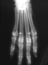
What digits and breed are commonly affected by paw pad corns?

Greyhounds (male>female)
90% thoracic digital pads 3&4 (40% had additional foot deformities)
mechanical = >50% return in 2m in Greyhounds, do not return in other species with excision and 1 layer closure
Stepped hybrid plates used for carpal arthrodesis. A, Single-step plate. B, Double-step plate.

ome ilial fractures are caudal to the body of the ilium, located directly adjacent to or extending dorsal to the acetabulum. This location requires a more dorsal plate position. A straight plate may be contoured with a simple twist to facilitate extension of the plate dorsal to the acetabulum. A, This is the dorsal view. B, Cranial and lateral view showing the dorsal position of the plate. C, End- on view of the twist contour needed for placement of a straight plate as used for the caudal ilial fracture

everal procedures can help in reduction of acetabular fractures. A, Kern bone-holding forceps applied to the ischiatic tuberosity or ischiatic table through a limited approach provide excellent control of the caudal acetabular fragment. A bone hook may assist in elevating the caudal fragment from its medially displaced location. B–C, Reduction can be maintained and compression of the articular surface achieved with point reduction forceps. Care is taken to avoid overcompression, which may alter articular shape and congruity. Care is taken to prevent crushing the sciatic nerve during retraction and reduction
note Bone hook

ixation of acetabular fractures. A, Oblique fracture types may be repairable with lag screws alone. Large dogs may need lag screws and a bone plate. B, Repair of a central transverse fracture with an acetabular plate. C, Small fragments of comminuted acetabular fractures are first held in reduction with Kirschner wires or small lag screws, and are reconstructed one at a time. D, After reconstruction of small fragments of a comminuted acetabular fracture, a bone plate is applied and must span the fracture, which requires a longer plate and careful plate contouring

As an alternative to plate fixation, pin, screw, wire, and polymethylmethacrylate composite fixation may be used for acetabular fracture repair. A, Fragments are held reduced with Kirschner wires and/or lag screws. One screw and washer is placed cranial and caudal to the fracture on the dorsal tension surface of the acetabulum. Figure-of-eight orthopedic wire connects the cranial and caudal screws. B, Polymethylmethacrylate is applied over the screw and wire fixation, creating the composite fixation
Doesn’t require perfect contour to maintain reduction

Fixation of sacroiliac luxation. A, Cranial view of the pelvis showing one lag screw deeply seated in the sacral body, at approximately 60% of sacral width. A second shorter screw is placed dorsal and cranial to the first, but must be short to avoid the spinal canal. B and C, Lateral views of the pelvis show the relationship between sacral screw position and the wing of the ilium. + marks the spot for the glide hole in the ilium and the thread hole in the sacrum. Craniocaudally in the ilial wing, the hole site is at the center of the caudal half. Proximodistally, the hole site is in the proximal one third of the ilial width. D, Lateral view of the sacral wing illustrating the location of the area for screw placement, as denoted by the clear area. This area is slightly larger than 1 cm × 1 cm in the average large dog. The cross-hatched area represents sites in which if placed here, screw depth would be short because of inadequate bone thickness or the presence of the spinal canal. The notch (a) and C-shaped cartilage (b) are used as visual landmarks in locating the site for screw placement. E, The first lag screw is inserted through the glide hole of the ilium and is directed by site into the thread hole previously drilled into the sacral body. A second short screw is located just cranial and dorsal to the first, and must stop short of the spinal canal. F, Dorsal view shows the positions of two lag screws. As an alternative, or if additional fixation is needed for stabilization, a transilial bolt, passing through the ilial wings and dorsal to the seventh lumbar vertebrae, may be used

Ischial body fractures are reduced with bone forceps, and fixation is achieved with a small contoured bone plate, taking care to preserve the sciatic nerve (Figure 57-21, A). Another fixation method using pin and tension band wire has been described.29 Avulsion fractures of the ischial tuberosity are seen and may be repaired with lag screws (Figure 57-21, B) or with pin and wire fixation
Ventral approach = hemicirclage

Radiographic view of a dog’s femur demonstrating a direct method of measuring the angle of femoral neck torsion on an axial fluoroscopic projection. Line A is drawn through the axis of the femoral neck, bisecting the femoral head. Line B is drawn parallel to the caudal surface of the distal femoral condyles. The angle formed by the intersection of these lines represents the angle of femoral torsion or anteversion.

Measurement of the Ortolani angles of reduction (left) and subluxation (right). The angles of reduction and subluxation of the hip joint, respectively, represent the maximal and minimal angles needed to provide stability for acetabular rotation

Location and orientation of pelvic osteotomies according to the procedure described by Slocum.122,123 The cranial ramus of the pubis is removed first (bottom inset), then an ischial osteotomy is performed (top inset); finally, a transverse ilial osteotomy is made immediately caudal to the caudal dorsal iliac crest (center) with the osteotomy oriented perpendicular to the long axis of the pelvis (dotted line)

Cranial view of the canine femur demonstrating two levels of osteotomy for canine total hip arthroplasty: a, region of isthmus; b, axis of femur; c, midcervical cut; d, lesser trochanter cut

Canine total hip replacement centralizer mold (A) and resulting polymethylmethacrylate centralizer (B) (BioMedtrix, Allendale, NJ)

What are treatments for CHD that have fallen out of favor?
Intertrocahnteric osteotomy
Pectineus myectomy
Shelf arthroplasty
Dorsal acetabular rim arthroplasty
Ventrodorsal radiographs of a dog with coxa valga and bilateral hip joint subluxation before (A) and after (B) intertrochanteric osteotomy. Coxa valga is accentuated by external rotation of the femur (noted by prominence of the lesser trochanter) in (A). Note how the hip subluxation is resolved in the postoperative view

Craniocaudal radiograph of the proximal femur illustrating the trabecular network of the femoral head and neck. This network is oriented to resist both tensile (A) and compressive (B) forces generated during activity. Note the orientation of the trabecular lines of the linea transversa extending from the greater trochanter to the femoral head. This presumably helps counter bending moments generated by eccentric loading of the femoral head

adiographic and gross images illustrating the angles of inclination and anteversion. Inclination, defined as the angle between the femoral neck and the anatomic axis of the femur in the frontal plane, is estimated from ventrodorsal and/or craniocaudal horizontal beam radiographs. Anteversion is defined as the angle between the femoral neck axis and the tangent to the femoral condyles. This is best illustrated using a proximal-to-distal projection of the femur; however, clinically, this projection is difficult to obtain without advanced imaging (CT). Lateral radiographs are traditionally used to estimate this angle

Radiographic appearances of the distal femur from a nonchondrodystrophic dog and a chondrodystrophic dog. In nonchondrodystrophic dogs, the distal metaphysis progressively widens from proximal to distal, which creates a smooth transition between the diaphysis and epiphysis. In contrast, the transition between the metaphysis and the epiphysis of chondrodystrophic dogs occurs at an acute angle (red line) and is associated with an isthmus proximal to the trochlea (arrowheads). This conformation is thought to predispose these breeds to supracondylar and condylar fractures

Radiographic appearance of the proximal femur of a nonchondrodystrophic dog and a chondrodystrophic dog. Note the altered contour of the femoral head, the shortened neck, and the more proximally located greater trochanter with respect to the femoral head (red X) in a chondrodystrophic dog compared with a nonchondrodystrophic dog.

Illustration of the blood supply over the cranial (radiograph) and caudal (dry bone specimen) surfaces of the proximal femur. The complex vascular network of the proximal femur includes the lateral (a) and medial circumflex (b) femoral arteries, the caudal gluteal artery (c), the extracapsular vascular ring (d), the ascending intracapsular arteries (e), the intraosseous arcuate network (f), the ligament of the femoral head (g), and the nutrient artery (h)

Classification of proximal femoral fractures with respect to their intracapsular (a through d) or extracapsular (e through g) locations. Intracapsular fractures include epiphyseal (a), physeal (b), subcapital (c), and transcervical (d). Extracapsular fractures include basic cervical (e), intertrochanteric (f), and subtrochanteric (g)

Classification of distal femoral fractures with respect to their anatomic location. These include supracondylar (a), physeal (b), and condylar (c and d) fractures. Condylar fractures are further classified as unicondylar (c) or bicondylar (d).

Radiographs showing different femoral neck fracture planes. Cervical fractures with steeper angles are associated with greater risk of postoperative instability than those with shallower angles

Craniolateral approach to the shoulder. A, Make an incision in the skin and subcutaneous tissue from just proximal to the acromion process to the proximal humerus. B, Incise the deep fascia along the cranial margin of the acromial portion of the deltoideus muscle and retract the muscle caudally. C, Isolate the infraspinatus tendon, place a stay suture in its proximal portion, and incise the tendon. D, Incise the joint capsule midway between the glenoid rim and the humeral head. Craniolateral approach to the shoulder. E, Internally rotate the humerus until the head subluxates and remove the cartilage flap. F, Curette the edges of the bony defect to ensure removal of all affected cartilage


Caudal approach to the shoulder. A, Make an incision in the skin, subcutaneous tissue, and deep fascia that extends from the midscapular spine to the midhumeral diaphysis. B, Incise the intermuscular septum between the caudal border of the scapular portion of the deltoideus muscle and the long head of the triceps muscle. C, Elevate and retract the teres minor muscle cranially, exposing the axillary nerve and joint capsule. D, Place a Penrose drain around the nerve and gently retract it caudally. E, Incise the joint capsule 5 mm from and parallel to the glenoid rim to expose the humeral head

Surgical stabilization of a medial shoulder luxation. A, Perform a craniomedial approach with biceps tendon transposition. Incise the skin and subcutaneous tissue over the greater tubercle and continue the incision medially to the midhumeral diaphysis. B, Incise the fascia along the lateral border of the brachiocephalicus muscle. C, Incise the insertions of the superficial and deep pectoral muscles from the humerus and retract them medially. Retract the supraspinatus muscle laterally. D, Transect the tendon of the coracobrachialis muscle to expose the subscapularis muscle tendon. Incise the tendon of the suprascapularis muscle. E, Incise the joint capsule to inspect the joint. F, To transpose the biceps tendon, incise the transverse humeral ligament. Make a small incision in the joint capsule under the biceps tendon to free it and move the tendon medially. Secure it to the humerus with a bone screw and spiked washer

To surgically stabilize a lateral shoulder luxation, perform a cranial approach with biceps tendon transposition. A, Expose the joint. B, Perform an osteotomy of the greater tubercle, including the supraspinatus muscle insertion. C, Incise the joint capsule and the transverse humeral ligament over the biceps tendon. D, Free the biceps tendon, and move it laterally across the osteotomy site. Hold the tendon in place, and reduce and stabilize the osteotomy with Kirschner wires and a tension band wire or lag screw

To expose the medial aspect of the elbow joint by means of transection of the pronator teres muscle, make a skin incision on the medial surface of the joint, starting at the medial epicondylar crest and extending distally over the medial epicondyle. B, Gently retract the median nerve and brachial artery and vein cranially. C, Transect the pronator teres tendon and make an incision parallel to the humeral condyle through the joint capsule and collateral ligament to expose the FCP. D, Remove the fragment

To expose the medial aspect of the elbow joint using a muscle-splitting approach, identify the demarcation between the flexor carpi radialis and the superficial digital muscles and separate and retract them. Expose the joint capsule and incise it parallel to the muscle-splitting incision to expose the coronoid process

For closed reduction of a laterally luxated elbow (B), flex the elbow and inwardly rotate the antebrachium to hook the anconeal process into the olecranon fossa. C, Then extend the elbow slightly and abduct and outwardly rotate the antebrachium while placing pressure on the radial head

To stabilize the elbow, replace collateral ligaments by using two screws and a figure-eight wire. C and D, To secure an avulsed fragment, use a lag screw with a spiked Teflon washer

Arthrodesis of the elbow. A, Make two osteotomies and remove a portion of the proximal ulna. Temporarily stabilize the elbow in the correct position with a pin placed through the ulna and into the humerus. B, Contour a plate to fit onto the caudal surface of the humerus and the caudal surface of the ulna. Place at least three screws in the humerus and three in the ulna. Use additional screws as lag screws to gain compression across the arthrodesis site

Radial lengthening of the radius using a transverse osteotomy

Cemented canine THR implants. B, Cementless canine THR implants

What complication is this?

Radiograph of aseptic loosening of a cemented THR
Coxofemoral joint stabilization by placement of a prosthetic capsule. Note the strategic placement of bone screws in the dorsolateral acetabulum. Suture material is passed from the screws through a predrilled tunnel in the dorsal femoral neck and tightened. The presence of suture in this position prevents craniodorsal reluxation. The same procedure may be performed using suture anchors

Intercondylar tubercles
cr/cd intercondyloid area - CrCL (cd-med lateral condyle & cd-lat intercondylar fossa, fibers spiral axially 90 degrees) and cr/cd meniscal ligs
Lateral area of politeal notch = CdCL (lat surface medial femoral condyle)

cr/cd meniscotibial ligs for MM and LM
intermeniscal lig
meniscofemoral lig
popliteal tendon
coronary lig (MM to MCL)
medial collateral lig - fused to MM and joint capsule
lat collateral lig - separated to LM dt popliteal tendion, loose attach to joint

Diagramatic representation of the collagen fiber orientations within the outer two thirds of the meniscus. The distribution of the type I collagen fibers is predominantly circumferential. Radial fibers are less numerous and act as a “tie” holding together the circumferential fibers. The illustration shows that a vertical longitudinal tear may develop along the circumferential fibers

Collagen fibrils - 3 layers
Collagen bundles on surface randomly oriented
Outter 2/3 = circumfrential, immermost budles in radial

Proximal aspect of the menisci showing the approximate extent of the three vascular zones of the meniscus. The red-red zone is characterized by greater vascularity and extends for 15% to 25% in the periphery of the meniscus. The rest of the meniscus is mostly avascular and is divided into the axial “white-white” zone and an intermediate zone called red-white, with only a small amount of vessels reaching this latter zone
Perimensical capillary plexus - medial and lateral genicular aa.

Figure 62-11 The Slocum active force model of the stifle joint. According to this model (A), the joint reaction force during weight bearing (magenta arrow) is approximately parallel to the longitudinal axis of the tibia, and can be resolved into a cranially directed shear force (cranially directed yellow arrow) and a joint compressive force (proximally directed yellow arrow, perpendicular to the tibial plateau). Tibial plateau leveling (B) results in a joint reactive force (magenta arrow) that is perpendicular to the tibial plateau. Thus it can only be resolved into a joint compressive force (yellow arrow); cranial tibial thrust is eliminated

The Tepic model of the stifle joint. According to this model (A), the joint reaction force (magenta arrow) is approximately parallel to the patellar ligament, and can be resolved into a cranially directed shear force (cranially directed yellow arrow) and a joint compressive force (proximally directed yellow arrow). Advancing the tibial tuberosity (B) such that the patellar ligament is perpendicular to the tibial plateau neutralizes the cranial tibial thrust force

Avulsion of the tibial attachment of the cranial cruciate ligament (A). Small fragments can be stabilized with stainless steel wire (B) placed through the ligament, around the fragment(s) and through two bone tunnels that exit on the medial cortex, where the wire is twisted. Large fragments can be stabilized with diverging Kirschner wires (C) or a bone screw placed in lag fashion

Alternatively can TX avulsion with epiphysiodesis - screw in cranial part of epiphysis - caudal continues to go and decreased TPA
In 22 dogs: reduction in TPA of 4-24 degrees
Eccentric screw = valgus

What are types of meniscal tears?
Proximodistal view of a right tibial plateau with illustrations of the classification of meniscal tears. A, intact; B, vertical longitudinal tear; C, bucket handle tear (oblique); D, flap tear; E, radial tears; F, horizontal tear; G, complex tear; H, degenerative tears

Mid-body meniscal release performed with an inside-to-outside technique (A) or with an outside-to-inside technique (

place blade caudal to the medial collateral lig and aim towards tuber of gerdi (30 degree angle)

What are the quasi isometric points?
F2 - caudal edge lateral femoral condyle immediately adjacent to distal pole of fabella
T3 - caual wll extensor groove

What is a triple tibial ostetomy?
- Similar to TTA in concept
- Partial frontal plan osteotomy with distal cortex intact
- Wedge angle = 2/3 or 0.6x angle between patellar ligament and a line perpendicular to the TP (CA) + 7.3
–Or TPA -5 degrees use if above formula <0
–Or TPA -12 degrees if small correction needed PTA close to 90 degrees
•Proposed advantages:
–Minimal change to articulating surface
–Small osteotomy gap
–No loss limb length
–“Low technical difficulty’
•Complication rate 23-36%
–Tibial tuberosity fracture, infection, postliminary tears
•Intra-op tibial tuberosity fracture occurred in 23%
Overall similar complications as TTA and TPLO combined

What are structural changes to the joint/bones that occur with MPL?
1, Coxa vara. 2, Distal femoral varus and genu varum. 3, Shallow trochlear groove with poorly developed or absent medial ridge. 4, Hypoplastic medial femoral condyle. 5, Medial displacement (torsion) of the tibial tuberosity associated with internal rotation of the tibia at the stifle joint. 6, Proximal tibial varus. 7, Internal torsion of the foot, despite external torsion of the distal tibia.

What are methods to repair a combination MPL and CCL?
If no tibial torsion is present:
TTT and LFS
TPLO with internal torsional correction combined with a de-rotational proximal tibial osteotomy
TTTA
lateral translation of the tibial tuberosity segment following TPLO/CCWO (in this case, a cranial closing wedge or a simple osteotomy at the exit of the tibial plateau leveling osteotomy caudally can be used to isolate the tibial tuberosity segment) (see Figure 62-47, A–D)373;
tibial closing wedge osteotomy and TTT

What are these?

Verbrugge bone clamp

Patellar fracture repair. Nondisplaced fissure fractures, or minimally displaced fractures can be reduced with an orthopedic wire placed circumferentially around the patella; the wire is tightened (twisted) to achieve reduction and compression. Further stability can be achieved by placing a second wire as a tension band from the tendon of insertion of the quadriceps muscle from proximal to the patella to distal to it (A). First placing a bent hypodermic needle in the desired location, and then inserting the wire through it can facilitate passage of wire through the tissue. Displaced transverse fracture of the patella (B) can be stabilized by a pin and tension band wire technique. A drill hole is made in a retrograde fashion from distal to proximal into the basilar fragment (C). The fracture is reduced, held in compression with pointed reduction forceps, and the drill hole is advanced from proximal to distal into the apical fragment (D). A Kirschner wire is placed in a normograde fashion from proximal to distal (E). The tension band wire is placed around the Kirschner wire and tightened (F). An additional tension band wire can be placed to augment the reconstruction (G). Multifragmentary patellar fractures can be reconstructed and stabilized with any combination of a Kirschner wire(s), tension band wire(s) and a wire placed circumferentially around the patella (H).

Rupture of the patellar ligament. The distracted ends of the patellar ligament (A) can be apposed by placing a tension band wire from a drill hole placed transversely across the patella, or into the insertion of the quadriceps tendon proximal to the patella to a drill hole placed in the tibial tuberosity (B–C). The wire is tightened to appose ligament ends, and a locking loop suture(s) is used to appose the ligament ends. The locking loop sutures can be augmented with additional simple interrupted or mattress sutures

tifle joint arthrodesis with bone plate fixation. Planning of the ostectomies (A). Kirschner wires 1 and 2 are placed perpendicular to the femoral and tibial shafts (dotted lines) respectively. In this case, the joint angle chosen is 140 degrees; the complementary angle is 40 degrees. Dividing this by two yields 20 degrees, thus pins 3 and 4 are placed at an angle of 20 degrees to pins 1 and 2, such that they are parallel to the planned femoral and tibial ostectomies, respectively. The tibial crest osteotomy has been completed, the tibial articular ostectomy has been completed parallel to pin 4, and the sagittal saw is placed parallel to pin 3 in preparation for the femoral ostectomy (B). The joint is temporarily stabilized by crossed pins (C). Prior to and during pin placement, ensure that Kirschner wires 1–4 are maintained in the sagittal plane to avoid tibial rotation at the stifle joint. In addition, Kirschner wires 3 and 4 should be parallel to optimize apposition of the femur and tibia at the ostectomies. Kirschner wires 1–4 are removed once the cross pins are placed, and the proper alignment and apposition of the femur and tibia are confirmed. A portion of the tibial crest is ostectomized to allow ample bone plate contact, and a compression plate is contoured and applied to the cranial surface of the femur and tibia (D). Two bone screws are applied in compression on each side of the stifle joint, and two plate screws are placed in lag fashion across the stifle joint.

A pin with a slightly smaller-than-normal diameter should be chosen, typically about 50% of the diameter of the medullary canal at the tibial isthmus

Methods to repair tibia fractures?

Malleolar fractures. A–B, Lateral malleolar fracture of the fibula with tibiotarsal luxation (4-year-old castrated male Maine Coon Cat); C, pin and tension band wire fixation; and D, screw fixation

tarsocrural (talocrural)
talcalcenal
talocalcaneaocentral
calcaneoquartal
centrodistal
tarsometatarsal

sustentaculumm tali - medial side
tuber calcanei

What are the ligaments of the Tarsus?
Thickended tarsal fascia = tendon sheath flexor helicus longus, saphenous, plantar n.
Medial tibial collateral (3)= long part (med malleous-1st tarsal: taut extension), tibiocentral (taut extension), tibiotalar (taut flexion, most substanial)
Lateral collateral ligment (fibular)(3) = long lateral (taut extension), c_alcaneofibular short_ (taut extension), talofibular short (taut flexion)
Plantar (3) = middle plantar (calcan - 4th tarsal - MT4/5), medial plantar (sustentaculum tali - tarsoMT joint), lateral plantar (cd calcaneus - long collateral -MT5)
(Long lig taut extension, underlined most improtant to fix)

Tarsal lig

Dental radiographs of a cat with separation of the mandibular symphysis. A, Before treatment. B, After circumferential wiring: note that two mobile incisor teeth were also extracted.

Final dissection of the right pelvic limb. 1, Adductor longus muscle; 2, quadrates femoris muscle; 3, external obturator muscle; 4, internal obturator muscle; 5, gemelli muscle; 6, superficial gluteal muscle; 7, middle gluteal muscle; 8, deep gluteal muscle; 9, articularis coxae muscle; 10, partially severed origin of the rectus femoris muscle; 11, tensor fasciae latae muscle; 12, biceps femoris muscle; 13, semitendinosus muscle; 14, semimembranosus muscle; 15, adductor muscle; 16, abductor cruris caudalis muscle; 17, vastus lateralis muscle; 18, sartorius muscle (cranial belly); 19, sartorius muscle (caudal belly); 20, sciatic nerve; 21, acetabulum; 22, head of the femur; 23, superficial circumflex iliac artery.
Final dissection of the right pelvic limb. 1, Adductor longus muscle; 2, quadrates femoris muscle; 3, external obturator muscle; 4, internal obturator muscle; 5, gemelli muscle; 6, superficial gluteal muscle; 7, middle gluteal muscle; 8, deep gluteal muscle; 9, articularis coxae muscle; 10, partially severed origin of the rectus femoris muscle; 11, tensor fasciae latae muscle; 12, biceps femoris muscle; 13, semitendinosus muscle; 14, semimembranosus muscle; 15, adductor muscle; 16, abductor cruris caudalis muscle; 17, vastus lateralis muscle; 18, sartorius muscle (cranial belly); 19, sartorius muscle (caudal belly); 20, sciatic nerve; 21, acetabulum; 22, head of the femur; 23, superficial circumflex iliac artery.

Labially mounted dental radiograph showing the left mandibular third and fourth premolars (teeth 307 and 308) and portions of the left mandibular second premolar and first molar (teeth 306 and 309). Moderate periodontitis is evidenced by loss of alveolar bone interdentally between teeth 306 and 307 and teeth 307 and 308, and also in the furcation region of tooth 308. AM, Alveolar margin; D, dentin; E, enamel; F, furcation; LD, lamina dura; PC, pulp cavity; PL, periodontal ligament.

Dental radiographs of the lower jaw of an 8-month-old dog. A, Before treatment: note fracture of the left mandibular body between the first premolar and the third premolar in the area of a missing second premolar (arrow). B, Interdental wiring and intraoral splinting: note that a modified Stout multiple-loop technique was used, with the wire twisted over the missing left mandibular second premolar. C, 5-week reexamination confirming fracture healing; splint and wire were removed, and root canal therapy of the endodontically affected left mandibular canine tooth (note slightly increased root canal width) was performed (not shown)

Clinical photograph of case in Figure 65-20, following interdental wiring and intraoral splinting. Note that the bis-acryl composite splint was applied primarily on the lingual surfaces of the mandibular teeth to allow closure of the mouth.

Occlusal alignment and stabilization of caudal mandibular fractures or chronic temporomandibular joint displacement may be achieved with bis-acryl composite bridges between the maxillary and mandibular canines (or other teeth)

Note that the tooth roots occupy a large portion of the mandible. Normal relationships of the maxillary and mandibular dental arcades are illustrated.

Bones of the skull, lateral aspect.

Bones of the skull, dorsal aspect.

Bones of the skull, ventral aspect

Left and right mandibles, dorsal lateral aspect

Bones of the skull, hyoid apparatus, and laryngeal cartilages, lateral aspect

Drawing of the intact mandible demonstrating normal lines of stress (red arrows) through the body of the mandible with muscular contraction and any external force applied to the rostral portion of the jaw (biting, chewing [small arrows]). B, Bending forces are the primary forces acting on the mandible during functional stress; with a fracture at this level, there is distraction along the alveolar surface of the bone; the point at C shows that the ventral portion of the bone is the only area where compressive stresses exist—if there is contact. (D, Digastricus muscle; M, masseter muscle; P, pterygoideus muscle; T, temporalis muscle.)

Cranial (A–B) and lateral (C) views of the canine skull, which is transilluminated to reveal the buttressing. The lines drawn indicate the horizontal (A) and vertical (B) buttresses of the maxillofacial region of the canine skull (compare with Figure 67-4). The three maxillofacial buttresses can be described as a combination of these horizontal and vertical buttresses (C). The nasomaxillary (medial) buttress is seen rostrally, the zygomaticomaxillary (lateral) buttress is seen caudally, and the pterygomaxillary (caudal) buttress is shown with the dashed line
Caudal = lacrimal, palatine and pterygoid bones
Can reconstruct facial frame with 2/3 butresses (rostal/medial and lateral)…if butresses not compromised does not need to be fixed

To facilitate appropriate tightening of the intraosseous wire, angled drill holes (obtuse angles [open arrows]) are placed in the bone such that after wire passage, all wire/bone contact is present with obtuse angulation, thus enabling the wire to slide easily (small arrows) during the early wire tightening process (large arrow, tension applied to the twist). B, If, for example, the drill holes are placed transversely through the bone, obtuse angles are present only at the wire/bone contact areas on the side where tension is applied (open arrows); on the opposite side of the bone, these bone/contact areas result in an acute angle of the wire as it passes through the drill hole (curved arrows). Despite appropriate tension applied to the twist, the wire cannot slide (at curved arrows), and this results in an inadequately tightened wire. C, Even in the case of appropriately angled drill holes, as the wire is tightened (as the twist moves toward the bone surface), wire/bone contact results in an acute angle of the wire (curved arrows), and the wire ceases to slide at this level; however, the obtuse angle at the wire/bone contact on the opposite bone surface (straight arrows) allowed this side to be appropriately tightened, as the wire will continue to slide more easily. D, This wire can be tightened further by levering an instrument (a periosteal elevator is shown) directly under the wire twist; this technique ensures a tight wire opposite the twist (large arrow) as it continues to slide easily with the obtusely angled corners. The twist then can be secured by taking up the remaining slack adjacent to the twist.

An intraosseous wire can adequately secure two opposing bone fragments as a rigid suture (and also compress the fragments together [arrows]). B, With an oblique fracture line or a curved section of bone, or when the cortex is thin, overriding of the bone fragments (arrows) often occurs. C, To prevent such malreduction, a Kirschner wire can be placed across the fracture site (as a “skewer-pin”) to prevent overriding, and a figure of eight wire is placed over the ends of the skewer-pin (compare with Figure 67-13, A–B). D, Alternatively, a Kirschner wire may be used as an external “splint,” where its position on the outer surface of the bone serves as an anchor to multiple wires fastened to each comminuted bone fragment (compare with Figure 67-13, C–D).

Artist’s rendition of the mandible and the relative size of the teeth to the bone. Miniplates can be contoured to match the alveolar bone surface and applied immediately adjacent to the gingival margin (as a tension band plate); the screws are directed so as to be placed in between, thus avoiding, the tooth roots. A second miniplate (or standard plate) is placed along the aboral (ventral) bone margin as a stabilization plate. The figure demonstrates appropriate plate position on the mandible and the ramus (in the latter, the tension band plate is located along the coronoid crest, and the stabilization plate is placed along the condyloid crest toward the condylar process).

Simplified schematic of proposed cytokine-mediated interactions in osteoarthritis. Chondrocytes respond to proinflammatory cytokines such as interleukin (IL)-1 and tumor necrosis factor (TNF)-α produced by activated macrophages and fibroblast-like synovial cells. In addition, chondrocytes produce these cytokines themselves to act in an autocrine/paracrine fashion. Activation of matrix metalloproteinases (MMPs) and aggrecanases leads to degradation of extracellular matrix components such as type II collagen and aggrecan. Neo-epitopes generated by such proteolytic activity are thought to lead to further proinflammatory cytokine production by synovial cells and chondrocytes. TIMPs, Tissue inhibitors of metalloproteinases; NO, nitric oxide.

Typical Total and Differential Cell Counts for Canine Synovial Fluid in Normal Joints and Joints With Osteoarthritis, Rheumatoid Arthritis, Nonerosive Immune-Mediated Polyarthritis (IMPA), and Infective Arthritis

How do you dx rhematoid arthritis?
Classical rheumatoid arthritis is diagnosed when seven criteria are satisfied, and definite rheumatoid arthritis when five are satisfied. With criteria 1 through 5, the joint signs should be present for at least 6 weeks. In addition, two of criteria 7, 8, and 10 should be satisfied; these three criteria have been considered probably the most specific for canine rheumatoid arthritis

Growth plate of a 17-week-old dog. EP, Epiphysis; EPC, epiphyseal cartilage; HZ, hypertrophic zone; MP, metaphysis; MZ, mineralization zone; PZ, proliferative zone; RZ, resting zone. Bar represents 100 µm.
Endrocondral Ossification = growth plate

1= superficial/tangential
2= transitiional
3= radiate
tide mark
4= calcified
