Development of the respiratory system Flashcards

1
Q

What does the ectoderm develop and what does this have?

A

develops a neural groove and the tips of the walls of this groove are called the neural crest

2
Q

What does the lateral plate mesoderm divide into?

A

2 layers

  • parietal (somatic)
  • Visceral (splanchnic)
3
Q

What does the parietal layer of mesoderm form?

A

body walls

4
Q

What does the visceral layer of lateral plate mesoderm form?

A

surround the organs

5
Q

What does the parietal layer of the lateral plate mesidern ‘merge’ with?

A

ecotderm

6
Q

What two processes occur during lateral folding of the trimlaminar embryo?

A
  1. primitive tolk sac becomes “pinched”. Primitive yolk sac will become the digestive tract or the gut tube. While this takes place, the visceral layer of the lateral plate mesoderm will envelope to the future gut tube
  2. The leading edge of the ectoderm and the pariteal layer of lateral plate mesoderm will move antero-medially
7
Q

Draw a diagram illustrating what happens during lateral folding of the trilaminar embryo

A
8
Q

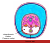
Draw a diagram of the trilaminar embryo

A
9
Q

What fuse together in the ventral mid line at the end of lateral folding?

A

parietal layer of lateral plate mesoderm and ectoderm

10
Q

What becomes the parietal pleura?

A

parietal layer of lateral plate mesoderm

11
Q

What are the main features of the embryo at the end of lateral folding

A
  1. a cavity (the coelom) which will become the thoracic abdominal cavity (the cavities are continuous cia the pericardio-peritoneal canals until the diaphragm forms)
  2. A body wall (thoracic wall) made out of a surface of ectoderm and pariteal layer of lateral plate mesoderm deep to it
  3. The gut tube, suspended by visceral layer of lateral plate mesoderm
12
Q

What also occurs while lateral folding is taking place?

A

cranio-caudal folding

13
Q

Draw what an embryo looks like at the end of lateral folding of the trilaminar embryo

A
14
Q

What happens when the embryo is about 4 weeks old?

A

In the cervical portion of the gut tube, in the ventral midline, the respiratory diverticulum starts to appear

15
Q

How does the respiratory diverticulum expand?

A

Ventrally and towards the chest, in front of the gut tube

16
Q

How is the respiratory diverticulum separated from the gut tube?

A

trans-eosphgeal ridges grow towards each other

17
Q

What does the diverticulum bifurcate into? And what subsequently happens?

A

2 branches

later these 2 branches expand and dilate. terminal ends of these dilations are called lung buds that will become lungs

18
Q

What does the gut tube into?

A

the digestive tract between the mouth and the anus

19
Q

When do pharyngeal arches start to appear?

A

4th-5th weeks of development

20
Q

What are pharyngeal arches separated by?

A

pharyngeal clefts

21
Q

How many pharyngeal arches are there? How are they numbered and what elements do they contain?

A

5

Numbered 1,2,3,4,6

  • cartilaginous element
  • an artery (aortic arch)
  • a nerve (Cranial)
22
Q

What is maintained even though the tracheo-esophgeal septum separated the gut tube from the upper respiratory tract

A

laryngeal opening

23
Q

What does the upper end of the respiratory diverticulum become?

A

larynx

24
Q

What does the middle section of the respiratory diverticulum become?

A

trachea

25
Q

What does the lower end of the respiratory diverticulum become?

A

tertiary bronchi, broncioles and the alveoli

26
Q

What does the bronchial tree push against as it grows and expands?

A

visceral layer of lateral plate mesoderm; which develops into visceral pleura

27
Q

Where is the epithelium of the respiratory system derived from?

A

endoderm

28
Q

The lung develops in 4 overlapping stages. What are these?

A
  1. Pseudoglandular
  2. Canalicular
  3. Terminal Saccular
  4. Alveolar
29
Q

What 2 stages are no compatible with life?

A
  1. Pseudoglandular
  2. Canalicular
30
Q

What secretes surfactant? And from when?

A

type 2 pneumocytes

from 20 weeks

31
Q

From when can the foetus survive?

A

28 weeks

32
Q

What are the four sources of origin of the diaphragm?

A
  1. pleura-peritoneal folds
  2. pleura-peritoneal folds fuse with septum transversum
  3. Mesentary of the oesphagus
  4. Muscular in-growth from the body wall
33
Q

What does the septum transversum form?

A

the central tendon of diaphragm

contains myoblasys frim the somites in C3,4,5 (phrenic nerve)

34
Q

What do the crura develop from?

A

mesentary of the oesphagus

35
Q

What causes a diaphragmatic hernia

A

failure of fusion

36
Q

Explain the 5 types of tracheo-esophgeal fistula

A

A + B = congenital abnormalities of the trachea and oesophagus, there is communication between the trachea and oesphagus, usually companied by esophageal atresia (gut = disconnected)

C, D + E = When the baby attempts to feed milk enters the trachea causing choking and possibly pneumonia. there is also the potential for retrograde passage of gastric acid from the GI tract into the respiratory system

37
Q

What is the most common congenital disorder of the respiratory system?

A

oesophgeal atresia

38
Q

What occurs in oesophgeal atresia?

A

During a normal pregnancy, the foetus swallows amniotic fluid which is resorbed from the gut and returned to the maternal circulation

If oesophageal atresia develops, this circulation of fluid is prevented and polyhydramnios develops (excess amniotic fluid)

39
Q

What are the 3 different types of diaphragmatic hernia?

A
40
Q

What causes respiratory distress syndrome?

A

deficiency in surfactant