Autonomic nervous system Flashcards
(39 cards)
What are general charecteristics of the Somatic Nervous System
- Voluntary or subconscious control single nueron pathway
- No ganglia involved in pathway
- Sensory input from general and special senses , motor output to skeletal muscle
- Excites using acetylcholine
- Axons are thick and myelinated=fast condution
- come conscious control
Autonomic Nervous System
- INvoluntary or unconscious control two neuron pathway
- Ganglia involved in pathway
- Sensory input from general and vsiceral senses motor output to cardiac ,smooth muscle and glands
- can excite or inhibit function using acetylcholine and norepinephrine
- axons are thin, some are myelinated , others are not=slower conduction
Autonomic Plexuses
- Collection of sympathetic postaganglionic axons(already synapsed aka grey) and parasympathetic preganglionic axons, as well as some visceral sensory axons
- Sympathetic(from spinal cord via sympathetic trunk) and parasympathetic(from cranial and caudal repositories) plexuses are close to one another, but they do not interact or synapse with one another
- Provide a complex innervation pattern to their target organs
Where do the prevertibral ganglia located?
How about hte paraverterbral ganglia?
- Prevertrebral ganglia only show up in the abdomen
- Paravertrebral ganglia run all the way from superior cervical ganglia down to the tailbone
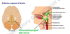
Cardiac plexus
- Increased sympathetic activity increases heart rate blood pressure,while
- Increased parasympathetic activity decreases heart rate
Pulmonary plexus
- Parasympathetic pathway causes bronchoconstriction and increased secretion from mucous glands of hte bronchial tree
- sympathetic innervation causes bronchodilation
- when you inhale you are acting on the sympathetic
Esophageal plexus
Abdominal aortic plexus
hypogastric plexus
- Parasympathetic axons control the swallowing reflex
- Consisst of the celiac plexus,superior mesenteric plexus, and inferior mesenteric plexus=autonomic control of digestion
- Innervates pelvic viscera=autonomic control of urinary and reproductive function
Two Nuerotransmitters used in the ANS
- Acetylcholine(ACh)
- norepinephrine(NE)
- Neurotrnsmitters released by the presynaptic cell
- bind to specific receptors in the postsynaptic cell membrane
- binding has eitehr an excitattoyr or an inhibitory effect on the effector depending on the specific receptor
Is parasympathetic division release acetycholine and if so which axons?
- Both preganglionic and postganglionic axons release acetylcholine and thus are called cholinergic
Does hte sympatehtic release acetycholine and norepinephrine
- Only the preganglionic axons in the sympatehtic division release acetycholine and are thus cholinergic
- Most of the postganglionic axons of the sympathetic division release norepinephrine and are called adrenergic
What is this ?

Parasympathetic
- Long preganglionic fibers
- myelinated
- very short postganglionic fiber
- many of postganglionic fibers are very short
What is this ?

Sympathetic
- Multiple axons from a single cell body-that makes it react a little quicker reaction
- short preganglionic,linger ,and multi axialted post ganglion
- One single cell can act on multiple axons
Autonomic(Visceral)nervous system explain

- Preganglionis autonomic motor neuron
- motor information is passed through preganglionic and ganglionic neurons
- Ganglionic autonomic motor nueron
- Sends nerve impulses to smooth msucle , cardiac muscle and glands
- Visceral sensory neuron
- Receives sensory information from blood vessels and visceral walls
- Follows the same pathway as the somatic sensation-goes through the dorsal root,has a cell boy in the dorsal root ganglia
Dual innervation
- Many visceral effectors(organs) are innervated by postganglionic axons from both ANS divisions
- Actions of the divisions usually oppose each other
- Opposing effects are also achieved by increasing or decreasing activity in one division(one division can up-regulate-or down regulate the other)
Autonomic Reflexes
- ANS helps maintain homeostasis through the involuntary ativity of autonomic reflexes or visceral refelxes
- Consist of smooth muscle contractions,cardiac muscle contractions ,or secretion by glands that are medaited by autonomic reflex arcs in response to a specific stimulus
- ex. Micrturition refelx,which partly controls the release of urine
- Other reflexes include alteration of heart rate ,cahgnes in respiratory rate and depth, regluation of digestive system activities , and alteration of pupil diamter
- comparable to spinal refelxes
- Classic autonomic relfex invovles the reduction of BP

CNS contorl of Autonomic function
- Autonomic function is influenced by the cerebrum, hypothalamus , brainstem,and spinal cord
- Sensory processing in the thalamus and emotional state controlled in the limbic system directly affect the hypothalamus
- the integration and command center for autonomic functions
- contains nuclei that conrol visceral functions in both divisions of the ANS
- communicates with other CNS regions,including the cerebral cortex, thalamus,brainstem,cerebellum,and spinal cord

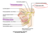
Organization and anatomy of the sympathetic divsion
- More complex thatn the parasympathetic division , both anatomically and functionally
- Sympathetic preganglionic neuron cell bodies are housed in the lateral horns of hte T1-L2 regions of the spinal cord
- CEll bodies in the brain are influencing these cell bodies in the spinal cord
- Only place you have the actual sympathetic preganglionic neuron cell bodies is in the spinal cord from T1-L2
- Preganglionic sympathetic axons travel with somatic motor neuron axons to exit the spinal cord and first enter the anterior roots and then the T1-L2 spinal nerves
- Its with the spinal neve until it goes into the sympathetic trunk
- Preganglionic sympathetic axons remain with the spinal nerve for a short distance before they branch off and leave the spinal nerve
Related to the sympatehtic division

- T1-T4: head heart lungs
- T5-T9: upper portions
- T11-L2: lower part of gut and pelvis

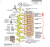
Left and Right sympathetic Trunks
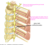
- Immediately anterior to the paired spinal nerves are the left and right sympathetic trunks
- EAch is loacted immediately alteral to the vertebral column
- A sympathetic trunk looks much like a string of beads
- Strin is composed of axons
- beads the sympathetic trunk(or paravertebral)ganglia, whch house sympathetic ganglion neuron cell bodies
- one sympathetic trunk ganglion is approximately associated with each spinal nerve
- Cervical portion of each sympathetic trunk is partitioned into only three sympathetic trunk ganglia– the superior, middle , and inferior cervical ganglia– as opposed to the eight cervical spinal nerves
White Rami
- Connecting the spinal nerves to each sympathetic trunk are rami communicantes
- connect spinal nerves to each sympathetic trunk
- Carry preganglionic sympathetic axons from T1-L2 spinal enrves to the sympathetic trunk
- Asoociated only with T1-L2 spinal nerves
- Preganglionic axons are myelinated
- White ramus has a whitish appearance
- similar to entrance ramps on a highway
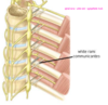
Gray Rami
- Carry postganglionic sympathetic axons from the sympathetic trunk to the spinal nerve
- Axons are unmyelinated
- gray rami have a grayish appearance
- similar to exit ramps on a highway
- connect to all spinal nerves,including the cervical,sacral, and cocygeal spinal nerves
- In this way sympathetic information that tarted out in the thoracolumbar region can be dispersed to all parts of the body
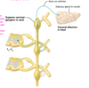
Splanchic nerves
- Composd of preganglionic sympatehtic axons
- they are myelinated come out ofthe sympathetic trunk unsynapsed to go to other ganglia elsewhere
- Run anteriorly from the sympathetic trunk to the most of hte viscera
- should not be confused with the pelvic splanchnic nerves associated with the parasympathetic division
- Larger Splanchnic nerves(all of theese sympathetic)have specific names
- Greater thoracic splanchnic nerves
- Lesser thoracic splanchnic nerves
- least thoracic splanchnic nerves
- lumbar splanchnic nerves
- sacral splanchnic nerves
- Terminate in prevertebral(or collateral) ganglia
- Called prevertebral because they are immediately anterior to the vertebral column on the anterolateral wall of the abdominal aorta
- Prevertebral ganglia typically cluster around the major abdmoinal arteris and are named for these arteries
- Piggyback on those arteries to get to their organs
- Sympathetic postganglionic axons extend away from the ganglionic neuron cell bodies in these ganglia and innervate many of hte abdominal organs

What are prevertebral ganglia and the types?
- Differ from the sympathetic trunk ganglia
- Are single strucutres ,rather then paired
- Anterior to the vertebral column on the anterior surface of the aorta
- Located only in the abdominopelvic cavity
- Prevertebral ganglia include the celiac ,superior mesenteric , and inferior mesenteric ganglia
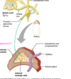
Sympathetic pahways
- Spinal nerve pathway
- postganglionics come out of the sympathetic system and the bulk fo them go to the skin,vessels, etc
- postganglionic sympatehtic nerve pathway
- Splanchnic nerve pathway
- they don’t synapse in the sympathetic trunk–they go to the gut and synapse in pre-vertebral ganglia
- Adrenal medulla pathway
- Essentially a splanchnic pathway
- Specific to a specific organ, the only time where there is no synapsing before it ges to the organ.
- take a myelenated track to go stratight in
- Don’t syanpse