Cornea Flashcards

1
Q

what keeps the cornea dehydrated

A

epithelium - barrier to tear film

endothelium - active pump and barrier to aqueous humor

2
Q

what 4 factors keep the cornea clear

A

avascular

non-mylinated nerves

dehydrated

ordered cell arrangement

3
Q

how long does epithelialization take to occur

A

7 days or less (even wirh complete epithelial loss)

4
Q

stromal healing results in _____

A

fibrosis/scarring

this takes days to weeks

5
Q

what is a facet

A

non-staining depression in the cornea

Often, the epithelium often slides over remodeled stroma before it becomes level with surrounding epithelium

6
Q

how long does it take a descemetocele take to heal

A

weeks to months

why referral and surgical repair is recommended

7
Q

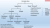
blue corneal opacity =

A

edema

8
Q

2 possible causes of blue corneal edema

A

epithelial barrier disruption - tear film entry into hydrophilic stroma

endothelial barrier/pump disruption

9
Q

3 ways endothelial barrier/pump can be disrupted

A

focal loss

generalized reduction in number

generalized reduction in function - glaucoma, uveitis

10
Q

what causes red corneal opacity

A

corneal neovascularization

11
Q

2 sources of red corneal opacity

A

superficial neovascularization

deep neovascularization

12
Q

superficial neovascularization occurs with

A

KCS, eyelid conformation or hair abnormalities, superficial corneal ulcers

13
Q

how long does it take for superficial neovascularization to occur

A

3 days from insult to start growing vessels

progress ~ 1mm per day

14
Q

what is an indication of chronic stimulation leading to superficial neovascularization

A

granulation tissue causing a dense raised collection of superficial vessels

15
Q

what are ghost vessels

A

non-perfused, empty vessels

occurs when stimulus/irritant has been removed

16
Q

what is ciliary flush and when does it occur

A

360º deep neovascularization

occurs with uveitis

17
Q

white corneal opacity with grey or wispy features indicates

A

fibrosis

18
Q

white corneal opacity with yellow or green hue indicates

A

white blood cell infiltration

19
Q

white corneal opacity that is crystalline or chalky indicates

A

mineral or lipid - dystrophy, degeneration

20
Q

WBC infiltrates detected or not detected

A
21
Q

characterisitics of WBC infiltrate

A

painful

associated with severe corneal disease

often associate with uveitis

signals corneal infection

22
Q

this corneal opacity is due to

A

corneal fibrosis

dull white; corneal scar from previous corneral laceration

23
Q

characteristics of corneal fibrosis

A

non painful

corneal scarring from previous keratitis

involves contracture of lamellar stromal collagen

24
Q

what is causing the opacities in the pictures below

A

cyrtalline white - corneal lipid degeneration

chalky white - corneal calcific degeneration

25
corneal degeneration
can be lipid or mineral secondary to: primary corneal disease or systemic disease
26
corneal dystrophy
most often lipid
27
2 causes of brown or black corneal opacities
pigment (melanin) - epi/endothelial feline corneal sequestrum
28
what dis?
feline corneal sequestrum
29
epithelial corneal pigment
origin - conjunctival melanin occurs as a result of chronic keratitis esp. in brachycephalic breeds
30
endothelial pigment
origin - uveal (iris) melanin occur congenitally (persistent pupillary membranes) or aquired (anterior synechial and uveal cysts)
31
what is the only cause of tan or greasy punctate in the cornea
keratic precipitates
32
what is the classic pattern of keratic precipitates
clear of precipitates dorsally small precipitates at mid cornea larger and larger precipitates as you move ventrally
33
this is helpful
but seriously...look at it
34
4 brachycephalic risk factors for corneal ulcers
ocular prominence decreased corneal sensitivity adnexal abnormalities tear film abnormalities
35
T/F topical steroids can be used in the treatment of ulcerative keratitis
**False!** *never use any types of topical steroid in cases of ulcerative keratitis, cause delayed healing and enhanced corneal destruction*
36
what consitutes a superficial corneal ulcer
loss of epithelium with no stromal loss
37
simple "uncomplicated" superficial corneal ulcers
superficial corneal ulcer that heals in 7 days or less
38
causes of superficial corneal ulcers
**irritants** - foreign body, tear film abnormalities, adnexal abnormalities **infection** - herpes (cats) **trauma**
39
treatment of simple superficial corneal ulcers
e collar! broad spectrum antibiotics 3-4x/day atropine 1-2x/day or until pupil stays dilated recheck in 5-7 days
40
3 possible causes of complicated/complex corneal ulcers
indolent ulcer persistent irritant (underlying cause has not been identified/treated) infection
41
Indolent ulcers (aka spontaneous chronic corneal epithelial defect) signalment/ history
middle age to older dogs - NOT CATS boxers corneal ulcer that won't heal - will take months is left untreated
42
diagnosis of indolent ulcers
test epithelium with cotton tiped applicator normal epithelium will not debride easily
43
treatment of indulent ulcers
**debridement!** - reset relationsho between epithelium and stroma once debrided - treat typucally similar to simple superficial corneal ulcers
44
what is the most important part of your approach to corneal ulcers
trying to find and treat the underlying cause (if possible)
45
3 possible infectious causes of complex corneal ulcers
FHV-1 - **Most Common** early bacterial (cats and dogs)\*\* early fungal (cats and dogs)\*\* \*\*This is where we transition into deep corneal ulceration as bacteria especially have affinity for stroma\*\*
46
what features indicate which complex category this dog's eye falls into
fluorescein "halo" - staining under the epithelial edge indicates indolent ulcer
47
what is the most likely cause of this cats complex ulcer
**Herpes!** *most common cause of non-healing ulcer in a cat is FHV-1*
48
why should you never perform ASP, GK, or DBD on cats with corneal ulcers
feline corneal sequestrum often occurs
49
T/F ASP, GK, or DBD should never be performed on infected ulcers
**True** *risks a severe progression of the infection*
50
what is the most common ophthalmic condition seen in practice that will require a decision of whether or not to refer to specialist
deep (stromal) corneal ulcer
51
99% of deep (stromal) corneal ulcers have \_\_\_\_\_\_
**corneal infection** bacterial or fungal (mostly horses)
52
collagenolysis
enzymatic destruction or corneal collagen
53
keratomalacia
softening or "melting" of corneal stroma due to collagenolysis from bacterial/fungal and neutrophil enzymes
54
3 most common bacteria involved in deep corneal ulcers
Staphylococcus spp. Streptococcus spp. Pseudomonas aeruginosa - most common cause of melting
55
what is an important identifying feature of stromal ulceration
reflux uveitis
56
clinical signs of reflex uveitis
miosis aqueous flare diffuse corneal edema hypopyon or hyphema
57
necessary diagnositics for deep corneal ulceration
STT - more important in normal eye fluorescein stain topical anthesia cytology aerobic culture tonometry - avoid in fragile eyes
58
treatment of deep corneal ulceration
e-collar antibacterial therapy anti-collagenase therapy reflux uveitis and pain management +/- surgical management and referral
59
antibiotics used to treat deep corneal infections
cephalosporins neomycin polymixin B Gtamicidin fluroquinolones and aminoglycosides high frequency of application (q1-2hrs)
60
3 indications for oral antibiotics with deep stromal ulcers
ulcer has become vascularized cornea is close to perforation or has perforated iatrogenic vascularization
61
anti-colagenase therapy options
autologous serum 1% EDTA solution 10% N-acetycysteine at lease every 2-4 hours
62
pain management from reflux uveitis
atropine - paralyzes ciliary body (tx until pupil is dilated; 1-2x per day) +/- oral anti-inflammatory and pain management (NSAID, tramadol)
63
3 guidelines for referral/surgical repair
ucler has 50% or greater stromal depth ulcer is failing aggressive medical therapy descemetocele or perforation
64
what dis?
Pannus aka chronic superficial keratitis ## Footnote *raised granulaton tissue*