Exam 1 Flashcards

(208 cards)

1
Q

Anatomy

A

The study of structure

2
Q

Physiology

A

The study of function

3
Q

Levels of organization: order

A
  1. Chemical 4. Cellular 5. Tissue 6. Organs 7. Organ systems 8. Organism
4
Q

Chemical level

A

includes atoms (C, O, H, N), the smallest units of matter. These 4 are essential to life. Atoms bond together to form MOLECULES.

5
Q

Cellular level

A

Molecules combine to form Organelles. Organelles function together to form a CELL, the smallest unit of life. There are different types of cells with different functions. A cell consists of about 70-95% water.

6
Q

Tissue level

A

Similar cells join together to perform a specific function. There are 4 different classifications of tissue: epithelial tissue, connective tissue, muscle tissue, nervous tissue.

7
Q

Organ level

A

2 or more different types of tissue come together to perform a specific function. Example = the STOMACH is composed of epithelial tissue + muscle + nervous + connective tissue.

8
Q

Organ system level

A

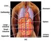
Organs work together . ex. The stomach joins with other organs like the intestines and the liver to digest food, forming the DIGESTIVE SYSTEM.

9
Q

Organism level

A

These SYSTEMS join together to form an ORGANISM, a living individual.

10
Q

HOMEOSTASIS & HOMEOSTATIC IMBALANCE

A

The term used to describe the body’s ability to maintain these stable conditions is HOMEOSTASIS. ( Ex. thermostat and heater and temp regulation in the hypothalamus.) Disease can be thought of as homeostatic imbalance.

11
Q

Negative feedback

A

“the diminution or counteraction of an effect by its own influence on the process giving rise to it, as when a high level of a particular hormone in the blood may inhibit further secretion of that hormone, or where the result of a certain action may inhibit further performance of that action.” Examples of negative feedback are B.P control

12
Q

Positive feedback

A

“the enhancement or amplification of an effect by its own influence on the process that gives rise to it.” Examples of positive feedback are aging, child birth, and lactation

13
Q

Anatomical position

A

That reference point for human A&P is the ANATOMICAL POSITION. This is the position of the skeleton, with the palms and feet forward. The terms are based on this position.

14
Q

Superior/cephalic/cranial

A

x

15
Q

Inferior/caudal

A

x

16
Q

Anterior/ventral

A

x

17
Q

Posterior/dorsal

A

x

18
Q

Medial

A

x

19
Q

Lateral

A

x

20
Q

Superficial

A

x

21
Q

Deep

A

x

22
Q

Proximal

A

x

23
Q

Distal

A

x

24
Q

Supine

A

x

25
Prone
x
26
Sagittal plane
vertical plane that divides the body or organ into a right and a left side.
27
Midsagittal plane/median plane
It’s Sagittal plane running through the nose & giving equal right & left halves
28
Parasagittal plane
PLANE:Its Sagittal plane that does not run in the middle and therefore does not gives equal right and left halves.
29
Frontal/coronal plane
divides specimen into an anterior and a posterior section.
30
Transverse plane
divides specimen into a superior and an inferior part.
31
Oblique
divides specimen into a diagonal section
32
Body cavity
A body cavity is a space within the body that contains internal organs.Cavities help to protect, separate and support organs.
33
Dorsal cavity
It consists of a) Cranial Cavity and b) Vertebral Cavity a) Cranial Cavity – it contains the brain b) Vertebral Cavity – contains the Vertebral column and spine
34
Ventral cavity
It consists of a) Thoracic cavity b) Abdominopelvic cavity
35
Abdominopelvic cavity
Abdominopelvic cavity is further divided into abdominal cavity and pelvic cavity by the Pelvic Diaphragm
36
Thoracic cavity
located superior to the DIAPHRAGM and contains areas that house the LUNGS which are separated by an area called the MEDIASTINUM which houses the heart.
37
DIAPHRAGM
x
38
MEDIASTINUM
The mediastinum is a region between the lungs, from the sternum to the vertebral column. The heart is located within the mediastinum (in
39
ABDOMINAL CAVITY
Exists right below the diaphragm and contains many of the digestive organs.
40
PELVIC CAVITY
Exists inferior to the abdominal cavity and contains the lower digestive organs, some of the organs of the urinary system as well as the reproductive organs.
41
MUCOUS MEMBRANE
It is a thin membrane that lines the cavities that open to the outside of the body. Examples are nose, mouth, Anus. This membrane produces a fluid called Mucus (pay attention to the spelling), that protects the surface of these cavities.
42
SEROUS MEMBRANE
A serous membrane is a thin sheet of cells that line cavities that do not open to the outside of the body. They produce a small amount of fluid called SEROUS FLUID
43
TYPES OF SEROUS MEMBRANE
Types of Serous Membrane: i) Pleura-(lung) ii) Pericardium- (heart) iii) Peritoneum- (abdomen)
44
CYTOLOGY
x
45
HISTOLOGY
x
46
PATHOLOGY
x
47
AXILLARY
x
48
BRACHIAL
x
49
CUBITAL
x
50
ANTEBRACHIAL
x
51
INGUINAL
x
52
PECTORAL
x
53
POPLITEAL
x
54
PEDAL
x
55
ANATOMICAL IMAGING
x
56
MAGNETIC RESONANCE IMAGING
x
57
CT
x
58
X-RAYS
x
59
COMPUTER TOMOGRAPHIC
x
60
ULTRASOUND
x
61
DYNAMIC SPATIAL RECONSTRUCTION
x
62
DIGITAL SUBTRACTION ANGIOGRAPHY
x
63
POSITION EMISSION TOMOGRAPHY
x
64
ATOMIC STRUCTURE
proton: subunits that are positively charged found within the NUCLEUS of the atom neutron: " " " neutral that are also " " " " " " " electrons: " " " negatively charged found moving in ENERGY SHELLS around the nucleus
65
ATOMIC NUMBER & MASS NUMBER
of protons # of protons + # of neutrons
66
ISOTOPE
same # of protons but diff. # of neutrons
67
HALF LIFE
the amount of time required by the element to reduce its strength of radioactivity to half
68
VALENCE SHELL
is the outside shell of the atom. When a chemical reaction occurs, the electrons in the outside orbital will be the only part of the atom that will participate.
69
INERT ELEMENT
is an element whose valence electron shell is full. EX. Noble gases in the periodic
70
REACTIVE ELEMENTS
is an element whose valence shell is empty.
71
MOLECULE
Atoms do not normally exist in nature, but join or BOND together in different ways to make a MOLECULE, a structure formed by atoms bound together by chemical bonds. Thus a molecule is formed when two or more atoms of the same kind or of different kind combine they form a molecule. When two or more different types of atoms chemically combine they form a compound.
72
IONIC BOND
1. IONIC BOND: bond formed by 1 atom losing an outside electron(s) to another, which gains the e-. These bonds are fairly strong. If you dissociate a molecule that has been held together by ionic bonds, ions results,
73
ION
A charged atom. Ions are also referred to as electrolytes. The human body is full of ions, resulting from chemical reactions. The charges on these ions are extremely important in the workings of the body. Some common ions include: Na+, K+, Cl+, etc
74
ELECTROLYTES
They dissociate in water and consist of cations (+) and anions (-). They conduct electric current and the currents is detected by the electrodes.
75
NONELECTROLYTES
They do not dissociate in water and do not conduct electricity.
76
COVALENT BOND
This type of bond is extremely important in life. Covalent bonds result when 2 or more atoms form a molecule by sharing of electrons ( these electrons will be found in the outer orbit). The bond that is formed is very STABLE and Strong and allows for DIVERSITY of molecules that can be formed. This is the way CARBON binds together.
77
POLAR VS NONPOLAR
bonds in a molecule so that the molecule is polarized in portions but neutral as a hole. molecule is not polarized Polar : unequal sharing of electrons between 2 different atoms held together by covalent Non-polar: equal sharing of electrons between 2 different atoms in a molecule so that the
78
HYDROGEN BONDS
Think of these more as an attraction than actual bonding between molecules, especially important in DNA structure (genetic material). It is important in maintaining molecule’s 3
79
CHEMICAL REACTIONS
When there is formation or breaking of bonds, chemical reactions are occurring. Thousands of reactions occur within the body per minute, rearranging atoms and leading to new molecules that have new chemical properties and new functions. Life is dependant upon these chemical reactions.
80
REACTANTS
These are substances that enter into a chemical reaction.
81
PRODUCTS
These are substances that result from the reaction.
82
REVERSIBLE REACTIONS
In these reactions the forward reaction equals the backward reaction
83
METABOLISM
is the sum of all chemical reactions occurring in the organism. This process can be divided into 2 subtypes: CATABOLISM and ANABOLISM.
84
CATABOLISM
1. CATABOLISM: reactions in which the chemical structures are broken down into smaller structures. ex. NaCl à Na+ + Cl-
85
ANABOLISM
2. ANABOLISM: reactions in which there is a combination of 2 or more chemical structures, resulting in a larger structure. ( protein synthesis)
86
HYDROLYSIS
Hydrolysis-add H2O and break (catabolism)
87
DEHYDRATION SYNTHESIS
Hydrolysis-add H2O and break (catabolism)
88
ORGANIC
Most organic molecules are large, contain carbon, and have covalent bonds. Organic chemicals that are biologically active are called biochemicals. There are 5 major organic molecules, all of them containing CARBON and HYDROGEN, as well as other elements. These 5 are CARBOHYDRATES (ex Glucose), LIPIDS (ex Fatty Acid), PROTEINS (ex Amino Acid), NUCLEIC ACIDS (ex Nucleotides)and ATP.
89
INORGANIC
molecules that do not contain CARBON. These inorganic molecules include water, acids, bases and salts. Exceptions are CO2, CO, and HCO3
90
CARBON
x
91
WATER
is a vital inorganic molecule. ( H2O) It makes up 50-60% of body weight and 70-95% of the cell a. It is an excellent solvent, dissolving many molecules (solutes) why? Because it is a polar, covalent molecule with hydrogen bonds and it can make hydration shell around each ion of the solute. b. It participates in many chemical reactions c. lubricant/protection d. temp regulation (high specific heat) e.adhesion attraction between water molecules and the container walls f. cohesion (capillary action) g. water can exist in all 3 states ie solid, liquid, gas
92
SALT
Molecule that when dissociates (ionizes) will not yield H or OH. The most plentiful salts in the human body are the calcium phosphates that are found in teeth and bones. When they become ions, they are important in nervous system and muscle contraction
93
ACID
Molecule that when ionizes (comes apart) in water, will yield H+ ion. It is a proton donor. ION = charged atom. HCL gives H+ and CL-
94
BASE
Molecules that when dissociates in water will yield OH ions (OH)- or is a proton acceptor. NaOH gives Na+ and OH-
95
BUFFER
A substance that has the capacity to maintain pH at a specific level (in other words, resists changes in pH). It is a combination of weak acid-base pair.
96
pH Strength
folds in multiples of 10.
97
ACIDOSIS
Acidosis is an increased acidity in the blood and other body tissue (i.e. an increased hydrogen ion concentration). If not further qualified, it usually refers to acidity of the blood plasma. Occurs below pH of about 7.35?
98
ALKALOSIS
Alkalosis refers to a condition reducing hydrogen ion concentration of arterial blood plasma (alkalemia). Generally, alkalosis is said to occur when pH of the blood exceeds 7.45. The opposite condition is acidosis (when pH falls below 7.35).
99
OXYGEN
O2
100
CARBON DIOXIDE
CO2
101
CARBON MONOXIDE
CO
102
BICARBONATE
HCO3
103
BIOCHEMICALS
Organic chemicals that are biologically active are called biochemicals.
104
MONOMER \< POLYMER \< MACROMOLECULE
x
105
CARBOHYDRATES & FUNCTIONS
Compounds that are made out of C, H and Oxygen. Formula is CH2O. They are of 3 types Monosaccharides, Disaccharides and Polysaccharides. FUNCTIONS: supply energy and structure
106
GLUCOSE
a monosaccharide
107
MALTOSE & SUCROSE
a disaccharide
108
POLYSACCHARIDES
Long chains of many monosaccharides bonded together. These are also too large to get through a cell membrane and are usually used as a storage molecule. ex. GLYCOGEN = a polysaccharide that is made up of bonded glucoses. This large structure serves to store glucose in the liver and muscles of animals. When needed the bonds are broken and free glucose is transported into the cells. Polysaccharides form part of cell surface markers.
109
MONOSACCHARIDE (6, 5, 3)
Simple sugars that are composed of 1 molecule. ex. glucose and fructose
110
DISSACHARIDES
double sugars that are composed of 2 monosaccharides, covalently bonded Examples of these would be SUCROSE (table sugar) and MALTOSE.
111
GLYCOGEN
polysaccharide in animals (starch and cellulose in plants).
112
ISOMER
These have same chemical composition but different arrangements ex is Glucose and Galactose
113
TYPES OF ISOMERS
Structural Isomers- switching of attached groups at the specificcarbon positions Stereoismers- switching of atoms at the same carbon position Enantiomers- mirror image
114
LIPIDS & FUNCTIONS
Lipids are substances that will not dissolve in water; made also from C, H, and O. FUNCTIONS: Energy source, food storage, structure (cell membrane), steroid formation, insulation and protection.
115
TRIGLYCERIDES (FAT)
Examples of lipids found in the body are: triglycerides (Fat), the most abundant form of fat in the human body, composed of 1 glycerol plus 3 fatty acid chains.
116
PHOSPHOLIPIDS
Phospholipids, an important part of the cell membranes and steroids (include sex hormones and cholesterol).
117
STEROIDS
Steroids are 4 ring C compounds and they give fluidity to the cell membrane EX of Steroids are: – Cholesterol – bile salts – Estrogen – testosterone. Functions of Steroids: – They are physiological REGULATORS – They give fluid mozaic model to cell membranes.
118
PROTEIN & FUNCTIONS
molecules made from C, H, O and N and sometimes I. Ex is insulin. FUNCTIONS: Energy (last resort), structure (cell membrane), hair and nail (Keratin), protection (antibody immunoglobulins), membrane transport (carrier proteins), regulate processes (enzymes), muscle contraction
119
ESSENTIAL 8 AMINO ACIDS
(not synthesized by the organism therefore essential in the diet)
120
NON ESSENTIAL 12 AMINO ACIDS
(synthesized by the organism).
121
TYPES OF PROTEINS
Dipeptide ….2 Amino Acid joined by a peptide bond Tripeptide …..3 Amino Acid joined by peptide bonds Polypeptide …4 or more Amino Acids joined by peptide bonds
122
LEVELS OF PROTEIN STRUCTURE
Dipeptide ….2 Amino Acid joined by a peptide bond Tripeptide …..3 Amino Acid joined by peptide bonds Polypeptide …4 or more Amino Acids joined by peptide bonds
123
ENZYMES
PROTEIN THAT INCREASES THE RATE OF CHEMICAL REACTIONS (ie:CATALYST). Enzymes are found inside of cells and in fluids outside of cells and are responsible for the thousands of chemical reactions that occur within the body. Their 3-D shape contains active site where reactants attach. This leads to Induced Fit Hypothesis meaning the enzyme changes it’s shape to accommodate the shape of the specific reactants. This is called the tight fit. – Enzymes are proteins that function as catalyst – catalyze reactions at the plasma membrane. – small intestine produces enzymes that break dipeptides to amino acids
124
COFACTORS
These are inorganic substances that can combine with active site on an enzyme and thus make nonfunctional enzymes functional
125
COENZYMES
are the Organic cofactors.
126
SICKLE CELL ANEMIA
serious genetically transmitted disease in which the protein that carries oxygen in red blood cells, HEMOGLOBIN, is abnormal. Just 1 of the amino acids that form hemoglobin ( a protein) are not correct in a sequence of 574 amino acids(Glutamic acid is replaced by Valine at Carbon # 6). This leads to malformed hemoglobin and malformed red blood cells. Some of these RBCs will become sickle shaped and cause the painful symptoms. This disease provides protection against malaria in African race. There is no cure for this disease.
127
NUCLEIC ACIDS
These are huge molecules that are important in inheritance. Main types of nucleic acids are DNA, RNA and ATP. Nucleic Acids are composed of C, H, O, N and P
128
DNA
double strand helix has genes structure: deoxyribose (5 C sugar) Nitrogen base combinations: adenine + Thymine guanine + cytosine
129
RNA
single strand makes protein structure: ribose (5 C sugar) Nitrogen base combinations: \*uracil\* + thymine guanine + cytosine
130
ADENOSINE TRIPHOSPHATE (ATP)
ATP is an important organic molecule found in all living organisms. It is made up of 1 adenosine molecule and 3 phosphate groups. There is energy stored in the bonds of this molecule and it is this energy that is used in the chemical reactions, muscle contraction and anabolism in the cells.
131
SUGARS
MONOMERS OR COMPONENTS: monosaccharides POLYMER OR LARGER MOLECULE: polysaccharides TYPE OF LINNKAGE: glycosidic linkages
132
LIPIDS
MONOMERS: fatty acids POLYMERS: triacylglycerols LINKAGE: ester linkages
133
PROTEINS
MONOMERS: amino acids POLYMERS: polypeptides LINKAGES: peptide bonds
134
NUCLEIC ACIDS
MONOMERS: nucleotides POLYMERS: polynucleotides LINKAGES: phosphodiester linkages
135
FUNCTION OF THE CELLS
Basic unit of life – Protection and support through production and secretion of various kinds of molecules (HCl) – Movement (microtubules - pseudopods). – Different kinds of cells occur because of specialized proteins produced in these cell (muscle, nerve cells) – Communication. Cells produce and receive electrical and chemical signals(nerve) – Cell metabolism and energy release (mitochondria) – Inheritance. Each cell contains DNA. Some cells are specialized to gametes for exchanging DNA during sexual intercourse
136
PROKARYOTE
Earliest type of cell, although still present on earth today. Lacks an organelle called the Nucleus. Examples are bacteria. Study these in microbiology.
137
EUKARYOTIC
Cells that have at least 1 nucleus at 1 part of their life (ex RBC). Plants and animal cells are examples. We will be concerned about eukaryotic cells in this class. Humans have 50 - 60 trillion eukaryotic cells, and there are 200 different types of cells.
138
PLASMA MEMBRANE
This is the outside layer of the cell as well as the boundaries around most organelles. Also referred to as cell membrane. According to the FLUID MOSAIC MODEL, the membrane is actually a PHOSPHOLIPID BILAYER. According to this model, the plasma membrane is not rigid or static, but is flexible and moving. \*On the outside of the Cell, the Plasma membrane is positively charged compared to the inside which is negative due to the gathering of specific ions along the outside and the inside of the membrane
139
GLYCOCALYX
Present on outer surface. It is a combination of : • carbohydrates and lipids (glycolipids) • Carbohydrates and proteins (glycoproteins)
140
MARKER MOLECULES (GLYCOPROTEINS AND GLYCOLIPIDS)
These molecules allow cells to identify one another or other molecules. They help with the following processes. – Immunity – Recognition of oocyte by sperm cell – Intercellular communication Plasma Membrane has a HYDROPHILIC side and a HYDROPHOBIC portion.
141
142
INTEGRAL PROTEIN
\*Extends from one surface of the plasma membrane to the other \* make channels
143
PERIPHERAL PROTEIN
• present on the inside or outside of the memb. ## Footnote * These proteins provide markers with an attachment site * May make channels * May make receptors * May make enzymes * May make carriers
144
CHANNEL PROTEINS (INTEGRAL)
Here the hydrophilic region faces inward; the charge will determine which molecule can pass through. ## Footnote The type of channels are as follows. * A) Nongated ion channels: always open * B) Gated ion channels can be open or closed. Types of gated channels: – Ligand gated ion channel: Opening of the channels depend on small molecules (ligands). These mol. bind to proteins or glycoproteins and open the gates – Voltage-gated ion channel: Opening of the channels depends on membrane charge
145
RECEPTOR MOLECULES
These are proteins present in the membrane. They have receptor sites that ## Footnote allow molecules to attach. These receptor molecules may help with intercellular communication
146
CELL JUNCTIONS
* Tight Junctions – (staples) * Desmosomes and Hemidesmosomes (thumb tacs) * Gap Junctions – (gaps act as passages)
147
SIMPLE DIFFUSION
Movement of a substance from an area of higher concentration to lower ## Footnote concentration, ie down the concentration gradient. Example: Oxygen and CO2(lungs)
148
PASSIVE TRANSPORT (method of movement) ACROSS MEMBRANE
\* Simple diffusion \* Faciliated diffusion \* Osmosis \* Filtration
149
FACILITATED DIFFUSION
Movement of a substance from area of higher to lower concentration with the ## Footnote help of carrier proteins (ie membrane transport integral proteins). Substances are often large and lipid insoluble. Example: glucose, Na+
150
OSMOSIS
Movement of WATER molecules through a semi-permeable membrane along the ## Footnote concentration gradient of the water. This concentration of solutes inside the cell compared to outside the cell will have an effect on the concentration of water molecules, and therefore on the movement of the water. Types of osmosis: ISOTONIC, HYPOTONIC, HYPERTONIC
151
FILTRATION
It works like a sieve. Here there is movement of a substance through the membrane from ## Footnote an area of higher PRESSURE to an area of lower PRESSURE. Example: used by kidneys in the formation of urine.
152
OSMOTIC PRESSURE
– force required to prevent osmosis ie (stop water from moving out of the solution or the ## Footnote environment over which the osmotic pressure is acting) – Greater the solute concentration greater is the osmotic pressure of the solution – Greater the osmotic pressure of a solution greater will be the movement of water into this solution from outside
153
SATURATION
1. When the concentration of x molecules outside the cell is low, the transport rate is low because it ## Footnote is limited by the number of molecules available to be transported. 2. When more molecules are present outside the cell, as long as enough carrier proteins are available, more molecules can be transported; thus, the transport rate increases. 3. The transport rate is limited by the number of carrier proteins and the rate at which each carrier protein can transport solutes. When the number of molecules outside the cell is so large that the carrier proteins are all occupied, the system is saturated and the transport rate cannot increase
154
REACTIONS
Are of two types ## Footnote i) Chemical - compounds ii) Physical – solutions
155
SOLUTIONS
Have two components: ## Footnote i) Solvent ii) Solute
156
CONCENTRATIONS OF SOLUTE
i) Hypo ## Footnote ii) Hyper iii) Iso
157
TONICITY
It is a comparative study between two solutions or environments. This relationship is very important for ## Footnote the normal functioning of all the cells. The cells react in the following manner in the following environments: – Isotonic: cell neither shrinks nor swells – Hypertonic: cell shrinks (crenation) – Hypotonic: cell swells (hemolysis) **.9% NaCl is best tonicity for animal cells**
158
DIFFUSION AT EQUILIBRIUM
Molecular movement still taking place ## Footnote * Thus equal movement of solute in both directions * Therefore no net movement of solute * Solution is homogenous at equilibrium (concentration of solute equal throughout the solution)
159
ACTIVE TRANSPORT ACROSS MEMBRANES
\* Active transport - energy \* secondary active transport or cotransport - energy \* Bulk tansport - endocytosis (phagocytosis, pinocytosis, receptor mediated endocytosis) and exocytosis
160
ACTIVE TRANSPORT
ATP is needed. Movement of a substance across the plasma membrane, against the ## Footnote concentration gradient. Example: Na/K pump (2 potassium in)
161
SECONDARY ACTIVE TRANSPORT / COTRANSPORT
Here one molecule provides the Energy for moving the other ## Footnote molecule. Ions or molecules move in the same (symport ie Cotransport –Na+ & Glucose ) or different (antiport – Na+ & K+) direction. 1. A sodium-potassium pump maintains a concentration of Na+ higher outside the cell 2. Na+ moves back into the cell by a carrier protein (by facilitated diffusion) that also moves glucose. The concentration gradient for Na provides the energy required to move glucose against the concentration gradient.
162
BULK TRANSPORT / ENDOCYTOSIS
Ingests substances through the formation of vesicles. Here there is ## Footnote movement of large substances across the plasma membrane into the cell by means of membrane invagination (vesicles).
163
PHAGOCYTOSIS (type of endocytosis)
cell eating. Surrounding of solid substance by PSEUDOPODS of cell membrane and ## Footnote drawing it into cell. Example WBC
164
PINOCYTOSIS (type of endocytosis)
Cell drinking Movement of fluid droplets into the cell by means of cell invagination.
165
RECEPTOR MEDIATED CYTOSIS (type of endocytosis)
specific receptor on the membrane allowing for endocytosis
166
EXOCYTOSIS
Exiting of substances from the cell through fusion of secretory vesicles ## Footnote with membrane. This process requires following – Pinching of vesicles – energy since it is a type of an active transport – Examples of Exocytosis are: -- Secretion of digestive enzymes by pancreas -- Secretion of mucous by salivary glands -- Secretion of milk by mammary glands
167
TYPES OF MOLECULES AND THEIR MOVEMENT
• Lipid soluble molecules – pass through the phospholipid bilayer ## Footnote * Large water soluble molecules- use carrier proteins * Small water soluble molecules- use membrane channel
168
GIVEN: A cell which has a potassium ( K+) concentration of 3.2% is in a solution that has a K+ ## Footnote concentration of 0.007%. The membrane is permeable to potassium. a. what type of solution is this cell in? (hypertonic, hypotonic or isotonic). b. Which way will the K+ move – in or out of the cell? c. What method is used to get K= across the membrane? d. Does this method need energy?
a. hypotonic b. out of the cell c. faciliated diffusion (high concentration to low concentration) d. no energy needed
169
GIVEN: Substance A has a higher concentration inside of the cell than outside, yet Substance A will ## Footnote continually be transported into the cell. a. What type of transport mechanism will be used by the cell? b. Will energy be needed? c. T/F Oxygen is transported across the membrane by this method.
a. active transport b. yes c. F
170
GIVEN: Substance B crosses the cell membrane following the concentration gradient. Substance B is ## Footnote composed of large molecules and is lipid insoluable. A transporter is needed. a. Name the method used to transport Substance B. b. Is Energy needed? c. Name a molecule that crosses the cell membrane in this fashion.
a. cotransport with carrier protein ? b. yes c. ?
171
CYTOPLASM & ORGANELLES
The cytoplasm is the fluid found between the nucleus and the plama membrane. It is the site of most of ## Footnote the chemical reactions of the cell. The cytoplasm is made up of Cytosol, Cytoskeleton, Cytoplasmic Inclusions(chemical molecules), Organelles. An organelle is a small structure that has a specific function.
172
CYTOSOL
• fluid portion ## Footnote * Has dissolved molecules ( like ions in water) * Has colloids (like proteins in water)
173
CYTOSKELETON
– Microtubules: hollow, made of tubulin protein(total 9 & are arranged in sets of 3). ## Footnote • Function: Internal scaffold(support structure), transport, cell division – Microfilaments: actin protein. • Fucntion: Structure, support for microvilli, contractility, movement – Intermediate filaments: keratin protein mechanical strength
174
CYTOPLASMIC INCLUSIONS
• chemicals ## Footnote • EX lipid droplets, melanin etc
175
ORGANELLES
• Small ## Footnote * Have specific structure * Perform specific function * Most organelles have membranes that separate interior of organelles from cytoplasm
176
RIBOSOMES
Most numerous of all organelles and the site of protein synthesis, each consist of 2 subunits. These are ## Footnote produced within a special structure of the nucleus called the NUCLEOLUS and they move into the cytoplasm once they are formed. These are the site for protein synthesis. They are made of a large and a small subunit. Ribosomes are of 2 types: Free and attached (attached to Rough Endoplasmic Reticulum)
177
ENDOPLASMIC RETICULUM (E.R.)
The outside plasma membrane is continuous with a series of channels inside of the cell called ## Footnote • Types – Rough Endoplasmic Reticulum (RER) * Has attached ribosomes * Function - Proteins produced and modified here and later shipped to the Golgi. – Smooth Endoplasmic Reticulum (SER) • No attached ribosomes
178
CISTERNAE
– Interior spaces of the ER and isolated from the rest of cytoplasm
179
GOLGI APPARATUS
It stores, modifies, packages and distributes proteins and lipids for secretion or for the cell’s own use. ## Footnote Found especially in cells that secrete material to the outside Ex. cells that are involved with making mucus or hormones). Golgi Apparatus has Cisternae which are flattened membrane sacs stacked on each other.
180
LYSOSOMES
Round structures that are found in the cytoplasm, and that contain hydrolytic digestive enzymes. These enzymes are able to digest material endocytized. In addition, they are responsible for autophagiadigesting organelles in the cell that are no longer functional. Also important in normal development formation of digits during gestation. Actually occurs when cells in the webbed fingers and toes of fetus produce a lot of lysosomes and they burst and destroy the web tissue. This is known as APOPTOSIS.
181
PEROXISOMES
They smaller than lysosomes. They contain enzymes that break down fatty acids and amino acids into hydrogen peroxide. The hydrogen peroxide is then broken down by CATALSE enzyme into water and oxygen.
182
PROTEASOME (enzyme complexes)
Consist of large protein enzyme complexes that break down and recycle proteins in the ## Footnote cell
183
MITOCHONDRIA
Organelles found in cytoplasm, especially in metabolically active cells. Surrounded by 2 membranes. Cristae is the infoldings of the inner membranes. Matrix is the substance located in the space that is enclosed by the inner membrane. They produce the high energy molecule, ATP ( adenosine triphosphate). Sometimes they are referred to as the POWERHOUSE of the cell. Mitochondria increase in number when energy requirements increase. Mitochondria contain their own DNA that codes for some of the proteins needed for mitochondria production.
184
CENTRIOLES & SPINDLE FIBERS
Organelles that occur in pairs in animal cells. They are located in Centrosome (space). The are found at right angles to each other and are actually shaped like cylinders made out of 27 microtubules (ie 9 triplet tubules). Right before cell division, the Centrosome and the centrioles divide, move towards ends of the cell and help organize the spindle fibers
185
EXTERNAL CELLULAR STRUCTURES (APPENDAGES)
Only found on some cells. ## Footnote A) CILIA: small hair-like structures that project from cell surfaces and will transport material over the cell surface. Ex: present in oviducts and Trachea. B) FLAGELLA (um) Long hair-like extension. Usually one per cell. It produces a wave like motion and is responsible for mobility of cell. Only human cell with flagellum = sperm.
186
NUCLEUS
This is a round structure found in eukaryotic cells at some point in their development. ## Footnote There is usually 1 per cell. However, in very active cells such as some muscle cells, they may be MULTINUCLEATED. The human red blood cell will not have a nucleus when mature and circulating in the blood, but will have had one during its development The nucleus is surrounded by a double membrane (nuclear envelope)and is the site that contains the genetic message of the cell. NUCLEAR PORES allow for passage of material into and out of the nucleus. There is a NUCLEOLUS inside of the nucleus where RIBOSOMES are produced. It alsocontains Nucleoplam.
187
CELL CYCLE
• Interphase ## Footnote – G1 – S – G2 – G0 * Mitotic Phase * Interphase
188
CELL DIVISION - MITOTIC PHASE
– Karyokinesis– Nuclear Division: Mitosis – Somatic Cells & Meiosis - Gametes – Cytokinesis - Cytoplasmic Division
189
KARYOKINESIS
There are 2 types of nuclear divisions that can occur in the human body - mitosis and meiosis. • Meiosis- – Gametes – Reduction division – Daughter cells different than parent cells – reproduction • Mitosis – Somatic Cells – Multiplication Division (Doubled & then Halved) – Daughter cells same as parent cell – Repair, growth, development
190
GENETIC MATERIAL
DNA ## Footnote – 23 pairs of chromosomes – Present in nucleus • Chromosome – In nucleus – Contains genes • Gene – Hereditary unit of a cell – Present on a Chromosome
191
What is Karyotyping?
arranging chromosomes from both parents on the basis of size and shape
192
What is a Loci?
postion of a gene on the chromosome
193
What is an Allele?
different forms of a gene
194
What are Homologous pair of chromosomes?
same shape and size chromosome coming form both ## Footnote parents
195
CELL CYCLE
196
MITOTIC PHASE
1. PROPHASE: chromosomes 2. METAPHASE: sister chromatids 3. ANAPHASE: daughter chromosomes 4. TELOPHASE: cleavage furrow; daughter chromsomes
197
COMPARISON BETWEEN MITOSIS and MEIOSIS
* Mitosis conserves the number of chromosome sets, producing cells that are genetically identical to the parent cell * Meiosis reduces the number of chromosomes sets from two (diploid) to one (haploid), producing cells that differ genetically from each other and from the parent cell * The mechanism for separating sister chromatids is virtually identical in meiosis II and mitosis
198
DIFFERENCES BETWEEN MITOSIS and MEIOSIS
Three events are unique to meiosis, and all three occur in meiosis l. These don’t happen in Mitosis: ## Footnote – Synapsis and crossing over in prophase I: Homologous chromosomes physically connect and exchange genetic information – At the metaphase plate (I), there are paired homologous chromosomes (tetrads), instead of individual replicated chromosomes – At anaphase I, it is homologous chromosomes, instead of sister chromatids, that separate
199
SIMILARITIES BETWEEN MITOSIS and MEIOSIS
* \*\*Meiosis has two nuclear divisions. In terms of names it is like going through mitosis twice (i.e., interphase I, prophase I-telophase I, interphase II, prophase II-telophase II). * Similarity of meiosis to mitosis are as follows (these stages are most alike): * --interphase and interphase I * --prophase and prophase II * --metaphase and metaphase II * --anaphase and anaphase II * --telophase and telophase II
200
CELLULAR ASPECT OF AGING
• Cellular clock. After a certain amount of time or certain number of cell divisions, cells die. ## Footnote * Death genes. Turn on late in life, or sometimes prematurely causing cells to deteriorate and die. Apoptosis(caused by lysosomes). * DNA damage. Telomeres at ends of chromosomes TTAGGG. During replication, nucleotides are lost. Telomerase enzyme ( protects telomeres) seem to be lost with aging. * Free radicals. DNA mutation is caused by free radicals (atoms or molecules with an unpaired electron that try to complete their shells by snatching e- from other healthy cells, thus destroying those cells. * Mitochondrial damage. Mitochondrial DNA is more sensitive to free radicals. Loss of ATP energy, cell death seen
201
STRUCTURE OF DNA
The chromosomes are composed of DNA molecules. In order to get the exact replica of a cell when it undergoes mitosis, the DNA must be copied. DNA = deoxyribonucleic acid, a large molecule that is made up of 2 backbones of deoxyribosen ( a sugar) and phosphate group that alternate with each other. Sticking out of these backbones, like the rungs on a ladder, are a series of bases. The 4 possible bases for DNA are: adenine (A), thymine (T), guanine (G), and cytosine (C). These bases go through COMPLIMENTARY BASE PAIRING = A-T and G-C. These are held together by hydrogen bonds. This DOUBLE HELIX structure, which resembles a spiral staircase. The signiuficance is that the diameter remains consistent throughout the molecule.
202
CANCEL CELLS
• Internal & External Cell Cycle Controls are lost ## Footnote * Cancer cells make their own growth factors * Cells divide excessively * Cancer develops * normal cell converted to a cancerous cell by Transformation process * Cancer cells form masses of abnormal cells called Tumors within a normal tissue * If abnormal cells remain at the original site, the lump is called benign tumor * Malignant tumors invade surrounding tissues and can metastasize, – Metastasize means exporting cancer cells to other parts of the body to form secondary tumors by the help of: • a) Blood vessels • b) Lymphatics
203
RNA = RIBONUCLEIC ACID
* Single stranded * Structure – Sugar-Ribose – Phosphorus Group – Nitrogen base pairs * Guanine – Cytosone * Adenine - Uracil * There are 3 types of RNA = messenger RNA (mRNA), transfer RNA (tRNA) and ribosomal RNA (rRNA).
204
TRANSCRIPTION
Occurs in the nucleus; making of RNA from DNA ## Footnote 1. A portion of the DNA molecule (bubble) opens up (not the complete molecule as on replication) and in that portion only 2. the bases are exposed. 3. RNA Polymerase reads bases on DNA (in3’-5’ direction) and supplies bases to mRNA strand. (RNA nucleotides within the nucleus line up with complementary bases on an opened area of the DNA on ONE STRAND ONLY.) 4. mRNA detaches from DNA and is further processed (cap and tail added) 5. mRNA leaves nucleus (nuclear pore) 6. mRNA enters cytoplasm 7. DNA bubble closes
205
STEPS IN TRANSLATION
* mRNA in cytoplasm now * Attach to ribosome (rRNA) * Ribosome reads mRNA (in CODONS from 5’-3’direction ie opposite to direction in which DNA is replicated) * CODONS – Nitrogen bases in sets of 3, coding for 1 amino acid (a. acids in cytoplasm) present on mRNA * 1 Start CODON: AUG (code for A.Acid Methionine) * 3 Stop CODON - UAA, UAG, and UGA (codes for nothing) * tRNA (clover leaf shaped) present in cytoplasm & has 2 ends – One end- specific amino acid – Other end – anticodon (anticodon is complimentary base pair that is paired with the * tRNA brings amino acids to the ribosome (that has mRNA attach to it) * Once a. acid is in place , tRNA detaches from a. acid and looks for more a. acid (cycle goes on…) codons on mRNA)
206
EXAMPLE FOR BASE PAIRING
* For example, the DNA sequence is T A C * Now the mRNA codon is A U G * Therefore the tRNA anticodon will be U A C * Thus the Amino Acid for protein reads A U G (start codon- methionine)
207
RECOMBINANT DNA
* Human mRNA sequence is introduced into bacterial DNA sequence (in the lab) * Bacterial DNA is then programmed to express human proteins * This type of DNA is called recombinant DNA (new type or new combination) * This process is called Genetic Engineering * Ex: Previously, Diabetics injected with insulin - derived from cows and pigs * Recently, Diabetics injected with insulin – (also called Humulin) derived from genetically engineered E. coli)
208
GENETIC CODE
* Genetic code (A. Acids codes) is universal for all organisms (whether animal or plants)ie using the same base pair rules but the sequencing is different. So, bacteria will have base sequences that code for bacteria protein and humans will have bases arranged that code for human proteins. * All somatic cells have same genetic code (whether skin, liver or hair cells) • But cells are different from each other and produce different proteins because certain genes are turned off in certain cells.