Exam 2 week 9 ppt 6 & 7 Motor Pathologies & Diseases of Motor System Flashcards
(37 cards)
What is spacticity?
What causes it?
When does it happen?
- Motor disorder characterized by a velocity-dependent increase in tonic stretch reflexes (muscle tone) and exaggerated tendon jerks
- Due to hyperexcitable spinal motor neurons (diminished TDIPs)
- Spasticity often develops in clinical disorders with UMN damage such as stroke, multiple sclerosis, and spinal cord injury
Spasticity is a Motor disorder characterized by a velocity-dependent increase in tonic stretch reflexes (muscle tone) and exaggerated tendon jerks. Due to hyperexcitable spinal motor neurons (diminished tonic descending inhibitory pathways) Spasticity often develops in clinical disorders with UMN damage such as stroke, multiple sclerosis, and spinal cord injury
Explain the Mechanisms of development of Spacticity (5 points)
- –Decreased pre & post-synaptic inhibition
- –Change in motor neuronal properties
- –Change in spinal neuronal circuitry
- –Change in muscle properties
- –NOT increased g-neuronal drive of spindle (known since early 70s)
Mechanisms of the development of spasticity are numerous. Prominent are the decreased pre & postsynaptic inhibition due to a decrease in tonic descending inhibitory pathway activity. A chronic decrease in inhibitory inputs can lead to changes in motor neuronal excitability secondary to changes in membrane properties of the motor neurons such as changes in threshold levels and changes in receptor distribution.
Changes in tonic descending inhibitory pathway activity can lead to changes in spinal circuitry with enhanced segmental connections and decreased descending synaptic connections – bad side of synaptic plasticity – changes in the pattern of firing with lead to changes in wiring. There are changes in the thixotropic properties of muscle (shear viscosity properties) making the muscle less compliant – more rigid due to the constant activation of muscle.
Spasticity is NOT due to increase gamma-neuronal drive of spindle. We have known this since early 70s with microneurography studies recording from spinal afference in spastic patients but still appears in textbooks.
What is Spinal Shock?
- •Spinal shock – following complete spinal cord transection or compression, the initial response is one of transient hypertonia and hyporeflexia
- –All sensation below the lesion is lost
- –All voluntary movement below the lesion is lost
- –Reflex activity above and below lesion is affected
- •In addition to motor and sensory losses, bowel and bladder function are lost, leading to fecal and urinary incontinence
Spinal shock – following complete spinal cord transection or compression, the initial response is one of transient hypertonia and hyporeflexia. All sensation and all voluntary movement below the lesion is lost. Reflex activity above and below lesion is affected. In addition to motor and sensory losses, bowel and bladder function are lost, leading to fecal and urinary incontinence
Explain the emergence of Spacticity after spinal shock
- •After the period of spinal shock, the intrinsic spinal cord circuits may begin to display autonomous activity
- •Minimal reflex activity and initially weak flexor responses to painful stimuli
- –Mass reflex, whole limb flexion
- •Eventually and gradually, extensor tone also increases
- •Due to altered suprasegmental influences
After the period of spinal shock, the intrinsic spinal cord circuits may begin to display autonomous activity
Minimal reflex activity and initially weak flexor responses to painful stimuli will be followed by possible development of Mass reflex, whole limb flexion. Eventually and gradually, extensor tone also increases Due to altered suprasegmental influences.
What is the pattern of involvement of muscle weakness due to damage to LMN?
- –Flaccid paralysis or flaccid paresis of individual muscles or groups of muscles
- –Ipsilateral to the lesion
LMN damage results in Flaccid paralysis or flaccid paresis of individual muscles or groups of muscles Ipsilateral to the lesion
What is the pattern of involvement of muscle weakness due to damage to UMN?
- –Spastic paresis of synergistic muscle groups
- –Contralateral to lesion if damage is rostral to decussation
- –Ipsilateral to lesion if damage is caudal to the decussation
UMN damage results in Spastic paresis of synergistic muscle groups Contralateral to lesion if damage is rostral to decussation and Ipsilateral to lesion if damage is caudal to the decussation
what are four pathological reflexes?
- •Babinski
- •Flexion
- •Clasp-knife
- •Clonus
Explain Babinski Reflex
- •Elicited by stroking sole of foot should produce flexion toes
- •Positive response (b) is dorsiflexion of the great toe and abduction of the other toes
Babinski Reflex is Elicited by stroking sole of foot with a Positive response (b) is dorsiflexion of the great toe and abduction of the other toes when negative response should produce toes flexion

Explain Triple Flexion Reflex
- •Normally elicited with noxious stimuli
- •Following UMN damage, the flexion response may be seen with innocuous stimuli
- •Due to disruption of TDIPs
- •May be combined with autonomic responses – Mass reflex
The LE triple flexion reflex is Normally elicited with noxious stimuli but Following UMN damage, the flexion response may be seen with innocuous stimuli. This is the result of the disruption of tonic descending inhibitory pathways. This flexion response May be combined with autonomic responses – Mass reflex

Explain Clasp-Knife Phenomenon including the pattern
- •Sometimes accompanies spasticity following UMN damage
- •Elicited with passive movement of a limb
- •Pattern
- –Limb moves freely for a short distance,
- –Followed by a rapid increase in resistance
- –Followed by a sudden giving way to movement
- •Hyperexcitability LMN & GTO reflex
The Clasp-Knife Phenomenon Sometimes accompanies spasticity following UMN damage and can be Elicited with passive movement of a limb.
Pattern of the Clasp-Knife Phenomenon involves the Limb moves freely for a short distance Followed by a rapid increase in resistance – spasticity which is Followed by a sudden giving way to movement
Hyperexcitability of LMN & GTO reflex

Explain Clonus
- •A clinical sign of spasticity
- •Classically elicited with abrupt and sustained ankle dorsiflexion
- •Series of rhythmic involuntary muscle contractions
Clonus is a A clinical sign of spasticity and is Classically elicited with abrupt and sustained ankle dorsiflexion
Series of rhythmic involuntary muscle contractions.

Explain Decerebrate Rigidity with the picture of the person’s position
- •Lesion upper pons
- •Decerebrate posturing- extension of back & extremities the arms are extended and limbs internally rotated
Decerebrate Rigidity results from damage in the upper pons. Decerebrate posturing- extension of back & extremities the arms are extended and limbs internally rotated

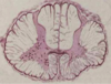
Explain Decerebrate Rigidity with the picture of neurons, etc.
- •Lesion upper pons
- •Decerebrate posturing- extension of back & extremities the arms are extended and limbs internally rotated
Lesion removes all cortical input to reticular formation upper motor neurons and lower motor neurons and loss of rubrospinal input to LMNs. This resutls in continued cerebellar driven vestibulospinal activation of extensor LMN which can be lessen by decreasing stretch reflex input (dorsal rhizotomy) as seen in this illustration

Explain Decorticate Rigidity with the picture of the person’s position
- •Lesion upper midbrain
- •Decorticate posturing- extension of back & lower extremities but the the arms are flexed
Decerebrate Rigidity results from damage in the upper midbrain. Decorticate posturing- extension of back & lower extremities but the the arms are flexed
Lesion removes all cortical input to reticular formation upper motor neurons and lower motor neurons but spares cerebellar driven rubrospinal input to LMNs that serve flexors of the UE. This resutls in continued vestibulospinal activation of extensor LMN and perhaps some reticulospinal input to extensors of L. So there is flexion of UE and extension of LE

Name four sites of the motor unite that can be attacked by specific disease entities:
- –Cell body of the LMN
- –LMN axon
- –Neuromuscular junction
- –Muscle fibers

what are two categories of diseases of the motor system (with examples)?
- •Neurogenic-affecting cell body or axons
- –(e.g., polio, neuropathies)
- •Myopathic-affecting striated muscle
- –(e.g., muscular dystrophy)
Often these causes are divided into Neurogenic and Myopathic conditions
Neurogenic conditions are those diseases affecting the affecting cell body or axons such as polio, and neuropathies
Myopathic conditions are affecting striated muscle directly such as muscular dystrophy
Explain characteristics of Neurogenic Disease
•Neurogenic Disease
- –Some neurons lost & others retained
- –Weakness & atrophy
- –Changes in EMG
- §Fibrillations & positive sharp waves
- §Reduced interference pattern – fewer motor units being activated
- §Change in MUAP shape
- –Polyphasic vs biphasic
- –Giant units as well as normal units seen with reinnervation of denervated muscle fibers
Neurogenic conditions involve Some loss of neurons lost while others are retained. Symptoms include Weakness & muscle atrophy. There are also changes in the EMG that we saw before with denervation, Fibrillations & positive sharp waves, changes in the motor unt action potentials and Reduced interference pattern due to having fewer motor units being activated. Neurogenic conditions are also accompanied by Change in MUAP shape with Polyphasic vs biphasic and the development of what are called Giant units as well as normal units which are seen with reinnervation of denervated muscle fibers.

Explain characteristics of Myogenic Disease
•Myogenic Disease
- –Death of muscle fibers
- –Also weakness & muscle atrophy
- –Changes in EMG
- §Change in MUAP shape
- –Polyphasic vs biphasic
- –Reduced amplitude
- §Reduced amplitude of interference pattern
- §Change in MUAP shape
Myogenic Disease is accompanied by death and dysfunction of muscle fibers. You also see the symptoms of weakness & muscle atrophy and changes in the recorded EMG activity. You see spontaneous potentials (Sharp positive and fibrillation potentials). You see a Change in MUAP shape with reduced amplitude Polyphasic vs the larger biphasic MUAPs and you see a Reduced amplitude of interference pattern.

Compare EMG: normal, neurogenic, and myopathic
- •EMG Comparison: normal, neurogenic (a) & myopathic (b)
Here we see the innervation and EMG Comparison of normal, neurogenic (a) & myopathic (b) disorders. You see normal muscle with normal EMG. Normal muscle has no spontaneous activity, normal biphasic or triphasic MUAPs with normal dense interference pattern with maximal activation of the muscle. With neurogenic you see denervation, spontaneous EMG activity with Sharp positive and fibrillation potentials and occasional giant potentials along with the normal MUAPs. Finally you see a normal amplitude but much reduced density of the interference pattern.
Myogenic Disease is accompanied by death and dysfunction of muscle fibers. You see spontaneous potentials (Sharp positive and fibrillation potentials). You see a Change in MUAP shape with reduced amplitude Polyphasic vs the larger biphasic MUAPs and you see a Reduced amplitude of interference pattern

what is Polio? what happens?
- •Viral infection, prevented by vaccination
- •Attacks cell bodies of LMNs
- –Loss of some LMNs to a muscle
- –Loss of all LMNs to a muscle
- –Loss of all LMN in a spinal segment (myotome)
Viral infection, prevented by vaccination. The virus attacks cell bodies of LMNs and depending upon the extent of the disease you can have Loss of some LMNs to a muscle, loss of all of the neurons going to a muscle (clustered together) or loss of Loss of all LMN in a spinal segment (myotome) or multiple spinal segemnts depending upon how much the disease progresses
Polio:
Cardinal Clinical Signs
what may reappear many years later?
- •Cardinal clinical signs:
- –Weakness/paralysis
- –Atrophy & deformities
- –Decreased reflexes
- •Postpolio syndrome may appear many years after acute infection
- –Surviving neurons have been overworked and now are failing
Following recovery you may see Postpolio syndrome may appear many years after acute infection
Surviving neurons have been overworked and now are failing. With polio vaccinations beginning the early 1950s there are fewer and fewer people who had polio still alive so you may never see polio or postpolio syndrome in your practice career. Of course that is assuming it does not return as a result of the no vaccination wacko

Neuropathies: explain pathelogical changes of the peripheral neruves with injury
- –Segmental degeneration – loss of myelin but intact axons
- –Axonal degeneration
- §If cut or crushed
- §Dying back of axon
- §Wallerian degeneration
- –Degeneration of peripheral axon
- –Chromatolysis/swelling of cell body
Then there are the peripheral neuropathies. In the classic neuropathy we see Pathological changes of the peripheral nerves with Segmental degeneration – loss of myelin but intact axons. However if the axon is If cut or crushed we see Axonal degeneration. There is a Dying back of axon to the most proximal node of Ranvier and all of the way to the distal axonal terminals. This pattern is classic Wallerian degeneration with demyelination, Degeneration of peripheral axon and Chromatolysis (loss of Nissl staining) and swelling of cell body

List 5 types of neuropathies
- Mononeuropathy
- Polyneuropathy
- Diabetic Neuropathy
- Acute Inflammatory Demyelinating Polyneuropathy (AIDP) - also called Guillian-Barre Syndrome
- Metabolic & Toxic Neuropathies
What is mononeuropathy?
–Damage to single peripheral nerve


