Final (Chs 13-14) Flashcards

(223 cards)

1
Q

The most common cell type in connective tissue such as pulp is the:

a. endothelial cell.
b. fibroblast.
c. white blood cell.
d. odontoblast.

A

b. fibroblast.

As in all forms of connective tissue, the fibroblasts are the largest group of cells in the pulp. The odontoblasts are the second largest group of cells in the pulp, but only their cell bodies are located in the pulp. The pulp contains white blood cells in its tissue and vascular supply, but levels are normally low, unless the cells are ready to be triggered by an inflammatory or immune reaction. Endothelial cells line the blood vessels within the pulp.

2
Q

Which of the following is the only sensation perceived by the brain from the pulp’s sensory nerves?

a. taste
b. pain
c. temperature
d. pressure

A

b. pain

The brain perceives all sensations directed to the pulp as sensations of pain. Therefore, changes in temperature, vibrations, and chemical changes that affect the pulp or dentin by way of the pulp’s nerves are perceived only as painful stimuli.

3
Q

During cavity preparation of a tooth, care must be taken to preserve the:

a. enamel rod fluid.
b. support of the dentinal tubules.
c. cemental vascularity.
d. vitality of the pulp.

A

d. vitality of the pulp.

Dental professionals must do their utmost to prevent injury and preserve the vitality of the pulp during preventive and restorative procedures. Such iatrogenic injury to the pulp can result from the heat or vibrations emitted by dental handpiece during cavity preparation.

4
Q

Which of the following pulp structures is formed when Hertwig epithelial root sheath encounters a blood vessel?

a. pulp horn
b. pulp chamber
c. apical foramen
d. accessory canal

A

d. accessory canal

Accessory canals form when Hertwig epithelial root sheath encounters a blood vessel during root formation. Root structure then forms around the blood vessel, forming the accessory canal. The coronal pulp is located in the crown of the tooth. Smaller extensions of coronal pulp into the cusps of posterior teeth form the pulp horns. The large mass of pulp is contained within the pulp chamber of the tooth. The apical foramen is the opening from the pulp into the surrounding periodontal ligament near each apex of the tooth.

5
Q

During tooth development, both the pulp and the dentin in the mature tooth are products of the:

a. dental papilla.
b. enamel organ.
c. dental sac.
d. epithelium.

A

a. dental papilla.

Dentin and pulp tissue have similar embryologic backgrounds because both are originally derived from the dental papilla of the tooth germ during tooth development.

6
Q

Secondary dentin usually forms within the tooth:

a. after the completion of the apical foramen.
b. before the completion of the apical foramen.
c. nearest to the dentinoenamel junction.
d. in response to tooth trauma.

A

a. after the completion of the apical foramen.

Secondary dentin is formed after the completion of the apical foramen(s) and continues to form throughout the life of the tooth. Primary dentin is formed in a tooth before the completion of the apical foramen(s) of the root, which is the opening in the root’s pulp canal. Mantle dentin is the first predentin that forms near the dentinoenamel junction. Tertiary dentin forms quickly in localized regions in response to a localized tooth trauma to the exposed dentin.

7
Q

Dentin in a mature tooth is on the average about ______ mineralized by weight.

a. 50%
b. 65%
c. 70%
d. 96%

A

c. 70%

Mature dentin is by weight 70% inorganic material or mineralized. The alveolar process is by weight 50% inorganic material. Mature cementum is by weight 65% inorganic material. Mature enamel is by weight 96% inorganic material.

8
Q

Which of the following terms associated with dentin can be used to correctly describe the type that makes up the largest part of the tooth’s dentin?

a. Tomes granular layer
b. mantle dentin
c. circumpulpal dentin
d. interglobular dentin

A

c. circumpulpal dentin

Deep to the mantle dentin is the layer of dentin around the outer wall of pulp, the circumpulpal dentin, which makes up the bulk of the dentin in a tooth. Tomes granular layer is most often found in the peripheral part of dentin beneath the root’s cementum. Mantle dentin is the first predentin that forms near the dentinoenamel junction. Interglobular dentin is found in those areas where only primary mineralization has occurred within the predentin, and the globules of dentin do not fuse completely and appear as dark arclike areas in a stained section of dentin.

9
Q

In which location is the cell body of the odontoblast found in a mature healthy erupted tooth?

a. along the dentinoenamel junction
b. along the outer pulpal wall
c. near the dentinocemental junction
d. near the pulpal core

A

b. along the outer pulpal wall

The odontoblasts are located only along the outer pulpal wall. Only their cell bodies are located in the pulp.

10
Q

With increased age, the pulp tissue can become:

a. displaced by primary dentin.
b. increasingly fibrotic.
c. filled with cementicles.
d. increasingly cartilaginous.

A

b. increasingly fibrotic.

With increased age, the pulp undergoes a decrease in intercellular substance, water, and cells as it fills with an increased amount of collagen fibers and thus becomes fibrotic.

11
Q

Predentin is the initial material laid down by the:

a. odontoblasts.
b. ameloblasts.
c. preameloblasts.
d. odontoclasts.

A

a. odontoblasts.

Predentin is a mesenchymal product consisting of nonmineralized collagen fibers produced by the odontoblasts. Ameloblasts form from preameloblasts and then later produce enamel. Odontoclasts are active during eruption, removing parts of the primary tooth.

12
Q

The dark, arc-like areas in histologic sections of a tooth are what type of dentin?

a. Tomes granular layer
b. mantle dentin
c. circumpulpal dentin
d. interglobular dentin

A

d. interglobular dentin

The dark, arclike areas in a stained histologic section in a tooth are considered interglobular dentin. In these areas, only primary mineralization has occurred within the predentin, and the globules of dentin do not fuse completely. Tomes granular layer is most often found in the peripheral part of dentin beneath the root’s cementum. Mantle dentin is the first predentin that forms near the dentinoenamel junction. Deep to the mantle dentin is the layer of dentin around the outer wall of pulp, the circumpulpal dentin, which makes up the bulk of the dentin in a tooth.

13
Q

Which of the following includes the tissue fluid surrounding the cell membrane of the odontoblast?

a. lymph
b. gingival crevicular fluid
c. dentinal fluid
d. synovial fluid

A

c. dentinal fluid

The dentinal fluid in the tubule includes the tissue fluid surrounding the cell membrane of the odontoblast. The lymph is found within the lymphatic system. The gingival crevicular fluid is located within the gingival sulcus. The synovial fluid is found within the temporomandibular joint, surrounding the disc of the joint.

14
Q

Which of the following are a number of adjoining parallel imbrication lines that are present in stained dentin?

a. lines of Retzius
b. reversal lines
c. arrest lines
d. contour lines of Owen

A

d. contour lines of Owen

The contour lines of Owen are a number of adjoining parallel imbrication lines that are also present in a stained section of dentin. These specific imbrication lines demonstrate a disturbance in body metabolism that affects the odontoblasts by altering their formation efforts, and they tend to appear together as a series of dark bands. The lines of Retzius are incremental lines noted in stained enamel. Both reversal lines and arrest lines are stained lines noted in both cementum and repair due to repair and apposition of the hard tissue, respectively.

15
Q

Lateral pulp canals within the pulp chamber extend:

a. from pulp tissue to the periodontal ligament.
b. vertically toward the cementum.
c. between two pulp canals, as a bridge.
d. from the chamber, parallel to another canal.

A

a. from pulp tissue to the periodontal ligament.

Accessory canals may also be associated with the pulp and are extra openings from the pulp to the periodontal ligament Accessory canals are also called lateral canals, because they are usually located on the lateral surface of the roots of the teeth, but this is not always the case because they can be found anywhere along the root surface.

16
Q

Dentin in the mature tooth is produced as a result of secretion by:

a. cementoblasts.
b. fibroblasts.
c. osteoblasts.
d. odontoblasts.

A

d. odontoblasts

Apposition of dentin by odontoblasts, unlike enamel, occurs throughout the life of the tooth, filling in the pulp chamber of both the crown and root.

17
Q

What are the smaller extensions of coronal pulp into the cusps of posterior teeth termed?

a. accessory canals
b. lateral canals
c. pulp horns
d. pulp canals

A

c. pulp horns

The coronal pulp is located in the crown of the tooth. Smaller extensions of coronal pulp into the cusps of posterior teeth form the pulp horns. Accessory canals may also be associated with the pulp and are extra openings from the pulp to the periodontal ligament; accessory canals are also called lateral canals. The radicular pulp, or root pulp, is the part of the pulp located in the root of the tooth; it is also called the pulp canal by patients.

18
Q

Which is the most common type of nerves associated with the pulp in a mature erupted tooth?

a. myelinated
b. unmyelinated
c. myelinated and unmyelinated are in equal numbers
d. dead ones

A

b. unmyelinated

Two types of nerves are associated with the pulp, which includes mainly unmyelinated nerves (70% to 80%) and in lesser amounts, myelinated nerves (20% to 30%).

19
Q

Which of the following zones in pulp is closest to the dentin?

a. odontoblastic layer
b. cell-rich zone
c. pulpal core
d. cell-free zone

A

a. odontoblastic layer

The first zone of pulp closest to the dentin is the odontoblastic layer. The next zone, nearest to the odontoblastic layer, inward from the dentin, is considered the cell-free zone. The next zone, nearest to the odontoblastic layer, inward from the dentin, is considered the cell-free zone. The final zone of pulp is the pulpal core, which is in the center of the pulp chamber.

20
Q

Which zone in the pulp contains a nerve and capillary plexus?

a. odontoblastic layer
b. cell-rich zone
c. pulpal core
d. cell-free zone

A

b. cell-rich zone

A nerve and capillary plexus are also located in the cell-free zone of the pulp. The odontoblastic zone consists of a layer of odontoblasts. The cell-rich zone has an increased density of cells compared with the cell-free zone but still does not contain as many cells as the odontoblastic layer. This zone also has a more extensive vascular supply than does the cell-free zone. The pulpal core, which is in the center of the pulp chamber, consists of many cells and an extensive vascular supply; except for its location, it is very similar to the cell-rich zone.

21
Q

Which of the following statements is correct when considering accessory canals?

a. Teeth have a standard number.
b. Radiographs always indicate the number and position.
c. Gingival recession may expose the opening.
d. They are examined by placing radiolucent materials.

A

c. Gingival recession may expose the opening.

Gingival recession may expose the opening of an accessory canal. Teeth have a variable number of these canals. Radiographs do not always indicate the number or position of these canals, unless they are examined with instruments using radiopaque materials.

22
Q

Which of the following is not associated with dentinal hypersensitivity?

a. enamel and cementum do not meet
b. dentin exposed due to caries process
c. blocking dentinal tubules
d. branching of dentinal tubules

A

c. blocking dentinal tubules

Dentinal hypersensitivity can be treated somewhat successfully with solutions applied either by professionals or within over-the-counter dentifrices available to patients; these desensitizing agents temporarily block the exposed open ends of the dentinal tubules. When dentin is exposed as a result of caries, the open dentinal tubules may be painful, causing dentinal hypersensitivity. However, many times it is the microscopic anatomy of the tooth that is the culprit; the enamel and cementum do not meet, leaving a gap with dentin exposed. Branching of the dentinal tubules containing the live odontoblastic processes throughout dentin adds to the overall level of exposure.

23
Q

The resorption process within the dentin can result in a clinically presentation of a:

a. silver-hued tooth
b. pulp with stones
c. pinkish crown color
d. carious tooth

A

c. pinkish crown color

Dentin can become resorbed in permanent teeth, but the cause is idiopathic and can involve either an internal or external resorption process. It can be noted radiographically, but it is hard to discern between the two processes. In contrast, when the process begins on the external surface of the root and then penetrates through the cementum into dentin, it can lead to a pinkish crown color noted clinically from the granulation tissue seen beneath the translucent enamel.

24
Q

What type of dentin occurs when odontoblasts in the area of the traumatized tubules may perish because of the injury, but neighboring undifferentiated mesenchymal cells of the pulp can move to the area and become odontoblasts?

a. reparative dentin
b. reactive dentin
c. sclerotic dentin
d. mantle dentin

A

a. reparative dentin

Odontoblasts in the area of the traumatized tubules may perish because of the injury, but neighboring undifferentiated mesenchymal cells of the pulp can move to the area and become odontoblasts, forming a type of tertiary dentin, reparative dentin. If the tertiary dentin is formed by existing odontoblasts, it is considered to be reactive dentin. A certain type of tertiary dentin, sclerotic dentin occurs when the odontoblastic processes die and leave the dentinal tubules vacant. Mantle dentin is the first predentin that forms.

25
In what direction do the imbrication lines of von Ebner run? a. at 45 degrees to the DEJ b. at 90 degrees to the dentinal tubules c. at 45 degrees to the inner pulpal wall d. at 90 degrees to the periodontal ligament
b. at 90 degrees to the dentinal tubules The imbrication lines of von Ebner are incremental lines or bands that stain darkly in a section of dentin. They run at 90 degrees to the dentinal tubules.
26
Sharpey's fibers are located in which of the following types of dental tissue? a. enamel b. dentin c. cementum d. pulp
**cementum** Sharpey fibers are a part of the collagen fibers from the periodontal ligament that are each partially inserted into the outer part of the cementum at 90 degrees or perpendicular to the cemental surface (as well as the alveolar process on their other end) as they are inserted on the other end, the alveolar process.
27
which of the following types of dental tissue provides the vascular nutrition for the tooth's cementum? a. dentin b. epithelial cells c. periodontal ligament d. alveolar process
c. periodontal ligament The canals of the cementocytes are oriented toward the periodontal ligament and contain cementocytic processes that exist to diffuse nutrients from the ligament because it is vascularized.
28
which of the following terms correctly describes the alveolar bone proper? a. lamina propria b. cancellous bone c. mucoperiosteum d. cribriform plate
d. cribriform plate * The alveolar bone proper makes up the lining of the tooth socket or alveolus. Although the alveolar bone proper is composed of compact bone, it may be called the cribriform plate because it contains numerous holes where Volkmann canals with their nerves and blood vessels pass from the alveolar process into the periodontal ligament. The lamina propria is the connective tissue component of the oral mucosa. A mucoperiosteum is a combined structure consisting of a mucous membrane, in the case of the oral cavity, the epithelium and lamina propria of the oral mucosa, with the periosteum of bone. The trabecular bone consists of cancellous bone or spongy bone that is located between the alveolar bone proper and the plates of cortical bone.*
29
the rests of Malassez are correctly described as: a. remnants of the outer enamel epithelium b. similar to incremental growth limes in dentin c. remnants of Hertwig root sheath d. similar to the incremental growth lines in enamel
c. remnants of Hertwig sheath The epithelial rests of Malassez, which represent groups of epithelial cells, are stranded in mature periodontal ligament after the disintegration of Hertwig epithelial root sheath during the formation of the root.
30
on radiographs, the periodontal ligament appears as a: a. radiopaque line covering the alveolar bone proper b. lamina dura surrounding the alveolus c. radiolucent space surrounding the alveolus d. spongy material between the alveolus and alveolar bone peoper
c.radiolucent space surrounding the alveolus The periodontal ligament appears on radiographs as a radiolucent area (or darker) located between the denser radiopaque (or lighter) lamina dura of the alveolar bone proper and the similar radiopaque (or lighter) cementum.
31
which of the following situations can be present at the cementoenamel junction in a mature tooth? a. cementum overlaps enamel b. enamel overlaps cementum c. dentin overlaps enamel d. pulp overlaps cementum or dentin
a. cementum overlaps enamel Three possible types of transitional interfaces may be present at the cementoenamel junction. The cementum may overlap the enamel at the cementoenamel junction. The cementum and enamel may meet end to end, presenting no problems for either the clinician or patient. Another possible interface at the cementoenamel junction is that a gap may exist between the cementum and enamel, exposing dentin.
32
which of the following fiber groups is a part of the alveodental ligament on a single- rooted adult tooth? a. circular ligamnet b. interdental ligament gingival fiber group d. apical group
d. apical group The main principal fiber group is the alveolodental ligament, which consists of five fiber subgroups: alveolar crest, horizontal, oblique, apical, and interradicular on multirooted teeth. The circular ligament is part of the gingival fiber groups and some consider these ligaments as a group to be part of the principal fibers but separate from the alveodental ligament. Another principal fiber other than the alveolodental ligament is the interdental ligament.
33
which of the following statements concerning the oblique group of the periodontal ligament is correct? a. part of the gingival fiber group b. found only in multi-rooted teeth c. resists intrusive forces to the tooth d. resists tilting forces to the tooth
c. resists intrusive forces to the tooth The function of the oblique group as part of the alveodental ligament of the periodontal ligament is to resist intrusive forces, which try to push the tooth inward, as well as rotational forces; it is found on both single and multirooted teeth.
34
which part of both of the jaws contains cancellous bone? a. alveolar bone proper b. cortical bone c. interdental septum d. alveolar crest
c. interdental septum The interdental septum consists of both the compact bone of the alveolar bone proper and cancellous bone of the trabecular bone. The alveolar bone proper is composed of compact bone. The cortical bone consists of a plate of compact bone on both the facial and lingual surfaces of the alveolar process. The alveolar crest is the most cervical rim of the alveolar bone proper.
35
the most common type of connective tissue fiber in the periodontal ligament is which of the following fiber groups? a. collagen b. elastic c. oxytalan d. collagen and elastic fibers
a. collagen All the fibers in the periodontal ligament are collagen in structure.
36
which of these fiber groups does not have any cemental insertion? a. interdental ligament b. circular ligament c. alveodental ligament d. Interradicular group
b. circular ligament The circular ligament encircles the tooth, as shown on a cross section of a tooth, interlacing with the other gingival fiber subgroups. Thus, it has no cemental insertion. The interdental ligament and alveodental ligament, which the interradicular group is part of on multirooted teeth, both have cemental insertions.
37
the periodontal ligament directly forms from which part of the developing tooth germ? a. enamel organ b. dental papilla c. dental sac d. both enamel organ and dental papilla
c. dental sac Similar to the alveolar process, the periodontal ligament develops from the dental sac of the tooth germ. The enamel organ gives rise to the enamel and the dental papilla gives rise to the dentin and pulp of the tooth.
38
the cementoblasts originate during tooth development from which of the following embryonic structures? a. enamel organ b. dental papilla c. dental sac d. both enamel organ and dental papilla
c. dental sac Cementum and its cells, the cementoblasts, develop from the dental sac and form on the root after the disintegration of Hertwig epithelial root sheath. The dental papilla gives rise to the dentin and its odontoblasts as well as the pulp and its fibroblasts. The enamel organ gives rise to the enamel and its ameloblasts.
39
the alveolar bone proper of the jaws in a mature adult refers to the: a. cancellous bone between the alveoli b. layer of compact bone lining the alveolus c. part of the jaw that houses the teeth d. done surrounding the lamina dura
b. layer of compact bone lining the alveolus The alveolar bone proper of the jaws makes up the lining of the tooth socket or alveolus, which is also considered to be the lamina dura on radiographs. The part of the jaws that contains the roots of the teeth is the alveolar process or alveolar bone. The supporting alveolar bone consists of both cortical bone and trabecular bone. The cortical bone consists of a plate of compact bone on both the facial and lingual surfaces of the alveolar process. The trabecular bone consists of cancellous bone that is located between the alveolar bone proper and the plates of cortical bone.
40
which of the following structures is not considered part of the periodontium? a. alveolar process b. cementum c. basal bone d. periodontal ligament
c. basal bone The part apical to the roots of the teeth is the basal bone, which then forms the body of the maxilla or body of the mandible and thus is not considered part of the periodontium. The alveolar process, cementum, and periodontal ligament are all parts of the periodontium.
41
which of the following tisssue types is most likely to source of cementogenic cells in adults who have undergone root planning? a. bone marrow b. periodontal ligament c. residual cementum patches d. alveolar process
b. periodontal ligament The periodontal ligament has a line of cementoblasts along the cemental surface of the root that can produce cementum throughout the lifetime of the tooth.
42
which of the following fiber groups of the periodontal ligament foes not contribute significantly to each tooth's anchorage in the jaw? a. gingival fiber group b. interdental fiber group c. alveodental fiber group d. interradicular fiber group
a. gingival fiber group The gingival fiber group does not support the tooth in relationship to the jaws, resisting any forces of mastication or speech; rather, this group supports only the marginal gingiva to maintain the relationship to the tooth. Instead, the interdental and alveodental, which the interradicular fiber group is part of, supports the tooth in relationship to the jaws.
43
which of the following is not a part of the gingival fiber group? a. interdental fiber group b. circular fiber group c. dentoperiosteal fiber group d. alveologingival fiber group
a. interdental fiber group Another principal fiber other than the alveolodental ligament is the interdental ligament. The gingival fiber group includes the fiber subgroups of the circular and dentogingival ligament, as well as both the alveologingival and dentoperiosteal ligaments.
44
the anchorage of the tooth to the alveolar process is mediated through which of the following? a. interdental ligament b. sharpey fibers c. oxytalan fibers d. both sharpey and oxytalan fibers
b. sharpey fibers Sharpey fibers in alveolar bone proper are each inserted at 90 degrees or perpendicular to the bone to mediate the anchorage of the tooth. A principal fiber of the periodontal ligament is the interdental ligament. Oxytalan fibers are a fiber type related to elastic fibers; they appear to consist of the microfibrillar component only, thereby resembling very immature elastic fibers.
45
cellular cementum is most likely found around which of the following root regions? a. cervical part of the root b. entire root of an unerupted tooth c. apical part of the root d. furcation region of the root
c. apical part of the root Cellular cementum consists of the last layers of cementum deposited over the acellular cementum, mainly in the apical one third of each root. At least one layer of acellular cementum covers the entire outer surface of each root with many more layers covering the cervical one third near the cementoenamel junction.
46
the lamina dura noted on radiographs corresponds to which of the following structures? a. basal bone of the both jaws b. buccal and lingual cortical plates of the jaws c. supporting cancellous bone between the teeth d. alveolar bone proper of the alveolar process
d. alveolar bone proper of the alveolar process The part of the alveolar bone proper of the alveolar process that is present on radiographs is considered the lamina dura, which is uniformly radiopaque (or lighter).
47
which of the following includes both the supporting soft and hard dental tissue between and including parts of the tooth and surrounding bone? a. periodontal ligament b. periodontium c. alveolar bone proper d. alveolar process
b. periodontium The periodontium consists of both the supporting soft and hard dental tissue between the tooth and the alveolar process, as well as parts of the tooth and alveolar process. Thus, the periodontium includes the periodontal ligament as well as the alveolar process, which includes the alveolar bone proper.
48
when many of the cementoblasts become entrapped by the cementum they produce, they become: a. odontoclasts b. odontoblasts c. cementocytes d. dental sac cells
c. cementocytes During the later steps within the stage of apposition of cementum, many of the cementoblasts become entrapped by the cementum they produce, becoming cementocytes. After the appositional growth of cementum in layers, the cementoblasts that do not become entrapped in cementum line up along the cemental surface along the length of the outer covering of the periodontal ligament. When the undifferentiated cells of the dental sac come into contact with the newly formed surface of root dentin, these cells are induced to become cementoblasts. Odontoblasts are cells that produce dentin. Odontoclasts resorb dentin, cementum, and enamel during tooth shedding and eruption.
49
the alveolar process between two neighboring teeth is specifically called the _____ bone a. interdental b. basal c. interradicular d. trabecular
a. interdental The alveolar process that separates two neighboring teeth is the interdental septum or interdental bone. The part apical to the roots of the teeth is the basal bone, which then forms the body of the maxilla or body of the mandible. The alveolar process that separates the roots of the same tooth is the interradicular septum or interradicular bone. The trabecular bone consists of cancellous bone that is located between the alveolar bone proper and the plates of cortical bone.
50
the most common cell in the periodontal ligament is the: a. cementoblast b. fibroblast c. odontoclast d. osteoclast
b. fibroblast Like all connective tissue, the fibroblast is the most common cell in the periodontal ligament, producing fibers and intercellular substance. The cementoblast and odontoclast as well as the osteoclast are present in the periodontal ligament. The cementoblast produces cementum and the odontoclast resorbs parts of the tooth during tooth shedding and eruption. The osteoclast resorbs the alveolar process.
51
with occlusal trauma, there can be a widening of the: a. periodontal space b. periodontal ligament c. lamina dura d. periodontal space, periodontal ligament, and lamina dura
d. periodontal space, periodontal ligament, and lamina dura Early occlusal trauma can be viewed on radiographs as a widening of the radiolucent (or darker) periodontal ligament space between the radiopaque (or lighter) lamina dura of the alveolar bone proper and the similar radiopaque (or lighter) cementum. Because the periodontal space represents the periodontal ligament, the ligament can double and the individual principal fiber bundles also become thicker. Thickening of the lamina dura, which represents the alveolar bone proper, is also possible with occlusal trauma in response to overall alveolar process loss.
52
which part of the periodontal ligament is noted to be wider in a healthy adult tooth? a. near the apex b. near the cervix c. both near the apex and cervix d. between the apex and cervix
c. both near the apex and cervix The periodontal ligament is wider near the apex and cervix of the tooth and narrower between these two end points.
53
in which part of the alveolar process is the loss of bone from periodontitis first evident? a. interradicular bone b. basal bone c. supporting alveolar bone d. alveolar crest
d alveolar crest The bone loss from periodontitis is first evident in the most coronal part of the alveolar bone proper, the alveolar crest, which looks moth eaten both microscopically and radiographically. The alveolar process loss of the jaws, which involves the presence of the roots of the teeth, progresses apically to involve the interradicular bone if the tooth is multirooted, as well as the surrounding supporting alveolar bone, but does not involve the inferior bone of the jaws, the basal bone.
54
which of the following is not bisible at least somewhat on bitewing radiographs? a. periodontal space b. lamina dura c. cortical bone d. trabecular bone
c. cortical bone The cortical bone is not visible on periapical or bitewing radiographs but only on occlusal radiographs as a uniformly radiopaque (or lighter) bony sheet, facial and lingual to the teeth. The periodontal space and lamina dura are both present somewhat. Only the parts of trabecular bone between the teeth and between the roots are ever present on any type of radiograph.
55
which type of bone is only present in an area of anodontia? a. cortical bone b. basal bone c. alveolar bone proper d. trabecular bone
b. basal bone The developmental dental anomaly of anodontia, in which tooth germs are congenitally absent, may affect the development of the alveolar process of either jaw. Proper development is impossible because the alveolar unit of each dental arch only forms in response to the tooth germs in the area. Only basal bone will be present in each one of the involved areas of the jaws. The alveolar process, which includes the alveolar bone proper and the supporting alveolar bone, the latter of which consists of the cortical bone and trabecular bone, will not have developed due to the anodontia.
56
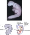
Frontonasal process
Forms as a bulge of tissue at the most cephalic end, cranial boundary of stomodeum
57
Lens placodes
Future eyes; initially located fishlike on each side of frontonasal process
59
Otic placodes
Future **internal ear**; lateral & posterior to lens placodes
60
Nasal placodes
Anterior part of frontonasal process; develop into olfactory epithelium for smell
61
9. The neural crest cells migrate from which embryonic structures during prenatal development? a. Stomodeum b. First branchial arch c. Neural folds d. Frontonasal process
c.Neural folds
62
18. Which of the following can occur that *mainly* involves the abnormal development of one or more structures from ectoderm within the embryonic period? a. Treacher Collins syndrome b. Ectodermal dysplasia c. Congenital syphilis d. Fetal alcohol syndrome
b.Ectodermal dysplasia
63
4. From which embryonic layer is mesoderm derived during prenatal development? a. Epiblast layer b. Hypoblast layer c. Endoderm d. Neuroectoderm
a.Epiblast layer
64
3. The mandibular arch during the embryonic period of prenatal development is the: a. fusion of the two paired mandibular processes. b. swelling found superior to the stomodeum. c. structure that gives rise to the lateral nasal process. d. posterior part of the hard palate.
a.fusion of the two paired mandibular processes.
65
Which of the following is the BEST term used for the embryonic layer located between the ectoderm and the endoderm? a. Mesenchyme b. Ectomesenchyme c. Mesoderm d. Mesiodens
c. Mesoderm
66
\_\_\_\_\_ is a layer in trilaminar embryonic disc derived from epiblast layer and lining stomodeum
Ectoderm
67
16. What *best* characterizes the first week of prenatal development so that the blastocyst splits into smaller and more numerous cells? a. Implantation b. Mitotic cleavage c. Meiosis d. Fertilization
b.Mitotic cleavage
68
17. What does the maxillary process form from during the fourth week of prenatal development? a. Lateral nasal processes b. Mandibular arch c. Intermaxillary segment d. Medial nasal processes
b.Mandibular arch
69
20. The development of the neck parallels the development of the face over time, beginning during the fourth week of prenatal development within the embryonic period and completed during the _____ period. a. preimplantation b. embryonic c. fetal d. both the embryonic and fetal
c.fetal
70
18. The fusion of the maxillary and medial nasal processes to form the upper lip is completed during the end of the _____ week of prenatal development, when the grooves between the processes are obliterated. a. fourth b. fifth c. sixth d. seventh
d.seventh
71
5. In which week of prenatal development does facial development begin in the embryo? a. Second b. Fourth c. Fifth d. Eighth
b.Fourth
72
13. The facial development that starts in the fourth week will be completed later in the _____ week within the fetal period. a. fifth b. sixth c. eighth d. twelfth
d.twelfth
73
4. A type of cleft lip can result during prenatal development from the lack of fusion between _____ processes. a. mandibular and maxillary b. medial nasal and maxillary c. lateral nasal and medial nasal d. two lateral nasal e. lateral nasal and maxillary
b.medial nasal and maxillary
74
Failure of fusion of the palatal shelves with the primary palate or each other will result in? A. Branchial cleft cyst B. Cleft palate C. Medial nasal process D.Zygote
**Cleft palate**
75
What area of the **mandible** articulates with the **temporal bone** at the **TMJ?**
The head of the **condyle** of the mandible
76
What **bones** are **articulated** with the **TMJ**?
The **temporal bone** and **condyle** of the **mandible**
77
78
Name the **3 major salivary glands**
1. Stensen's duct (Parotid) 2. Bartholin's duct (Sublingual) 3. Wharton's duct (Submandibular)
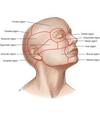
79
**region of the face** that includes the **forehead** and area **above eyes**
**frontal**
80
**region of the face** that includes the **eyeball, supporting structures,** and **bony eye socket**
**orbital**
81
**region of the face** which the main feature is the **external nose**
**nasal**
82
What structure is located **between the eyes**? What is another name for the **tip of the nose**?
**root of nose** **apex of nose**
83
**region of the face** located **inferior** to the **orbital region** and **lateral** to the **nasal region**
**infraorbital**
84
**Region** of the face **lateral** of the **infraorbital region**?
**zygomatic region**
85
**inferior** to the **zygomatic arch** and just **anterior** to the **external ear**
**temporomandibular joint**
86
the **region of the face** composed of **soft tissue** of **cheeks** which is a broad area **between nose, mouth, ear**
**buccal region**
87
On the **lateral aspect** of the **mandible**, the **stout, flat plate** of the \_\_\_\_\_\_\_\_\_\_ that extends **upward** and **backward** from the **body of the mandible** on each side
ramus, rami
88
**anterior border** of the **ramus** is a **thin, sharp margin** that **terminates** in the\_\_\_\_\_\_\_\_\_ \_\_\_\_\_\_\_\_\_\_
**coronoid process**
89
\_\_\_\_\_\_\_\_\_ ________ is the main part of the **anterior border of ramus** that forms a **concave forward curve**
**Coronoid notch**
90
\_\_\_\_\_\_\_\_\_\_\_ ___________ is a **projection** at the **posterior border** of the **ramus** that is **thickened** and **extends** from the **angle of the mandible**
**Mandibular condyle**
91
**strong muscle** that forms the **cheek** along with upper flesh and fat
**masseter muscle**
92
In the **superficial** and **deep** part of the **masseter**, where is the **origin** and **insertion** points?
**Superficial part** ORIGIN: **maxillary process** of **zygomatic bone** and **anterior 2/3** of the **zygomatic process** of the maxilla INSERTION: **angle of the mandible** and **posterior part** of **ramus** **Deep part** ORIGIN: **medial aspect** of the **zygomatic arch** INSERTION: **central** and **upper** part of **ramus** as high as the **coronoid process**
93
Where is the **origin,** **insertion,** and **action** of the **temporalis**?
**ORIGIN**: the **floor** of **temporal fossa** and **temporal fascia** **INSERTION**: the apex and deep surface of the **coronoid process** and along the **anterior border of the ramus** **ACTION: Anterior fibers elevate** the mandible, while the **posterior fibers retract** the mandible
94
Where is the **origin** and **action** of the **lateral pterygoid muscle**?
ORIGIN: **Superior** portion - **infratemporal surface** of **greater wing of sphenoid.** **Inferior** portion - **lateral surface** of **lateral pterygoid plate** Action: **Depress, protrude** and **move** the **mandible** from **side to side**
95
What is the **origin** and **action** of the **medial pterygoid muscle**?
ORIGIN: **medial surface** of the **lateral pterygoid plate**, the **posterior surface** of the tubercle of **palatine bone** and **tuberosity of maxilla** Action: **elevates** and **protracts** the **mandible** also moves the jaw from **side to side**
96
97
4. The line of demarcation between the attached gingiva and the alveolar mucosa is the: a. mucogingival junction. b. interdental gingiva. c. mucobuccal fold. d. marginal gingiva.
a.mucogingival junction.
98
The line of demarcation between the firmer and pinker attached gingiva and the movable and redder alveolar mucosa is the scallopp-shaped \_\_\_\_\_\_\_\_
Mucogingival junction
99
There are 4 muscles of mastication: 1. Masseter 2. Temporalis 3. Medial pterygoid 4. Lateral pterygoid
All attach to the mandible and are innervated by **V3** Form in the **tenth week** of development **First arches (mandibular arches)**
100
Which of the following muscles is involved in the lateral deviation of the mandible? a. masseter muscle b. medial pterygoid muscle c. lateral pterygoid muscle d. temporalis muscle e. digastric muscle
c. lateral pterygoid muscle
101
The ______ of the hyoid arches helps form the muscles of facial expression, the middle ear muscles, and a suprahyoid muscle.
mesoderm
102
Which specific muscle can become clinically enlarged in patients who habitually clench or grind (with bruxism) their teeth and in those who constantly chew gum? a. Masseter muscle b. Lateral pterygoid muscle c. Medial pterygoid muscle d. Temporalis muscle
a. Masseter muscle
103
Gingival margin ## Footnote .
the edge of the gingiva nearest to the incised surface of the tooth
105
**Lateral pterygoid** The **superior head** originates from the **greater wing of the sphenoid**. The **inferior head** originates from the **lateral pterygoid plate** of the sphenoid. The two heads converge into a tendon, which attaches to the **neck of the mandible.**
short thick almost conical muscle of mastication superior to the medial pterygoid. **Two separate heads (superior an inferior).** They are separated anteriorly by a slight interval but fuse together posteriorly. The entire muscle lies within the **infratemporal fossa deep to the temporalis muscle.** The **only** muscle of mastication that **ASSISTS in depressing the mandible**, **lowering the lower jaw**! Depression of the mandible occurs during the opening of the jaws. Protrusion also occurs when the jaws are opened The muscle is innervated by the **lateral pterygoid nerve**, a branch of the **mandibular nerve** (V3) **Pterygomandibular space** is formed by the **lateral pterygoid muscle (roof), medial pterygoid muscle (medial wall), and the mandibular ramus (lateral wall).** It is the injection site for the inferior alveolar block.
106
22. On what structure do BOTH heads of the masseter muscle originate? a. Zygomatic process of the maxilla b. Coronoid process c. Zygomatic process of the frontal bone d. Zygomatic arch
d.Zygomatic arch
108
Gingival sulcus
The crevice between the free gingiva and the tooth.
109
Embryonic layers (germ layers)
increased number of **embryonic cells** within the **blastocyst**.
110
1. The folding of the embryo during prenatal development causes _____ to be on the \_\_\_\_\_. a. endoderm; inside of the embryo b. ectoderm; inside of endoderm c. endoderm; outside of the mesoderm d. mesoderm; outside of the ectoderm
a.endoderm; inside of the embryo
111
Alveolar mucosa
The thin, moveable, loosely attached tissue **covering the alveolar bone**. non keratinized
113
**Medial pterygoid**
Rectangular form has 2 heads due to differing depth (deep and superficial, smaller head). The **superficial head** originates from the **maxillary tuberosity** and the pyramidal process of **palatine bone**. The **deep head** originates from the **lateral pterygoid plate of the sphenoid bone**. Both parts attach to the **ramus** of the mandible, near the angle of mandible. **DEEPEST MUSCLE OF MASTICATION** **Elevates the mandible** during the closing of the jaws Innervated by V3
114
4. A type of cleft lip can result during prenatal development from the lack of fusion between _____ processes. a. mandibular and maxillary b. medial nasal and maxillary c. lateral nasal and medial nasal d. two lateral nasal e. lateral nasal and maxillary
b.medial nasal and maxillary
115
**Masseter**
**Most superficial and strongest**. Thick flat rectangular muscle on each side of the face, **anterior to the parotid gland**. **2 heads** (superficial and deep) Origin- Both heads **originate from the zygomatic arch.** Superficial =**angle of the mandible**. Deep = **ramus** When the patient **clenches their teeth** is when the **elevation of the mandible** is taking place (**closing of the jaws**) **Innervated by the Masseteric nerve (V3)**
116
How does cleft lip form?
**Failure of fusion** of the maxillary process with the medial nasal process
118
**Temporalis**
broad **fan-shaped muscle** of mastication on each **side of the head** that fills the temporal fossa, **superior to the zygomatic arch**. Temporal space is formed by the **temporal fascia** covering the temporalis muscle. **Infratemporal space is bordered laterally by the medial surface of the mandible and the temporalis muscle.** originates from the **temporal fossa** – a shallow depression on the lateral aspect of the skull. Inserts on **coronoid process** Action- **retraction of mandible, elevating the mandible**. Also allows **freeway space** at its physiologic rest position Innervated by V3
119
15. On which of the following orofacial tissue is the linea alba located? a. Attached gingiva b. Marginal gingiva c. Labial mucosa d. Buccal mucosa
d.Buccal mucosa
120
Free gingiva
closely adapted around each tooth but **not attached** keratinized
121
Interdental papillae
Extensions of unattached gingiva between adjacent teeth keratinized fills embrasure spaces
123
What’s the diff b/w meiosis and mitosis
Meiosis – The process of reproductive cell production that ensures the **correct number of chromosomes;** meiosis that takes place during **fertilization**. Mitosis – **cell division** that occurs in phases and results in **two daughter cells;** Mitosis is a process that **takes place during tissue growth or regeneration**
124
Mucogingival junction
The line that marks the **connection between** the **attached gingiva** and the **alveolar mucosa** on the facial surfaces of all quadrants and the lingual surfaces of the mandibular arch
126
Attached gingiva
Part of the gingiva that is **tightly connected** to the cementum on the root (cervical third) and to the connective tissue cover of the alveolar bone
127
15. Which of the following muscles can show enlargement due to repetitive muscle contraction associated with clenching of the teeth in a patient? a. Buccinator muscle b. Masseter muscle c. Temporalis muscle d. Zygomatic muscle
b.Masseter muscle
128
23. Which of the following muscles when contracted allows for the retraction of the mandible? a. Masseter muscle b. Temporalis muscle c. Lateral pterygoid muscle d. Medial pterygoid muscle
b.Temporalis muscle
130
2. The philtrum of the upper lip forms during sixth week of prenatal development from the: a. mandibular processes. b. medial nasal processes. c. lateral nasal processes. d. nasal placodes.
b.medial nasal processes.
133
The mucogingival junction is a line of demarcation between the attached gingiva and the: a. buccal mucosa. b. marginal gingiva. c. gingival sulcus. d. alveolar mucosa.
d. alveolar mucosa.
134
18. Which of the following muscles when unilaterally contracted deviates the mandible to one side? a. Masseter muscle b. Lateral pterygoid muscle c. Medial pterygoid muscle d. Temporalis muscle
b.Lateral pterygoid muscle
136
2. From the following list of muscles, select which are considered muscles of mastication. (*Select all that apply*.) a. Buccinator b. Risorius c. Platysma d. Temporalis e. Masseter
d. Temporalis e. Masseter
138
21. Surrounding the teeth in the alveoli and covering the alveolar processes is the \_\_\_\_\_, which is composed of a firm pink tissue. a. gingiva b. minor salivary glands c. Fordyce spots d. linea alba
a.gingiva
139
Which of the following muscles has two bellies, giving the muscles two different origins? a. lateral pteryoid b. geniohyoid c. thyrohyoid d. stylohyoid
a. lateral pteryoid
140
17. The pink labial mucosa or buccal mucosa meets the redder _____ at the mucobuccal fold. a. marginal gingiva b. attached gingiva c. alveolar mucosa d. interdental papilla
c.alveolar mucosa
141
What do the muscles of mastication work with to accomplish movements of the mandible? a. Maxillae b. Temporal bone c. Temporomandibular joint d. Zygoma e. Hard palate
c. Temporomandibular joint
142
8. Which of the following muscles inserts onto the coronoid process? a. Lateral pterygoid muscle b. Masseter muscle c. Medial pterygoid muscle d. Temporalis muscle
d.Temporalis muscle
143
Which muscle listed below is MOST superficial in regard to location? a. masseter muscle b. medial pterygoid muscle c. lateral pterygoid muscle d. superior pharyngeal constrictor muscle
c. lateral pterygoid muscle
144
Which of the following descriptions concerning the masseter muscle is CORRECT? a. most superficial muscle of the facial expression b. originates from the zygomatic arch c. insert on the medial surface of the mandible's angle d. depresses the mandible during jaw movement
b. originates from the zygomatic arch
145
Which of the following muscles is considered a muscle of mastication? a. buccinator b. risorius c. mentalis d. masseter e. corrugator supercilii
d. masseter
146
How many **X-type chromosomes** are necessary for the **embryo** to become of the **female gender** when present during **prenatal development**? a. One b. Two c. Three d. Four
**Two** ***A person is born with 23 pairs of chromosomes - a total of 46.** The photographic analysis of a person’s **chromosomes** is done by orderly arrangement of the pairs in a karyotype, with the **sex** known by the presence of either **XX chromosomes for females** or **XY for males.***
147
148
20. The development of the neck parallels the development of the face over time, beginning during the fourth week of prenatal development within the embryonic period and completed during the _____ period. a. preimplantation b. embryonic c. fetal d. both the embryonic and fetal
c.fetal
149
17. What does the maxillary process form from during the fourth week of prenatal development? a. Lateral nasal processes b. Mandibular arch c. Intermaxillary segment d. Medial nasal processes
b.Mandibular arch
150
1. The folding of the embryo during prenatal development causes _____ to be on the \_\_\_\_\_. a. endoderm; inside of the embryo b. ectoderm; inside of endoderm c. endoderm; outside of the mesoderm d. mesoderm; outside of the ectoderm
a.endoderm; inside of the embryo
151
4. A type of cleft lip can result during prenatal development from the lack of fusion between _____ processes. a. mandibular and maxillary b. medial nasal and maxillary c. lateral nasal and medial nasal d. two lateral nasal e. lateral nasal and maxillary
b.medial nasal and maxillary
152
9. The neural crest cells migrate from which embryonic structures during prenatal development? a. Stomodeum b. First branchial arch c. Neural folds d. Frontonasal process
c.Neural folds
153
24. The developmental disturbance of cleft lip is *more* commonly found: a. in males. b. bilaterally. c. on the right side. d. on the lower lip.
a.in males.
154
What’s the diff b/w meiosis and mitosis
Meiosis – The process of reproductive cell production that ensures the **correct number of chromosomes;** meiosis that takes place during **fertilization**. Mitosis – **cell division** that occurs in phases and results in **two daughter cells;** Mitosis is a process that **takes place during tissue growth or regeneration**
155
3. The mandibular arch during the embryonic period of prenatal development is the: a. fusion of the two paired mandibular processes. b. swelling found superior to the stomodeum. c. structure that gives rise to the lateral nasal process. d. posterior part of the hard palate.
a.fusion of the two paired mandibular processes.
156
12. The _____ are rounded areas of specialized, thickened ectoderm found at the location of developing special sense organs. a. placodes b. branchial arches c. branchial pouches d. processes
a.placodes
157
2. The philtrum of the upper lip forms during sixth week of prenatal development from the: a. mandibular processes. b. medial nasal processes. c. lateral nasal processes. d. nasal placodes.
b.medial nasal processes.
158
Process of reproductive cell production that ensures correct number of chromosomes
Meiosis
159
19. The maxillary processes on each side of the developing face partially fuse with the mandibular arch on each side to create the: a. philtrum. b. tubercle. c. labial commissures. d. lower lip.
c.labial commissures.
160
in week 4 (embryonic period), name the events that occur within the organism as it develops (in order): (Hint/trick to remember-**fold, feed, face**)
1. ) trilaminar embryonic disk undergoes anterior (cephalic) and lateral embryonic folding (endoderm inside ecto+meso) 2. ) formation of tube that becomes future digestive tract from cephalic (oropharyngeal membrane) to caudal (cloacal membrane) end. (Foregut [anterior], Midgut [posterior], Hindgut[posterior]) 3. ) Development of face and neck (primitive eyes, ears, nose, oral cavity, and jaw areas)
162
4. From which embryonic layer is mesoderm derived during prenatal development? a. Epiblast layer b. Hypoblast layer c. Endoderm d. Neuroectoderm
a.Epiblast layer
163
18. Which of the following can occur that *mainly* involves the abnormal development of one or more structures from ectoderm within the embryonic period? a. Treacher Collins syndrome b. Ectodermal dysplasia c. Congenital syphilis d. Fetal alcohol syndrome
b.Ectodermal dysplasia
164
6. Which of the following facial structures is formed from the mandibular arch during prenatal development? a. Forehead b. Lower face c. Philtrum d. Nose
b.Lower face
165
24. What is the *exact* term for the process that affords the development of specific tissue structure or differing form due to embryonic cell migration and inductive interactions? a. Folding b. Morphogenesis c. Proliferation d. Appositional or interstitial growth
b.Morphogenesis
166
13. The facial development that starts in the fourth week will be completed later in the _____ week within the fetal period. a. fifth b. sixth c. eighth d. twelfth
d.twelfth
167
**What condition** develops from the **neural tube failing to fuse properly**, characterized by **defects** in the **vertebral arches** and **various degrees of disability**?
**Spina Bifida**
168
During which **week** of **prenatal development** does the **neural plate differentiate**? a. Second week b. Third week c. Fourth week d. Fifth week
b. **Third week** * A **specialized group of cells differentiates** from the **ectoderm** and is now **considered neuroectoderm**. These **cells are localized to the neural plate** of the **embryo**, a central band of cells that extends the length of the embryo, from the **cephalic end** to the **caudal end**, during the **third week of prenatal development. This plate undergoes further growth** and thickening, which causes it to deepen and invaginate inward, forming the **neural groove.***
169
By the end of the **first week** of **prenatal development**, the **blastula stops traveling** and undergoes: a. implantation. b. migration. c. disintegration. d. amniocentesis.
a. **Implantation** By the end of the **first week**, the **blastocyst stops traveling** and undergoes **implantation**; thus it becomes embedded in the prepared endometrium, the innermost lining of the uterus on its back wall.
170
Found in **early prenatal development**, the **neural tube** will **form in the future** which of the following **structures**? a. Heart b. Spinal cord c. Face d. Digestive tract
**Spinal cord** *As further growth of the neuroectoderm occurs, the **neural tube** is formed during the **fourth week** by the **neural folds undergoing fusion** at the most superior part. The **neural tube forms the future spinal cord** as well as other **neural tissue** of the **central nervous system.***
171
Within **fully mineralized bone** are **osteocytes**, which are **entrapped mature** **_osteoclasts_**. **Osteoblasts** never undergo **mitosis** during tissue formation, and thus **only one** **osteocyte** is ever found in a **lacuna.**
The **first** statement is **false** the **second** statement is **true.**
172
What are the **3 Embryonic Layers** involved in **facial development**? During **what week** does the **face begin to form** and during **what week** does the **face development end**?
**Ectoderm, Mesoderm,** and **Endoderm** **Begins in the 4th** Week and **ends** in the **12th Week**
173
174
What are the **dentin** levels **(% inorganic, organic, water**)
**70% inorganic (mineralized)** **20% organic** 10% water
175
During which week of prenatal development is the **palate completed**? a. Fifth b. Eighth c. Tenth d. Twelfth
d. ## Footnote **Twelfth**
176
The **tongue develops** from multiple swellings derived from the: a. outer parts of the maxillary process. b. inferior growth of the intermaxillary segment. c. palatal shelves of the maxillary process. d. first four branchial arches.
d. ## Footnote **first four branchial arches.**
177
The fusion of the **two palatal shelves** with the primary palate is dependent on the: a. ingrowth from the maxillary arch. b. change in position of the paranasal sinuses. c. continued vertical growth of all the processes. d. change in the position of the tongue.
d. change in the position of the **tongue**.
178
During **tongue development**, what does the **epiglottic swelling** develop from? a. Outer parts of the maxillary process b. Inferior growth of the intermaxillary segment c. Palatal shelves of the maxillary process d. First four branchial arches
d. First four branchial arches * posterior swelling that develops from the mesenchyme of the fourth branchial arches marking the development of future epiglottis*
179
Which of the following statements concerning tongue disturbances is *correct*? a. Abnormalities of the tongue are common. b. Ankyloglossia results from a long attachment of the lingual frenum. c. Ankyloglossia may be associated with other craniofacial abnormalities. d. Lingual frenum is never surgically cut anymore to free it.
c. **Ankyloglossia** may be associated with other **craniofacial abnormalities**.
180
During the **sixth week** of prenatal development, within the embryonic period, the **bilateral maxillary processes** give rise to **paired**: a. lateral nasal processes. b. medial nasal processes. c. palatal shelves. d. mandibular processes.
c. **palatal shelves**.
181
Which teeth are associated with the further development of the **primary palate**? a. Maxillary incisors b. Mandibular incisors c. Maxillary canines d. Mandibular posterior teeth
a. ## Footnote **Maxillary incisors**
182
What happens to the specific cells from the **enamel organ** that will differentiate into **preameloblasts**? a. Their length shortens. b. Their nuclei repolarize. c. Their cellular contents mineralize. d. They line up along the basement membrane.
b.Their **nuclei repolarize**.
183
Which embryonic structure is specifically responsible for the **development of the root**? a. Stellate reticulum b. Enamel organ c. Stratum intermedium d. Cervical loop
d. **Cervical loop**
184
During root development, the **Hertwig epithelial root sheath** is entirely composed of: a. all layers of the enamel organ. b. enamel organ and dental papilla. c. inner and outer cells of the dental papilla. d. inner and outer enamel epithelium.
d. ## Footnote **inner and outer** **enamel epithelium.**
185
The **dental lamina** is an embryonic structure that **develops from** the: a. oral epithelium. b. mesenchyme. c. neural crest cells. d. endodermal layer.
a. **oral epithelium**.
186
Enamel **hypo**calcification is a type of **enamel dysplasia** that involves: a. an increased number of ameloblasts. b. a reduction in the quantity of enamel matrix. c. grooves and pitting on the enamel surface. d. interference in the metabolic processes of ameloblasts.
d. **interference** in the **metabolic processes** of **ameloblasts**.
187
Which of the following statements concerning **active eruption** of a **permanent succedaneous** tooth is *correct*? a. The tooth erupts when the root is completed. b. The shedding of the associated primary tooth is intermittent. c. The first junctional epithelium is from the surrounding dental sac. d. The tooth actively erupts as gingival recession occurs.
b. The **shedding** of the associated **primary tooth** is **intermittent**. (**irregular intervals**)
188
How many **buds** in the **dental lamina** appear along each **dental arch** during **odontogenesis** of the primary dentition? a. 5 b. 10 c. 15 d. 20
b. ## Footnote **10**
189
**Nonsuccedaneous permanent teeth** develop from **buds** that grow off an extension of: a. each successional dental lamina. b. both paired palatal shelves. c. the posterior part of the intermaxillary segment. d. each primary second molar’s dental lamina.
d. each primary **second molar’s dental lamina**.
190
During the **cap stage** of tooth development, the **tooth germ** consists of: a. enamel organ and dental sac. b. enamel organ and dental papilla. c. enamel organ, dental sac, and dental papilla. d. dental sac and dental papilla.
c. **enamel organ**, **dental sac**, and **dental papilla**.
191
The **outer cells** of the **dental papilla** are **induced** to differentiate during tooth development into: a. pulp tissue. b. preameloblasts. c. odontoblasts. d. cementoblasts.
c. **odontoblasts**.
192
Which of the following embryonic structures becomes the **dentinoenamel junction** during the completion of tooth development? a. Inner cells of the dental papilla b. Outer cells of the dental papilla c. Outer enamel epithelium of the enamel organ d. Basement membrane between the enamel organ and dental papilla
d. **Basement membrane** between the **enamel organ** and **dental papilla**
193
Which of the following structures is ***not*** part of the **enamel organ**? a. Stellate reticulum b. Stratum intermedium c. Odontoblastic layer d. Outer epithelial cells
c. **Odontoblastic layer3`**
194
The **initiation stage** of **odontogenesis** *mainly* involves the process of: a. differentiation. b. proliferation. c. induction. d. morphogenesis.
c. **induction (6-7th week)** ## Footnote **I -- I** **B--P** **C--PDM** **B--PDM** **A--IP** **M--M**
195
Where does the **dental lamina** begin to form *first* in the developing dental arches? a. Near the midline b. Adjacent to the mental foramen c. Over the region of the future molars d. Adjacent to the developing temporomandibular joint
a. **Near the midline** **(incisors)**
196
Which **permanent teeth** undergo **partial** **anodontia** the *most* within the developing oral cavity? a. Third molars b. Mandibular second premolars c. Maxillary lateral incisors d. Maxillary first premolars
a. **Third molars** ***Anodontia**- the lack of initiation within the **dental lamina** results in the **absence** of a **single tooth** or **multiple teeth (partial)** or an entire dentition (complete),*
197
What is the known **etiology (cause)**of **supernumerary teeth**? a. Hereditary factors b. Occlusal trauma involvement c. Interatogen considerations d. Disintegration of associated membranes
a. **Hereditary factors** ## Footnote **(6th-7th week Initiation stage)**
198
Which of the following permanent teeth can be considered **nonsuccedaneous**? a. Incisors b. Canines c. Premolars d. Molars
d. **Molars** **Primary Molars → Premolars** **Permanent molars grow on their own**
199
Which stage of tooth formation occurs for the primary dentition during the **eleventh and twelfth week** of prenatal development? a. Bell stage b. Initiation stage c. Cap stage d. Bud stage
a. **Bell stage** Initiation stage (6-7th week) Bud stage (8th) Cap stage (9-10th week) Bell stage (11-12th week)
200
What is the **angled part** of the **ameloblast** that **secretes** the **enamel matrix**? a. Inner enamel epithelium b. Repolarized preameloblasts c. Tomes process d. Disintegrating basement membrane
c. **Tomes process** ***(Ameloblast) = Tomes process → secretes enamel matrix***
201
Where does the **initial teeth** for **both dentitions** develop? a. Anterior maxillary region b. Anterior mandibular region c. Posterior maxillary region d. Posterior mandibular region
b. **Anterior mandibular region** ***mandibular incisors***
202
During what period of prenatal development is ***most*** of the **permanent dentition formed**? a. Preimplantation period b. Embryonic period c. Fetal period d. Both embryonic and fetal period
c. **Fetal period**
203
**Lack of initiation** within the **dental lamina** results in the **absence of a single tooth or multiple teeth**, producing: a. supernumerary teeth. b. anodontia. c. dens in dente. d. germination.
b. **anodontia.** ## Footnote **(6-7th week)**
204
When does **tongue development** occur during prenatal development? a. During the fourth to fifth week b. During the fourth to eighth week c. During the eighth to tenth week d. During the eighth to twelfth week
b.During the **fourth** to **eighth week** the **body of the tongue** develops from the **first branchial arch**, and the **base of the tongue** originates later from the **second., third, and fourth** branchial arches.
205
As the tongue develops still further, the **copula** of the **tongue base**, after overgrowing the **second branchial arch**, merges with the **anterior swellings** of the **first branchial arch** of the tongue body, with the fusion is **superficially demarcated** by the: a. sulcus terminalis. b. tuberculum impar. c. copula. d. median lingual sulcus.
a. **sulcus terminalis.**
208
Which part of the **tooth germ** is the primary source for the **periodontal ligament (PDL)** of the tooth? a. Dental sac b. Hertwig epithelial root sheath c. Stratum intermedium d. Central cells of dental papilla
a. **Dental sac** **Mesoderm → Dental sac = PDL, cemuntum and some alvelor bone**
209
What term describes the **movement of the nuclei within the inner enamel epithelium** as the tissue forms into **preameloblasts**? a. Dentinogenesis b. Repolarization c. Disintegration d. Maturation
b. **Repolarization** **movement of nuclei = repolarization​**
210
Which of the following functions are performed by the **Golgi complex** within the cell? a. Metabolizes foreign compounds b. Produces ribosomes c. Packages protein compounds d. Produces energy for the cell
c. **Packages protein compounds** * Golgi complex: **Responsible for** the packaging, segregation, and **transporting proteins into secretory vesicles** without losing or mixing of the cytoplasm within the cell*
211
The **specialized, metabolically active structures** within the cell are *generally* considered to be: a. nuclear pores. b. organelles. c. chromosomes. d. vacuoles.
b. **organelles**.
212
Which of the following basic tissue types is usually composed of **fewer cells spaced farther apart** and **containing larger amounts of matrix between the cells**? a. Epithelium b. Nerve tissue c. Muscle tissue d. Connective tissue
d. **Connective tissue**
213
The **macrophages** found in the **connective tissue** are derived from: a. neutrophils. b. monocytes. c. basophils. d. mast cells.
b. **monocytes.** Other cells found in **connective tissue** include migrated white blood cells from the blood supply, such as **monocytes (macrophages),** **basophils (mast cells)**
214
During the cell cycle, **interphase** involves the cells engaging in: a. organelle replacement. b. substance destruction. c. chromatin removal. d. centrosome reduction.
a. **organelle replacement.** Interphase: Period when a cell is between divisions but is growing and functioning. Mitosis: Interphase→Prophase→Metaphase→Anaphase→Telophase **IPM AT**
215
How many **phases** are within **mitosis** or cell division? a. One b. Two c. Three d. Four
d. **Four** **1. Prophase** **2. Metaphase** **3. Anaphase** **4. Telophase**
216
Which of the following **organelles** in a cell can appear microscopically either **smooth or rough**? a. Golgi complex b. Lysosomes c. Cytoskeleton d. Endoplasmic reticulum
d. **Endoplasmic reticulum (ER)** **ER** = **Membrane-bound organelle with** **channels** that is either **rough or smooth**
217
Which of the following tissue types **provides nutrition** for the underlying **compact bone**? a. Cartilage b. Periosteum c. Compact bone d. Cancellous bone
b. **Periosteum**
218
Which of the following structures **separates the epithelium** from the **connective tissue**? a. Submucosa b. Basement membrane c. Periosteum d. Endosteum
b. **Basement membrane**
219
Which of the following tissue types **covers and lines** *both* the **external and internal body surfaces**? a. Adipose b. Connective tissue c. Cartilage d. Epithelium
d. **Epithelium**
220
Which type of **blood cell** is *directly* responsible for **immunoglobulin** **production**? a. Neutrophils b. Basophils c. Plasma cells d. Mast cells
c. **Plasma cells** **lymphocytes = plasma cells**
221
What is the term used to **describe the amount of time** it takes for **newly divided cells** to be **completely replaced** throughout a tissue? a. Fusion b. Mitosis c. Turnover d. Meiosis
c. **Turnover**
228
Which of the following is a **projection** of the **basal or secretory** end of each **ameloblast** that faces the **dentinoenamel junction**? a. Enamel spindle b. Tomes process c. Tomes granular layer d. Hunter-Schreger bands
b. Tomes process
229
Which of the following **cell types** are ***not* renewable** during the lifetime of the tooth? a. Ameloblasts b. Odontoblasts c. Odontoclasts d. Cementoblasts
a. **Ameloblasts**
230
**Enamel matrix** is a(n) _____ product because **ameloblasts** are derived from the **inner enamel epithelium** of the enamel organ. a. ectodermal b. endodermal c. mesodermal d. neural crest cell
a.**ectodermal = outer**
231
The **attached gingiva** of the **oral cavity** consists of which type of **oral mucosa**? a. Alveolar mucosa b. Masticatory mucosa c. Specialized mucosa d. Lining mucosa
b. **Masticatory mucosa** *masticatory mucosa is a type of oral mucosa noted for its **rubbery surface texture and resiliency.** Masticatory mucosa includes that of the **hard palate, attached gingiva, and dorsal surface of the tongue**. Mucosa associated with**keratinized stratified squamous** epithelium.*
232
What type of tissue makes up the **bulk** of the **lamina propria** of the **oral mucosa**? a. Connective tissue b. Muscle tissue c. Nerve tissue d. Epithelium
a.**Connective tissue**
233
The **interdental gingiva** assumes a **nonvisible concave** form **between** the **facial and lingual gingival surfaces** called the: a. col. b. papilla. c. free gingival crest. d. gingival apex of the contour.
a.**Col.**
234
**Cell renewal** of the **junctional epithelium** takes place in the: a. internal basal lamina. b. external basal lamina. c. lamina propria. d. basal layer of the tissue
d.**basal layer** of the tissue
235
Which of the following has **no stippling** & is **free** from **underlying tooth.** A. attached gingiva B. free gingival groove C. Marginal Gingiva/Free Gingiva D. free gingival width
Marginal Gingiva/Free Gingiva
236
What structure is responsible for **root development**? (A) Cervical Loop (B) Hertwig Epithelial Root Sheath (C) Cementocytes (D) Cementogenesis
**Cervical Loop**; most cervical part of enamel organ
237
Both the **attached gingiva** and **buccal mucosa** are ***mainly* pinkish** in color and ***not* reddish** due to the: a. vascularity of the lamina propria. b. closeness to bone tissue. c. decreased number of melanocytes. d. increased thickness of the epithelial layers.
d.**increased thickness** of the **epithelial layers**
238
Which of the following occurs with the **deposition of minerals** into **mature enamel**? a. Demineralization b. Erosion c. Remineralization d. Abfraction
c. **Remineralization**
239
**Enamel matrix** is produced by the _____ cells. a. ameloblasts b. odontoblasts c. odontoclasts d. cementoblasts
a.**Ameloblasts**
240
Which of the following has the ***highest*** rate of **turnover**? a. Oral epithelium b. Sulcular epithelium c. Junctional epithelium d. All have an equally high rate
c.**Junctional epithelium** ***JE turnover = 4-6 days​​***
241
The **lining mucosa** is **keratinized**. True or False
**False** **Lining mucosa = mucosa** associated with **nonkeratinized stratified squamous epithelium.**
242
The **outer cells** of the **dental papilla** are induced to **differentiate** during **tooth development** into: a. pulp tissue. b. preameloblasts. c. odontoblasts. d. cementoblasts.
c. **odontoblasts.**
243
\_\_\_\_\_\_; the function of this membrane is to **shape the root.** (A) Cervical Loop (B) Hertwig Epithelial Root Sheath (C) Cementocytes (D) Cementogenesis
**Hertwig Epithelial Root Sheath (HERS)**
244
**Dentin forming cells**? (A) Ameloblasts (B) Odontoblasts (C) Outer cells (dental papilla) (E) Preameloblast
**Odontoblasts**