Leg & Ankle Flashcards

(58 cards)

1
Q

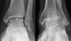
What bones are involved in forming the ankle joint?

A

it is formed by the articulation between:

  1. distal tibia
  2. distal fibula
  3. talus
2
Q

What are the only 2 movements present at the ankle joint?

A

it is a synovial hinge joint that permits:

  1. dorsiflexion (extension)
  2. plantarflexion
3
Q

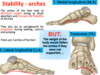
What factors help to contribute to the stability of the ankle joint?

A
  1. there is good congruity between the malleolar mortice and trochlea of the talus

the malleoli grip the talus and keep it in place

  1. very strong ligaments
4
Q

Label the components of the ankle joint

A
5
Q

Why is dorsiflexion the most stable position of the ankle joint?

A
  1. the trochlea of the talus is wider anteriorly than posteriorly
  2. during dorsiflexion, the anterior part of the trochlea moves between the malleoli
  3. this spreads the tibia and fibula slightly and increases their grip on the talus
6
Q

What are the 2 ligament complexes that stabilise the ankle joint?

A

lateral ligament complex

medial ligament complex (or deltoid)

7
Q

What are the 3 ligaments of the lateral ligament complex?

A
  1. anterior talofibular ligament
  2. posterior talofibular ligament
  3. calcaneofibular ligament
8
Q

label the lateral ligaments of the ankle joint

A
9
Q

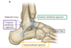
What are the attachments involved with the medial ligament?

A

it is attached to the medial malleolus and fans out to attach to the talus, navicular and calcaneus

10
Q

What movements of the ankle joint are shown?

A
11
Q

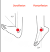
What movements are shown?

A
12
Q

Which joint is responsible for eversion and inversion?

A

subtalar joint

this is the between the talus and the calcaneus (heel bone)

13
Q

Which muscles are involved in dorsiflexion?

A

anterior compartment of the leg

tibialis anterior (TA)

extensor hallucis longus (EHL)

extensor digitorum longus (EDL)

14
Q

What is the artery and the nerve of the anterior compartment of the leg?

A

deep fibular (peroneal) nerve

anterior tibial artery

15
Q

What are the muscles involved in plantarflexion?

A

posterior compartment of the leg

  1. tibialis posterior
  2. flexor hallucis longus
  3. flexor digitorum longus
16
Q

What is the artery and the nerve of the posterior compartment of the thigh?

A

tibial nerve

posterior tibial artery

17
Q

What are the 3 muscles of the anterior compartment of the thigh?

What is their function?

A
  1. tibialis anterior (TA)
  2. extensor digitorum longus (EDL)
  3. extensor hallucis longus (EHL)

they are involved in dorsiflexion of the ankle and extension of the toes

18
Q

What is the insertion of the tibialis anterior?

A

it originates from the lateral surface of the tibia and interosseous membrane

it inserts on the base of the first metatarsal bone

19
Q

What is the origin and insertion of extensor hallucis longus?

What is significant about the insertion of its tendons?

A

it arises from the middle portion of the fibula and the interosseous membrane

its tendon inserts on the distal phalanx of the big toe

this allows it to extend the big toe

20
Q

What is the origin and insertion of extensor digitorum longus?

What is significant about the insertion of its tendons?

A

it originates from the lateral condyle of the tibia and interosseous membrane

its tendons insert onto the middle and distal phalanges of digits 2-5

this allows it to extend the toes

21
Q

label the anterior leg muscles

A
22
Q

How are the posterior leg muscles divided?

A

there are 3 deep compartment muscles and 2 superficial compartment muscles

23
Q

What are the 3 muscles of the deep group of the posterior compartment?

A
  1. tibialis posterior (TP)
  2. flexor hallucis longus (FHL)
  3. flexor digitorum longus (FDL)

they are involved in plantarflexion and flexion of the toes

24
Q

what is the origin and insertion of tibialis posterior?

A

it originates from the tibula and fibula

it inserts onto the navicular and medial cuneiform bone

it can cannot flex the toes

25
What is the origin and insertion of flexor digitorum longus? What does the insertion of its tendons allow it do?
it originates from the posterior surface of the tibia it inserts onto the plantar surface of the base of the distal phalanges of digits 2-5 this allows it to flex the toes
26
What is the origin and insertion of flexor hallucis longus? What does the insertion of its tendon allow it to do?
it originates from the fibula it inserts onto the plantar surface of the distal phalanx of the big toe (hallux) this allows it to flex the big toe
27
Why does flexor hallucis longus play such a crucial role?
plantar flexion is crucial to walking as the final stage in walking is pushing off of the ground with the big toe (toe-off) (going on tip toes is also plantar flexion when the foot is fixed on the ground)
28
label the deep muscles of the posterior leg
29
What are the 2 superficial muscles of the posterior leg?
gastroenemius is the most superficial muscle soleus is attached to the soleal line and covers all of the deep muscles
30
What are the functions of the superficial muscles of the posterior leg?
they are plantarflexors of the ankle soleus contracts to squeeze the underlying deep veins of the leg to force the blood back to the heart
31
What is the Achilles tendon? Where does it insert?
both soleus and gastroenemius insert onto the calcaneum via the Achilles tendon
32
33
Label the superficial muscles of the posterior leg
34
What are the 2 muscles of the lateral compartment of the leg? What is their function?
**fibularis longus** and **fibularis brevis** they are involved in eversion
35
What are the 2 divisions of the sciatic nerve? Which compartments of the leg are innervated by which divisions?
**tibial division:** * this innervates all the muscles in the posterior compartment **common fibular division:** * this divides into the superficial fibular nerve that innervates the lateral compartment * and the deep fibular nerve that innervates the anterior compartment
36
What action are most ankle injuries caused by?
most ankle sprains are inversion injuries with twisting of a plantar-flexed foot this causes tearing and stretching of the lateral ligaments
37
Why do ligaments heal slowly?
they are relatively avascular
38
what happens if a ligament is detached from bone?
fibres do not grow back into the bone cortex as extensively this means that the healed ligament is usually weaker
39
How do torn ligaments affect the ankle joint? What ligaments of the ankle joint are weaker?
they predispose to dislocation the lateral ligament is weaker than the medial ligament, particularly the anterior talofibular part
40
What injury is shown? What usually causes this?
**ankle fracture** this is caused by fracture of the lateral malleolus it may be coupled with fracture of the medial malleolus
41
What causes an eversion injury? What injuries are associated with this?
they are caused by the foot being forced into an eversion position this pulls the medial ligament and causes avulsion of the medial malleolus this causes the talus to rotate laterally and fractures the fibula
42
What injury is shown here?
fracture dislocation this tends to be obvious as there is not much fat or subcutaneous tissue around the ankle
43
what injury is shown here?
osteoarthritis of the ankle joint
44
What are the tarsal bones?
45
Why is the foot a series of small bones and ligaments?
this allows the foot to deform to absorb shock and adapt to walking on uneven surfaces if the foot was rigid, each impact with the ground would generate large forces
46
What are the roles of the tarsal bones?
1. to support and transmit body weight 2. acts as a lever to propel body forward during motion 3. acts as a resilient spring to absorb shock
47
What are the roles of the arches of the feet?
**_STABILITY_** they help to distribute weight, act as shock absorbers and increase the flexibility of the foot they also act as springboards for propulsion during walking, running and jumping
48
What are the 3 arches of the foot?
1. medial longitudinal 2. lateral longitudinal 3. transverse
49
What are the methods of passive and dynamic support of the foot arches?
**passive support:** ligaments and the shape of the bones **dynamic support:** intrinsic and extrinsic muscles of the foot
50
What would happen if the arches of the foot were not supported?
they would flatten out due to the weight of the body when standing
51
What condition is shown?
**pes planus** this is seen in adolescents or adults it is usually caused by loose of degenerating ligaments
52
53
What are the 2 types of pes planus?
flexible and rigid flexible is more common - this is where the arch is present when not bearing weight but absent when standing
54
What are the exacerbating factors in pes planus?
gaining weight and/or spending a long time standing
55
Why must pes planus be treated even if it is asymptomatic?
it can result in displacement of the talus infero-medially this causes misalignment of the ankle and knee causing pain in these areas this can also decrease shock absorption by the foot leading to lower back pain
56
What is the treatment for pes planus?
orthotics (specialised insoles) that support the arches of the foot
57
What movements are present at the toes? Which joints are involved?
**flexion and extension:** acheived by the metatarsophalangeal joints, PIPJs, DIPJs **abduction and adduction:** limited abduction and adduction occurs at the metatarsophalangeal joints
58