Midterm I - Website Photos [Better Quality] Flashcards

1
Q
A

Babesia canis: Merozoites

Blood smear

2
Q
A

Cryptosporidium baileyi oocysts

On the ciliated cell surface of the trachea

3
Q
A

Babesia canis merozoites

Blood smear, Giemsa-stained

4
Q
A

Unsporulated Eimeria oocysts from a lamb

5
Q
A

Eimeria acervulina macrogamonts & developing oocysts

6
Q
A

Eimeria maxima: Sporulated oocyst

Iodine stain

7
Q
A

Eimeria bakuensis: Unsporulated oocysts

Floatation method, methylene-blue stain

8
Q
A

Eimeria: Unsporulated oocyst

E. robusta, methylene-blue stain

9
Q
A

Eimeria stiedai: Oocysts

In destroyed biliary epithelium

10
Q
A

Leishmania donovani: Promastigotes

Giemsa stain

11
Q
A

Toxoplasma gondii: Tissue cysts

Mouse brain, HE stain

12
Q
A

Trypanosoma equiperdum: Trypomastigotes

Giemsa stain

13
Q
A

Encephalitozoon cuniculi: Focal encephalitis (see macrophages)

Rabbit brain, HE stain

14
Q
A

Encephalitozoon cuniculi: Interstitial nephritis

Rabbit kidney, HE stain

15
Q
A

Encephalitozoon cuniculi: Interstitial nephritis + CT proliferation

Rabbit kidney, HE stain

16
Q
A

Eimeria bovis (larger) & Eimeria zuernii (smaller): Oocysts

Floatation, native

17
Q
A

Leishmania donovani: Amastigotes in macrophages

Giemsa stain

18
Q
A

Sarcocystis tenella: Tissue cyst

Sheep oesophagus, HE stain

19
Q
A

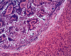
Eimeria stiedai: Biliary hyperplasia

Rabbit liver, HE stain

20
Q
A

Trypanosoma cruzi: Pseudocyst

In a section of heart muscle, HE stain

21
Q
A

Trypanosoma equiperdum (Dourine): Dollar-spots on the horse

22
Q
A

Giardia intestinalis: Cyst

Faecal smear, Lugol solution (iodine)

23
Q
A

Giardia intestinalis: Trophozoite

24
Q
A

Engorged tse tse fly

Responsible for the transmission of African Trypanosomes E.g:

  • T. vivax
  • T. congolensis
  • T. brucei brucei
25
*Histomonas meleagridis*: Ameboids ## Footnote *Liver tissue, PAS stain*
26
Kissing bug: Biological vector *Transmission of Leishmania chagasi (Chagas disease)*
27
*Leishmania*: Amastigotes ## Footnote *Inside a macrophage*
28
Cutaneous Leishmaniosis
29
Clinical signs of Nagana (African Trypanosmosis) ## Footnote *Transmitted by the Tsetse fly*
30
*Trichomonas* infection (Hepatic form) ## Footnote *Pigeon head & liver*
31
Sandfly: Biological vector ## Footnote *Transmission of Leishmania*
32
*Trichomonas*: Trophozoites (Most likely: poultry) ## Footnote *Giemsa stain*
33
*Trypanosoma brucei*: Trypomastigotes ## Footnote *Blood smear, Giemsa stain*
34
*Trypanosoma cruzi*: Trypomastigote ## Footnote *+ a large kinetoplast, Giemsa stain*
35
Lesions in the caeca & liver associated with *Histomona meleagridis* ## Footnote *Samples from a turkey*
36
Lesions in the surface of the duodenum associated with *Eimeria acervulina* ## Footnote *Sample from a chicken*
37
*Eimeria brunetti*: Bloody mucus in the lumen of the hindgut ## Footnote *Sample from a chicken*
38
Macrogamonts with granular cytoplasm One-nucleated oocysts surrounded by a wall in the destroyed wall of the intestinal mucosa
39
*Eimeria maxima*: Small haemorrhages & whitish spots of schizonts in the mucosa of the small intestine ## Footnote *Not to be confused with "salt & pepper" spots in E. necatrix infections*
40
*Eimeria necatrix*: Haemorrhages in the jejunal wall
41
Nests of schizonts ## Footnote *Destroyed intestinal mucosa of chicken, HE stain*
42
*Eimeria tenella*: Lesions of the caeca ## Footnote *Sample from a domestic chicken*
43
*Eimeria tenella*: Lesions of the caeca ## Footnote *Sample from a domestic chicken*
44
*Eimeria truncata*: Macrogamonts ## Footnote *Goose kidney tissue, HE stain*
45
*Eimeria*: Sporulated oocyst ## Footnote *Stained with iodine*
46
Three schizonts & free merozoites in a native mucosal smear
47
*Coccidium* *spp.*: Thick-walled, unsporulated oocysts
48
*Eimeria stiedai*: Biliary hyperplasia Rabbit liver, HE stain
49
Clinical signs of acute bovine coccidiosis Either *E. zuernii or E. bovis*
50
Clinical signs of Isosporosis of piglets (*Isospora suis)*
51
*Eimeria stiedai*: Unsporulated oocysts ## Footnote *These are often stained yellow by the bile*
52
*Eimeria bovis* (large) & *E. zuernii*: Unsporulated oocysts
53
Section of a globidium ## Footnote *Located in the top layer of interstitial mucosa*
54
Bovine coccidiosis (*E. zuernii* or *E. bovis*) * Pinhead-sized white spots in the thickened mucosa of the intestine * Covered with bloody mucus
55
*Eimeria flavescens*: Dilated caecum of a rabbit
56
*Eimeria intestinalis*: Thickened wall of small intestine ## Footnote *Sample from a rabbit*
57
*Eimeria magna*: White groups of macroschizonts ## Footnote *Wall of the small intestine, rabbit*
58
*Eimeria piriformis*: Thickened & whitish wall of large intestine ## Footnote *Sample from a rabbit*
59
*Eimeria stedai*: * Broadened parts of biliary ducts * Yellowish nodules can be seen on the surface *Rabbit liver sample*
60
*Isospora*: Sporulated oocyst
61
*Isospora*: Oocyst from: 1. A dog 2. A cat
62
*Cryptosporidium baileyi*: * Thick-walled oocysts (red) * Thin-walled oocysts (colourless) *Mucosal cell debris of bursa fabricus, Kinyoun stain*
63
*Neospora caninum*: Thick-walled tissue cyst ## Footnote *Brain tissue, HE staining*
64
*Sarcocystis*: Haemorrhagic diatheses on visceral serosa ## Footnote *Dalmeny disease (bo. only), Calf*
65
*Besnoitia besnoiti*: Large cysts ## Footnote *Subcutaneous tissue of bo., HE stain*
66
*Sarcocystis gigantea*: Cysts ## Footnote *Muscle layer of the oesophagus, sheep*
67
*Sarcocystis*: Cyst (longitudinal section) ## Footnote *Skeletal muscle, HE stain*
68
*Besnoitia besnoti*: Clinical symptoms * Top row: "Anasarca" stage * Bottom row: "Scleroderma" stage
69
*Sarcocystis*: Cyst (cross-section) ## Footnote *Skeletal muscle, HE staining*
70
*Sarcocystis*: Cyst ## Footnote *Compressed skeletal muscle, native*
71
*Sarcocystis*: Thin-walled oocyst ## Footnote *Faecal debris*
72
*Toxoplasma*: Cysts ## Footnote *Brain tissue*
73
*Toxoplasma gondii*: Tachyzoites ## Footnote *Giemsa stain*
74
*Toxoplasma*: Cysts ## Footnote *Brain tissue, HE stain*
75
*Toxoplasma gondii*: Oocysts (Sporulated & unsporulated)
76
*Theileria annulata*: Enlarged lymph nodes ## Footnote *(Bovine theileriosis)*
77
*Babesia canis*: Clinical signs
78
*Dermacentor reticulatus* (brown dog tick/kennel dog tick) The vector of *Babesia canis*
79
*Hepatozoon canis*: 1. In a mononuclear cell 2. In a polymorphonuclear cell
80
*Encephalitozoonosis*: Clinical sign ## Footnote *Interstitial nephritis*
81
1. *Babesia bigemina* (large) 2. *Babesia divergens* ## Footnote *In blood cells*
82
*Theileria annulata*: Schizonts ## Footnote *Taken from bovine lymphocytes*
83
*Theileria equi*: Merozoites ## Footnote *Blood smear*
84
*Encephalitozoon cuniculi*: Torticollis (clinical sign) ## Footnote *Rare occurance*
85
*Histomonas meleagridis*: Amoeboids ## Footnote *Liver tissue, PAS stain*
86
*Giardia intestinalis*: Trophozoites
87
*Eimeria truncata*: Macrogamonts ## Footnote *Goose kidney tissue, HE stain*
88
*Trypanosoma brucei*: Trypomastigotes ## Footnote *Blood smear, Giemsa stain*
89
*Entamoeba histolytica*: Trophozoite ## Footnote *Faecal smear + Saline + Lugol*
90
*Entamoeba histolytica*: Cyst ## Footnote *Floatation method*