Muscular System Flashcards
(21 cards)
A muscle cell that consumes very little energy.
A) Smooth muscle
B) Skeletal muscle
C) Cardiac muscle
D) All of the above
Smooth muscle
When compared to skeletal and cardiac muscles cells, smooth muscles cells have the ability to maintain a force or contraction for long periods of time with a little use of very little energy.
Which type of connective tissue links bones and muscles together?
A) Tendon
B) Ligament
C) Cartilage
D) Aponeurosis
Tendon
Choice A. Tendon is a cord or band of dense connective tissue that connects a muscle to a bone or another structure.
Choice B. Ligament is a fibrous connective tissue that connects bones or cartilages.
Choice C. Cartilage is a firm, smooth, resilient, nonvascular connective tissue.
Choice D. Aponeurosis is a sheath of white-silvery tissue that resemble a flattened tendon. It serves to bind muscles and connect them to a bone or fascia.
This muscle is superficial and located directly lateral to the tibial shaft.
A) Peroneus Longus
B) Peroneus Brevis
C) Extensor Digitorum Longus
D) Tibialis Anterior
Tibialis
Anterior Tibialis anterior is superficial and located directly lateral to the tibial shaft.
Extensor Digitorum Longus is located between TA and Peroneus Longus.
The Peroneus longus and brevis can be found between the lateral malleolus and head of the fibula.
Which type of muscle fibers are red, have a small diameter, contain a large amount of myoglobin, a rich blood supply, split ATP aerobically and are resistant to fatigue, suiting them to endurance activities and maintaining posture?
A) Slow oxidative
B) Fast oxidative
C) Slow glycolytic
D) Fast glycolytic
Slow oxidative
Slow twitch oxidative fibers are designed for long run (endurance) and need more oxygen to sustain their movement. They higher oxygen supply gives them their red appearance.
Which of the following is the basic functional unit of a muscle fiber and is made up of interlocking contractile proteins?
A) Sarcoplasm
B) Sarcolemma
C) Sarcomere
D) Sarcoplasmic reticulum
Sarcoplasm
Sarcoplasm refers to the muscle fibers’ cytoplasm without the myofibrils.
Choice B. Sarcolemma refers to the plasma membrane of a muscle fiber.
Choice C. The sarcomere is considered as the basic functional unit of a muscle. It consists of the protein filaments actin and myosin.
Choice D. Sarcoplasmic reticulum refers to the specialized smooth endoplasmic reticulum situated in the sarcoplasm between the T tubules.
Three of the four rotator cuff muscles insert onto the greater tubercle of the humerus. Which one does not?
A) Subscapularis
B) Supraspinatus
C) Infraspinatus
D) Teres Minor
Subscapularis
Subscapularis inserts onto the lesser tubercle of the humerus and is responsible for medial/internal rotation of the shoulder. Supraspinatus, infraspinatus and teres minor all insert onto the greater tubercle of the humerus.
A muscle that is circular in shape:
A) Anconeus
B) Orbicularis
C) Peroneus
D) Occiput
Orbicularis
Orbis is Latin for “circle or disk.” The orbicularis oris muscle is a sphincter muscle that encircles the mouth. Another circular shaped muscle is the orbicularis oculi muscle. It is a sphincter muscle that encircles the eye.
A muscle that separates the thoracic and the abdominal cavity.
A) Peritoneum
B) Diaphragm
C) Cranial
D) Thoracic
Diaphragm
Just inferior to, or below, the thoracic cavity is the abdominal cavity. The two are separated by the diaphragm.
What is the insertion of the trapezius?
A) Clavicle
B) Acromion
C) Spine of scapula
D) All of the above.
All of the above.
ORIGIN Occiput, ligamentum nuchae, and the spinous processes of C1 – C7.
INSERTION Lateral 1/3 of the clavicle, acromion, and spine of the scapula.
ACTION Elevate, retract, and rotate scapula upward.
Which of the following is not a type of muscle tissue? A) skeletal
B) cardiac
C) smooth
D) connective
connective
Choice A. Skeletal muscles are those attached to bones and where one can palpate them. Their cells are very long and are cylindrical in shape and have multiple nuclei that are located in the periphery. Skeletal muscles have striations and voluntary contraction is possible. This type of muscle cells are the only ones not capable of producing spontaneous contractions.
Choice B. Cardiac muscles are found specifically in the heart only. Their cells are cylindrical and branched and have a single, centrally located nucleus. The cells are attached to one another through the intercalated disks (a type of intercellular junction specialized for impulse conduction to contract heart). Cardiac muscle cells have striations and voluntary control is not possible. If the heart cells do not beat in unison, heart arrhythmias can occur.
Choice C. Smooth are found in the walls of hollow organs, blood vessels, eyes, glands, and skin. They are spindle-shaped and have a single, centrally located nucleus.Smooth muscle cells do not have striations and gap junctions join some visceral smooth muscle cells together. Voluntary contraction is not possible with this type of cells.
Choice D. Connective tissues are abundant in the human body and makes up every organ. It is unique from other tissue types since it consists of cells separated from each other by abundant extracellular matrix. Connective tissues connect, support, or separate tissues or organs. They are not a type of muscle tissue. Examples of this type include ligaments, cartilage, bone and blood.
Which is an example of a strap muscle?
A) Biceps brachii
B) Sartorius
C) Pectoralis major
D) Deltoid
Sartorius
Sartorius is an example of a strap muscle. Biceps brachii is a fusiform muscle. Pectoralis major is a convergent, fan-shaped muscle. Deltoid is multipennate.
How many flexor tendons are in one carpal tunnel?
A) 4
B) 5
C) 9
D) 10
9
The carpal tunnel is a narrow passageway located on the palmar side of the wrist. This tunnel contains the median nerve and the nine tendons that flex the wrist: one - flexor pollicis longus four - flexor digitorum superficialis four - flexor digitorum profundus
_______________ cannot be consciously controlled: A) Voluntary skeletal muscles
B) Smooth muscle tissue
C) Both A and B
D) None of the above
Smooth muscle tissue
Skeletal muscle is under voluntary control. Smooth muscle is not under voluntary control. Cardiac muscle, although striated like skeletal muscle, is also not voluntary.
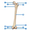
All of the following muscles insert to #10, except:
A) Biceps femoris
B) Adductor longus
C) Adductor brevis
D) Adductor magnus

Biceps femoris
The biceps femoris belongs to the posterior compartment of the thigh. It has two heads: the long head originates from the ischial tuberosity while the short head originates from the lateral supracondylar ridge of the femoral shaft and from the linea aspera. Both heads then insert distally to the head of fibula. The muscle functions by flexing and externally rotating the leg at the knee joint. The long head specifically, can extend the hip. Adductor longus belongs to the medial compartment of the thigh and as its name implies, participates in hip adduction and assists is lateral rotation as well. It originates from the body of pubis and inserts to the linea aspera.
Adductor brevis belongs to the medial compartment of the thigh and as its name implies, participates in hip adduction and assists is lateral rotation as well. It originates from the inferior ramus of pubis and inserts to the linea aspera.
The adductor magnus is a powerful adductor of the hip and can be found at the posterior and medial compartments of the thigh. It has two portions: the adductor portion originates from the inferior ramus of the pubis and from the ramus of the ischium and then inserts to the linea aspera. The hamstring portion of the muscle originates from the ischial tuberosity and inserts to the adductor tubercle of the femur.
The muscle functions as a hip adductor and lateral rotator. The hamstring portion specifically, act as a hip extensor.
Where is the location of the thenar eminence?
A) Beneath the little finger on the palm
B) The radial side of the palm
C) The ulnar side of the palm
D) Beneath the middle digit on the palm
The radial side of the palm
The thenar eminence refers to a group of muscles located on the palm of the hand at the base of the thumb (which is the radial side).
Also referred to as visceral muscle:
A) Smooth muscle
B) Skeletal muscle
C) Cardiac muscle
D) All of the above
Smooth muscle
Visceral muscle tissue, or smooth muscle, is tissue associated with the internal organs of the body, especially those in the abdominal cavity. Visceral manipulation is a massage technique used to manipulate the soft tissues in the abdominal cavity.
Which muscle does not attach to the sternum?
A) Sternocleidomastoid
B) Pectoralis Minor
C) Pectoralis Major
D) Sternohyoid
Pectoralis Minor
SCM originates at the manubrium of the sternum. Pectoralis Major originates at the sternum. Sternohyoid is a thin, narrow muscle that attaches the hyoid bone to the sternum. It depresses the hyoid bone. Pectoralis Minor originates at the 3rd, 4th, and ribs near their costal cartilages and does not have a sternal attachment.
Which is not part of the triceps surae?
A) Gastrocnemius
B) Soleus
C) Plantaris
D) Achilles Tendon
Plantaris
The triceps surae is a group of muscles in the posterior lower leg. This group includes gastrocnemius, soleus, and their common tendon of insertion, the Achilles tendon.
The quadriceps are located on which side of the body?
A) Medial
B) Posterior
C) Anterior
D) Lateral
Anterior
The quadriceps are on the anterior, or front, side of the body.
Which of the following muscle groups are referred to as the rotator cuff?
A) Trapezius, rhomboids, levator scapulae
B) Biceps femoris, semitendinosus, semimembranosus
C) Rectus femoris, vastus lateralis, vastus medialis, vastus intermedius
D) Subscapularis, supraspinatus, infraspinatus, teres minor
Subscapularis, supraspinatus, infraspinatus, teres minor
The SITS muscles make up the rotator cuff. This includes: Subscapularis Infraspinatus Teres minor Supraspinatus


