Neoplasms of Bone and Joint Pathology Flashcards

1
Q

How does the diagnosis of bone tumors differ from other tissues?

A

histology comes last

2
Q

What is osteochondroma?

A

benign cartilaginous lesion and most common benign tumor

contains marrow space contiguous with the underlying medulla of the bone

mutation in suppressor gene EXT1 that affects growth plate kinetics

3
Q

Multiple Hereditary Exostosis

A

AD disorder resulting in multiple osteochondromas

caused by biallelic inactivation of EXT1 and EXT2

malignant transformation (2-3% of cases) mostly into chondrosarcomas

4
Q

What is the clinical presentation of osteochondroma?

A

peak is in 1st and 2nd decades

male-female ratio is 2:1

located in the distal femur, proximal humerus, and proximal tibia (>50%)

5
Q

What are the x-ray findings of osteochondroma?

A

cartilaginous cap on bony stalk that is continuous with medulla of bone

tumor grows away from growth plate (cap size>3cm. suggests malignant transformation)

“arc and ring” calcifications

6
Q

What is the treatment and prognosis of osteochondroma?

A

no treatment unless symptomatic or there is evidence of possible secondary chondrosarcomatous transformation of the cartilaginous cap

1-2% recur, 1-3% of solitary and up to 5% of multiple osteochondroma undergo malignant transformation

mostly chondrosarcoma

7
Q

What are the histological features of osteochondroma?

A

cartilaginous “cap” shows features of normal growth plate cartilage

stalk of tumor contiguous with intramedullary portion of bone and composed of normal marrow elements

8
Q

What is osteoid osteoma?

A

small benign bony process < 2 cm

9
Q

What is the clinical presentation of osteoid osteoma?

A

increasing nocturnal pain relieved by ASA/NSAID (80% of patients)

any bone except skull/sternum, 50% involve femur and tibia

mostly in 2nd and 3rd decades, rare after 30 years of age

male-female ratio is 2:1

10
Q

What is the radiological findings in osteoid osteoma?

A

zone of sclerotic bone surrounding a well-demarcated lucent focus (the “nidus”) composed of woven bone in a vascular stroma

11
Q

What is the prognosis of osteoid osteoma?

A

excellent, but must remove nidus

12
Q

What are the histological features of osteoid osteoma?

A

the “nidus” (arrows) is the radiolucent central focus of woven bone within a vascular stroma

the surrounding cortical bone is thickened (top)

nidus consists of woven bone with osteoblastic rimming and a highly vascular stroma (right, bottom)

13
Q

What is enchondroma?

A

benign hyaline intramedullary cartilaginous neoplasm

**periosteal chondroma is located on the cortical surface under the periosteum

14
Q

What is the clinical presentation of enchondroma?

A

2nd to 4th decades, M=F

located in the finger and toes in 50% of cases

proximal humerus, proximal and distal femur

15
Q

What are the radiologic findings of enchondroma?

A

well-circumscribed, lucent with cartilage matrix calcification

“arc and ring” due to enchondral bone formation at periphery of tumor nodule

16
Q

What is the prognosis of enchondroma?

A

observation with serial radiographs

curettage (<5% recurrent state)

17
Q

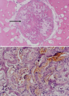
What are the histological findings of enchondroma?

A

nodules of mature cartilage within a fatty bone marrow (top)

note low cellularity or lack of pleomorphism (bottom)

benign chondrocytes evenly distributed or arranged in clusters

18
Q

What is multiple enchondromata?

A

non-hereditable disorder characterized by multiple enchondromas (usually affects same region of body)

incidence of malignant transformation is 15%-50%

unilateral distribution in the appendicular skeleton

19
Q

Ollier’s disease

A

multiple enchondromata

20
Q

Maffuci’s syndrome

A

multiple enchondromata + soft tissue/visceral vascular tumor (mainly, hemangiomas)

21
Q

What is giant cell tumor of bone?

A

locally aggressive neoplasm

tumor composed of osteoclasts and neoplastic spindle cells

22
Q

What is the clinical presentation of giant cell tumor of bone?

A

peak in 3rd decade (skeletally-mature), F>M (~3:2)

located in the distal femur, proximal tibia, distal radius, proximal humerus (single/multiple GCT in odd sites -> hyperparathyroidism)

23
Q

What are the radiologic features of giant cell tumor of bone?

A

eccentric, lytic lesion that may involve soft tissue (extends from metaphysis into epiphysis once growth plate closes)

24
Q

What is the prognosis of giant cell tumor of bone?

A

en bloc excision is prefferred

high recurrence rate (25%-60%) if curetted

<5% of GCT metastasize to lung (histology can’t predict clinical course)

sarcomatous transformation is rare (usually follows radiotherapy)

25
Q

What are the histological findings in giant cell tumor of the bone?

A

lytic meta- and epiphyseal lesion, ill-defined cellular margins and osteoclast-like giant cells

microscopically, evenly dispersed osteoclasts with numerous nuclei (INSET) and intervening mononuclear stromal cells

26
Q

What is fibrous dysplasia?

A

developmental fibro-osseous defect due to mutations in GNAS1 gene that encodes the alpha subunit of the G receptor protein (cause decreased GTPase activity)

increased cAMP (affects normal maturation of osteoblasts)

27
Q

What is the clinical presentation of fibrous dysplasia?

A

mostly diagnosed in first three decades of life (M=F)

located in the femur/tibia, craniofacial, ribs, pelvis

monostotic (80%)

polyostotic (20%)

28
Q

McCune-Albright syndrome

A

polyostotic disease + cafe au lait spots + endocrinopathies, mostly isosexual precocity

29
Q

What are the radiological findings of fibrous dysplasia?

A

expansile, lesion with a variable internal density (“ground glass”)

30
Q

What is the prognosis of fibrous dysplasia?

A

monostotic FD usually stabilizes at puberty

symptomatic lesions treated with biphosphonates

sarcoma is rare (<0.5%; mostly gnathic bones in adults)

may recur if not completely excised

31
Q

What are the histological features of fibrous dysplasia?

A

expansile, circumscribed lesion within the mid-portion of the rib

irregularly contoured (“C” and “S” shaped) spicules of woven bone with no osteoblastic rimming (INSET) within a spindled and collagenous stroma

32
Q

What is osteosarcoma?

A

primary malignant tumor in which malignant neoplastic cells produce osteoid and/or bone, even if only in small amounts

develops in metaphysis of long bones of the young

in older patients, presents more often in caniofacial bone or axial skeleton and is usually associated with a predisposing bone condition

33
Q

What is the clinical presentation of osteosarcoma?

A

peak in second decade, 60% < 25 yrs old

male-female ratio is 3:2 (particularly patients < 20 years)

located in metaphysis of long bones, particularly distal femur, proximal tibia, and proximal humerous

incidence of OS involving pelvis and craniofacial bones increase with age reflecting malignant transformation of benign process like Paget’s disease or fibrous dysplasia

34
Q

What are the radiographical findings of osteosarcoma?

A

most cases are mixed lytic/blastic + cortical destruction + soft tissue extension

35
Q

What is the prognosis of osteosarcoma?

A

depends mostly on response to pre-operative chemotherapy (>90% tumor necrosis = survival 80-90%)

whereas <90% necrosis and no change in chemo = survival < 15%

36
Q

What are the histological findings on osteosarcoma?

A

gross speciment shows OS in metaphyseal region with soft hissue extension

OS defined as a sarcoma in which malignant cells directly produce osteoid/woven bone

note highly pleomorphic cells

37
Q

What is the therapy for osteosarcoma?

A

preoperative chemotherapy -> 2-6 cycles of chemotherapy -> histologic examination of excised tumor -> fit for prosthesis -> postoperative chemotherapy

if < 90% necrosis after chemo, consider changing drugs and local therapy if margins positive

if >/= 90% necrosis, continue therapy and use local therapy if margins are positive

38
Q

What is the clinical presentation of primary chondrosarcoma?

A

peaks in 5th to 7th decades

male-femal ratio is 2:1

mostly central (within medullary cavity) and less often peripheral (surface of bone)

pelvis (ilium), proximal femur, proximal humerus, distal femur, and ribs

39
Q

What are the radiologic findings of primary chondrosarcoma?

A

lobulated, lytic lesion with cartilage matrix calcification

cortical destruction/thickening, no/minimal periosteal reaction

“arc and ring” calcification

40
Q

What is the treatment and prognosis of primary chondrosarcoma?

A

depends mostly on grade and surgical adequacy

5 yr survival for G1 = 90%, G2 and G3 = 53%

metastases are rare and occur late

41
Q

What are the histological features of primary osteosarcoma?

A
42
Q

What is Ewing Sarcoma/primitive neuroectodermal tumor (ES/PNET)?

A

primitive neuroectodermal tumor of bone/soft tissue characterized by a t(11;22) in 90% of cases or t(21;22) in 5-10% of cases

second most common primary malignant bone tumor (behind OS) in children

43
Q

What is the clinical presentation of Ewing Sarcoma/primitive neuroectodermal tumor?

A

about 80% <20 years

male-femal ratio is 2:1

most common in femur, tibula, fibula, and humerus

followed by pelvis and ribs

44
Q

What are the radiological features of Ewing Sarcoma/primitive neuroectodermal tumor?

A

Ill-defined lytic lesion in diaphysis or meta-diaphysis

cortical destruction

+/- “onion-skin”/”sunburst” periosteal reaction

soft tissue extension frequent

45
Q

What is the treatment and prognosis of Ewin sarcoma/primitive neuroectodermal tumors?

A

treated with neoadjuvant multiagent chemotherapy (like osteosarcoma) followed by wide excision and/or radiation

current overall survival for non-metastatic sarcoma is 60% to 80%

metastases in 20-25% of cases and associated with 80% mortality

46
Q

What are the histological features of Ewing sarcoma/primitive neuroectodermal tumor?

A

sheets/nests of small polygonal cells

metadiaphyseal location of sarcoma with soft tissue component and periosteal reaction

diffuse proliferation of small neoplastic cells with focal necrosis

cells have rounded nuclei and cleared cytoplasm (INSET)

due to glycogen deposition

47
Q

What are type A synoviocytes?

A

macrophage-like cells found in the synovium of diarthrodial joints

48
Q

What are type B synoviocytes?

A

cells found in synovium that secrete hyaluronic acid (a large viscoelastic mucopolysaccharide) into the synovial space

49
Q

Where in a joint do inflammatory processes like rheumatoid arthritis first manifest?

A

the pocket/recess formed by the synovium on one side of the joint space

the bone lacks cartilage here

50
Q

What is the clinical presentation of osteoarthritis?

A

older aged caucasians with a male:female ratio of 2:1

most commonly affects hip, knee, DIP and PIP of fingers, and spine

Heberden’s nodes in DIP in mostly females

symptoms include pain, stiffness after use (“evening stiffness”), joint effusion, and loss of joint mobility

majority of individuals over age 65 have radiologic and/or clinical evidence of osteoarthritis

5% to 20% of patients have a predisposing condition that compromises integrity of joint and leads to early development of osteoarthritis (secondary osteoarthritis)

51
Q

What happens in the initiation stage of the pathogenesis of osteoarthritis?

A

chondrocyte injury/death caused by repetive abnormal mechanical loading (ability for joint cartilage to properly dissipate mechanical stress depends on genetic factors, age-related chagnes to cartilage, and anatomy/alignment of the joint)

52
Q

What happens during early OA?

A
  • deeper chondrocytes proliferate and synthesize matrix proteins, proteinases, and growth factors in attempt to remodel the matrix
  • endochondral bone formation by by deeply located chondrocyte (with potential to form bone or cartilage)

leads to osteophyte formation, subchondral bone thickening, and bone cyst formation

53
Q

What happens in late OA?

A

imbalance in matrix synthesis and breakdown during remodeling process eventually leads to loss of cartilage

54
Q

What are the pathologic findings of osteoarthritis?

A

pale articular cartilage due to loss of glycoaminoglycans and increased water content

vertical cracks in surface cartilage that parallel bundles of collagen type II (fibrillation)

complete loss of cartilage (eburnation)

early thickening of subarticular bone and osteophyte formation result form deepest chondrocytes forming new bone by echondral ossification

microfractures occur in subarticular bone as vessels penetrate bone and thin cartilage during process of echondral bone formation

this allows seepage of synovial fluid and the formation of subarticular (synovial fluid filled cysts)

55
Q

What are the common seropositive noninfectious inflammatory arthritis?

A

rheumatoid arthritis

juvenile rheumatoid arthritis (ANA-positive)

56
Q

What are the common seronegative (HLA-B27 associated arthridities?

A

ankylosing spondylitis

reactive arthritis (Gi/GU bacterial and IBD associated)

arthritis (urethritis/cervicitis + uveitis/conjunctivitis = REITER syndrome)

psoriatic arthritis (10%)

57
Q

What is the clinical presentation of rheumatoid arthritis?

A

affects patients mostly in 5th to 8th decades of life, a lot more females than males (over 2:1)

joints involved include hands (MCP and PIP), feet, wrists, ankles, upper spine, and knees

arthritis is symmetrical , plus the pain and morning stiffness

rheumatodi nodules (palisaded granulomas) in 25% of patients

serology positive for rheumatic factor and/or cyclic citrullinated peptide antibody - 20% of patients remain negative for both

58
Q

What disease are some diseases assocated with rheumatoid arthritis?

A

Sjogern’s syndrome

amyloidosis

pleuritis and pericarditis

lung fibrosis

aortic valvulitis

vvasculitis

splenomegaly (Felty syndrome)

59
Q

What are the steps in the pathogenesis of rheumatoid arthritis?

A

1) CD-4 T cells respond to arthritogen and stimulate synoviocytes and macrophages to produce pro-inflammatory molecules and allow for future production of proteases
2) RANKL prootes osteoclastogenesis resulting in bone erosions/osteoporosis
3) B cells produce antibodies that cause imune complex-mediated tissue damage
4) activation of vascular adhesion molecules promoting migration of inflammatory cells into joint

60
Q

What are the usual arthritogen?

A

infectious organism

collagen

proteoglycans

citrullinated proteins such as fibrin

61
Q

What is the pathology behind rheumatoid arthritis?

A

1) proliferative synovitis with increased T cells, B cells, plasma cells, macrophages, and synoviocytes
2) increased vascularity (angiogenesis)
3) osteoclastogenesis (osteoporosis)
4) surface fibrin deposition and formation “rice bodies”
5) pannus (inflammatory cells, granulation tissue, macropahges, and fibroblastic synoviocytes - causes chondrolysis that resolves by fibrous ankylosis of joint (scarring of joint space)

62
Q

What are some common radiologic findings of rheumatoid arthritis?

A

osteoporosis and “ulnar drift”

opera glass deformity