Nervous System - Peripheral Nervous System Flashcards
(21 cards)
Peripheral Nervous System
- all of the nervous tissue outside of the CNS
- primarily consists of nerves that function to communicate between the body and the CNS
Cranial nerves - PNS nerves that connect to the brain
Spinal Nerves - PNS nerves that connect to the spinal cord
Cranial Nerves
- allow direct communication between the body and the brain
- one exception - accessory nerve (CN XI) arises from the spinal cord
- 12 pairs (CN I - CN XII)
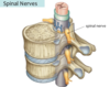
Spinal Nerves
- allow communication between the body and the spinal cord
- 31 pairs
- connected to the spinal cord by rootlets (dorsal root and ventral root)
- attached to spinal cord at regular intervals (reflection of segmental development)
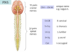
8 Cervical
12 Thoracic
5 Lumbar
5 Sacral
1 Coccygeal
Cranial Nerves and # Spinal Nerves

- 12 pairs cranial nerves
- 31 pairs spinal nerves
Spinal Cord Segment
- each individual portion of the spinal cord that is derived from one somite (blocks of mesoderm that lead spinal cord to develop in segmental fashion)
- each spinal cord segment is associated with one spinal nerve pair (one nerve on right and one left)

Somites
- derived from paraxial mesoderm
- repeating “blocks” of mesoderm that lie on either side of the neural tube
- develops into adult structures in a segmental pattern (i.e. vertebrae)
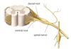
Dorsal Root
- the posterior root (roots connect spinal nerves to spinal cord)
- sensory root - contains sensory neurons
- you’re “sensitive” when someone talks behind you “back”

Ventral Root
- the anterior root (roots connect spinal nerves to spinal cord)
- motor root - contains motor neurons
- you drive a “motor” vehicle facing “front”

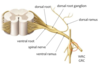
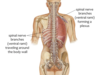
Spinal Nerve
- collection of axons in the PNS
- connected to the spinal cord by rootlets (dorsal and ventral roots)
- divides into two branches (dorsal ramus and ventral ramus)
Dorsal Ramus
- one of two primary branches of the spinal nerve
- innervates the muscles and the skin of the back

Ventral Ramus
- one of two primary branches of the spinal nerve
- innervates structures of the ventrolateral neck/trunk and limbs (everything the dorsal does not)
- typical - travels around the body wall and innervates muscle and skin of the trunk
- atypical - forms nerves plesuses
Dorsal Root Ganglion
- a collection of neuron cell bodies in the PNS
- enclosed by a connective tissue covering
- each dorsal root has a ganglion associated with it (dorsal root ganglion) - contains cell bodies for all the afferent neurons that travel in a particular spinal nerve and its dorsal root

Cauda Equina
- collection of dorsal and ventral roots in the inferior portion of the dural sac that have not exited the vertebral canal through intervertebral foramina
- named for its resemblance to a “horses tail”

Distribution of cranial vs. spinal nerves
Cranial nerves - primarily innervate structures in the head and neck
Exceptions - CN X (Vagus Nerve) travels to the lower abdomen
Spinal nerves - primarily innervate structures below the head
Exceptions - the first few cervical nerves do send some branches to the head

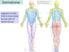
Dermatome
- the area of skin innervated by a particular spinal nerve pair
- due to segmental development from somites
Nerve Roots and Rami


Spinal Nerve Innervation
Dorsal Ramus - travels to the back region
Ventral Ramus - travels around the body wall
Thoracic Region (T2-T12) are “typical” spinal nerves because their ventral rami do not for plexuses

Nerve Plexus
- a network of mixing nerves
- T2-T12 do not tend to form plexuses
“Atypical” Spinal Nerves
- form plexuses
- only ventral rami
- some plexuses travel out into the limbs/extremities
- Spinal nerves that tend to form plexuses:
Cervical
T1
Lumbar
Sacral

How spinal nerves exit the vertebral column
- through the intervertebral foramen
- Cervical spinal nerves exit superior to the vertebra they are named for (i.e. C4 nerve exits between C3 and C4 vertebrae)
- C8 passes inferior to C7 and superior to T1
- All remaining spinal nerves below pass through inferior to the vertebra they are named for (i.e. T4 nerve passes between T4 and T5)

Distance bw spinal nerve and corresponding vertebra
- increases as you move inferiorly down the spinal column
- consequently, the spinal nerve roots must travel further to exit the vertebral canal
- creates cauda equina since the spinal cord ends at L2 (conus medullaris)

