Ophthalmology Flashcards
(34 cards)
What is the definition and epidemiology of cataract?
Cataracts is the opacification of the crystalline lens. It is estimated that50% of the world’s reversible blindness is caused by cataracts (and therefore is the leading cause of blindness worldwide).
What is the aetiology of cataracts?
Although the most common cause of cataracts is the normal ageing process (>90%), it can also be caused by trauma, metabolic disorders (diabetes mellitus), infections and glucocorticoids. <1% of cataracts are congenital, as part of a hereditary condition, or congenital conditions such as rubella
What are the clinical features of cataracts?
The classic presentation is a gradual decrease in vision over many years. Patients are usually slow to notice, and only complain when they have significant difficulty. The patient may complain of:
- Blurry vision (decrease in visual acuity) which is painless and bilateral
- Glare in daylight and often when driving (by headlights). This is associated with halos around lights.
- Washed-out colours are also a common complaint.
On examination there would be a defect in red reflex.

Describe the diagnosis and management of cataracts
Cataract visible on dilated fundus examination.
Measurement of intraocular pressure is normal or may be elevated if associated with glaucoma.
The most frequent indication for cataract surgery is to improve vision. Surgery of choice globally is extracapsular cataract extraction (ECCE). Cataract surgery does no require anticoagulative measures, and complications are rare.
What are the types of retinal vessel occlusion?
Retinal vessel occlusion causes retinal ischaemia. Based on the site of occlusion, retinal vessel occlusion can be classified into the following entities:
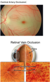
- Central retinal artery occlusion (CRAO)
- Branch retinal artery occlusion (BRAO)
- Central retinal vein occlusion (CRVO), either ischaemic (haemorrhagic retinopathy) or non-ischaemic (venous stasis retinopathy)
- Branch retinal vein occlusion (BRVO)
Retinal vein occlusion is much more common than artery occlusion. Retinal vein occlusion is the second most common vascular disease of the retina (after diabetic retinopathy)!
What is the aetiology of retinal artery occlusion/amaurosis fugax?
Retinal artery occlusion (stroke of the eye) is either due to emboli from carotid artery atherosclerosis (most common) or atrial fibrillation, or due to thrombosis of the retinal vessels as a result of atherosclerosis. Rarer causes include vasculitis (e.g. temporal arteritis) or fibromuscular dysplasia.
- These are also the causes of amaurosis fugax (sudden painless loss of vision followed by spontaneous recovery) which should therefore warrant investigation and treatment of the underlying cause.
What is the aetiology of retinal vein occlusion?
Retinal vein occlusion is most commonly due to systemic diseases such as atherosclerosis, hypertension and diabetes mellitus.
Central retinal vein occlusion (CRVO), either ischaemic (haemorrhagic retinopathy) or non-ischaemic (venous stasis retinopathy).
What are the clinical features of retinal artery occlusion?
General physical examination may reveal:
- A bruit over the carotid artery is a sign of carotid atherosclerosis.
- An irregular pulse may indicate atrial fibrillation.
- Scalp tenderness and/or jaw claudication is a sign of temporal arteritis.
What are the clinical features of retinal vein occlusion?
Branched vein occlusion is usually asymptomatic, but more common than central retinal vein occlusion.
Only ∼25% of patients with CRVO present with ischemic CRVO right at the outset. Of the rest who present with non-ischemic CRVO, a third eventually develop ischemic CRVO.

Describe the diagnosis and investigation of retinal vessel occlusion
Retinal vessel occlusion is primarily a clinical diagnosis (based on thepatient’s history and fundus examination). Additional investigations are usually performed to identify underlying risk factors, to differentiate between subtypes (e.g., in the case of CRVO).
CRAO: Greyish-white discolouration of the entire retina. May also be a cherry-red spot on fovea centralis. Narrowing of all retinal vessels.
CRVO: Many dot-and-blot and/or flame haemorrhages, cotton wool spots, severe macular oedema and papilloaedema.
For retinal artery occlusion, evaluation for cardiovascular risk factors:
- Carotid doppler (to look for atherosclerotic plaques)
- Echocardiography (to look for potential sources of emboli)
- Tests to rule out temporal arteritis: ESR, temporal artery biopsy
Retinal vein occlusion: Fluorescein angiography: in order to differentiate ischemic from non-ischemic forms of retinal vein occlusion
What is the management of retinal artery occlusion?
Retinal artery occlusion is an ophthalmologic emergency. Treatment should be initiated ASAP, as permanent damage occurs within 1.5 hours of CRAO. Treatment options include:
- Eyeball massage
- Carbogen therapy: inhaling a mixture of 5% CO2 and 95% O2
- Decrease intraocular pressure with drugs and/or surgical therapy (e.g., paracentesis of the anterior chamber)
- Vasodilators (e.g., calcium channel blockers, sublingual nitroglycerine)
- High-dose glucocorticoid therapy if temporal arteritis is suspected
What is the management of retinal vein occlusion?
Ischemic CRVO must be treated with:
- Laser therapy
- Panretinal photocoagulation
- If macular oedema is present: grid photocoagulation
- Intravitreal injection of VEGF inhibitors and/or steroids to prevent neovascularisation of the retina or iris.
BRVO and non-ischemic CRVO usually do not require treatment.
What are the potential complications of CRVO (Central Retinal Vein Occlusion)?
Ischaemic CRVO is most commonly associated with neovascularisation due to release of VEGF.
- Neovascularization of the iris (rubeosis iridis) → neovascular glaucoma
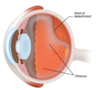
- Neovascularization of the retina → vitreous haemorrhage → retinal detachment
What is the definition and epidemiology of Glaucoma?
Glaucoma is a group of eye-diseases that result in damage to the optic nerve and visual loss. Usually caused by an increase in intra-ocular pressure.
Glaucoma is the second leading cause of blindness in the world, causing permanent vision loss.
What is the aetiology of and risk factors for Open-Angle Glaucoma?
Open-angle glaucoma is caused by slowed drainage of the trabecular meshwork, but an anatomically open angle. It is the most common cause of glaucoma, affecting 2/3 of patients.
Risk factors for POAG (primary open-angle glaucoma) include:
- Age - is present in around 2% of people older than 40 years.
- Genetics: first degree relatives of an open-angle glaucoma patient have a 16% chance of developing the disease
- Black patients
- Myopia (short-sightedness) as opposed to hypermetropia which is associated with closed-angle glaucoma.
- Hypertension
- Diabetes mellitus
- Corticosteroids
What are the clinical features of Open-Angle Glaucoma?
Patients often have a diagnosis of intra-ocular pressure (50%).
Patients rarely experience symptoms, rather discovered during comprehensive ophthalmic examination. This is in contrast to closed-angle glaucoma which has many symptoms and signs.
On Examination
- Peripheral vision loss in advanced disease, shown by missing areas in the field of vision.
- Scotomas also found on visual field testing.
- Cupping of the optic disk (increased cup:disk ratio of >0.7).
- Retinal haemorrhages are uncommon
What are the investigations for Open-Angle Glaucoma?
Tonometry shows elevated intra-ocular pressure above the normal range (>21mmHg).
Ophthalmoscopy shows cup-to-disk ratio over 0.7. Asymmetry of greater than 0.2 between the two is also a sign of glaucoma.
Visual field testing should be done in all patients.
Describe the management of Open-Angle Glaucoma
Management of ocular hypertension (OH), suspected primary open angle glaucoma (POAG), and confirmed POAG are normally under the direction of an ophthalmologist. The aim is to prevent progression of visual field loss.
- First-line treatment includes topical prostaglandin analogues such as latanoprost. Adverse effects include brown pigmentation of the iris, increased eyelash length. Alternative second-line options include:
- Beta-blockers (e.g. timolol, betaxolol) reduces aqueous humour production but should be avoided in asthmatics and patients with heart block.
- Sympathomimetics such as brimonidine reduce aqueous production and increase outflow.
- Carbonic anhydrase inhibitor reduce aqueous production.
Surgery in the form of a trabeculectomy may be considered in refractory cases.
What are the clinical features of Closed-Angle Glaucoma?
Acute
Patients present with an acute onset headache, with aching eye or brow pain and nausea/vomiting.
Also complain of reduced visual acuity, blurred vision and halos around lights and eye redness.
Chronic
Most patients are asymptomatic, and usually found on ophthalmic examination.
Describe the management of Closed-Angle Glaucoma
If acute angle closure is suspected, admit immediately for specialist ophthalmology assessment and treatment.
Let the person lie flat with their face up and head not supported by pillows, as this may relieve some of the pressure on the angle. If the drugs are available, give:
- Pilocarpine (muscarinic receptor agonist causing miosis and allowing drainage of aqueous humour) eye drops, one drop of 2% in blue eyes or 4% in brown eyes;
- Acetazolamide 500 mg orally to reduce production of aqueous humour (provided that there are no contraindications)
- Analgesia
- Anti-emetic if required.
What is the definition and aetiology of Anterior Uveitis?
Anterior uveitis is one of the important differentials of a red eye. It is also referred to as iritis. Anterior uveitis describes inflammation of the anterior portion of the uvea - iris and ciliary body. It is associated with HLA-B27 and may be seen in association with other HLA-B27 linked conditions (see below).
Associated conditions
- Ankylosing spondylitis
- Reactive arthritis
- Ulcerative colitis, Crohn’s disease
- Behcet’s disease
- Sarcoidosis: bilateral disease may be seen
What are the clinical features of Anterior Uveitis?
Anterior uveitis is a cause of red eye and is usually of acute onset but presents with progressive ocular discomfort & pain (may increase with use). Patients also complain of:
- Photophobia (often intense)
- Blurred vision
On examination, pupil may be irregular and small, there may be lacrimation and ciliary flush. Hypopyon; describes pus and inflammatory cells in the anterior chamber, often resulting in a visible fluid level.

What are the investigations for Anterior Uveitis?
Slit lamp examination shows:
- Leukocytes in the anterior chamber
- Protein in the aqueous humour → vitreous haze
- Signs of inflammation of the iris (e.g., red eye, hypopyon).
Conjunctival smear and cytology if infectious cause is suspected.
Tonometry to exclude secondary open-angle glaucoma. Cells in the anterior chamber may cause impaired drainage of aqueous humour.
Describe the management of Anterior Uveitis
Urgent review by ophthalmology.
Cycloplegics (dilates the pupil which helps to relieve pain and photophobia) e.g. Atropine, cyclopentolate
Steroid eye drops