Pharmacology Flashcards

1
Q

What are the 3 princpiple voltage and time dependent conductances in the heart?

A

• Voltage active sodium channels (depolarise) • Voltage activated calcium channels (depolarise) • Voltage activated potassium channels (repolarise)

2
Q

What fundamentally differs nodal tissue for cardiac muscle tissue?

A

Pacemaker potential - diastolic potential/phase 4 slope (rather than horizontal line in cardiac muscle tissue at phase 4 - resting potential). Also atrial/ventricular cells have a plateau on the AP which nodal cells do not.

3
Q

What is over-drive suppression?

A

SA node discharges APs at the highest rate and overrides the spontaneous discharge in the AV node and purkinje system and so is the dominant pacemaker

4
Q

**What 4 conductances underlie the diastolic/phase 4 potential in pacemaker nodal cells?

A

1) Background sodium current (Ib) - leaky and present all of the time 2) Funny current (If) - hyperpolarisation mediated 3) Delayed rectifier potassium current (Ik) - switched off in phase 4, therefore contribute to depolarisation by inhibiting hyper polarisation) 4) Transient Voltage Activated Calcium Conductor (Icat) - kicks in at the end of phase 4 to give final kick to threshold => these all contribute to depolarisation and allow pacemaker cells to reach threshold

5
Q

**Once threshold has been reached, which channel kicks in to bring the upstroke in nodal cells?

A

5) Long Calcium Channel (Ical) - inward movement of calcium underlies nodal AP (unlike in atrial/ventricular cells where it is sodium mediated)

6
Q

**Which channels underlie repolarisation in nodal cells?

A

Delayed rectifier potassium current (Ik) which were previously switched off, now switch on and cause the outward flow of potassium

7
Q

Why is there not a gradual depolarising drift in atrial/ventricular cells, like there is in nodal cells?

A

Because there is a constant trickling of potassium out of the cell by Ik1 - an inward rectifying conductor which helps maintain the resting membrane potential with slight hyper polarisation

8
Q

What therefore causes an AP in atrial/ventricular myocytes, if not a gradual depolarising drift?

A

The arrival of depolarising influence from an adjacent cardiac muscle cell.

9
Q

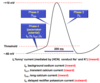
**What causes Phase 0: Depolarisation in atrial/ventricular myocytes?

A

Arrival of a depolarising AP from adjacent cardiac muscle cells causes voltage-activated sodium channels- INa channels to open and allow a large current of Na influx, activating rapidly.

10
Q

**What causes Phase 1: Early repolarisation in atrial/ventricular myocytes?

A

Rapid opening/closing of transient outward K current – It0, which combined with INa channel closure results in a slight repolarisation. However, don’t get complete repolarisation, as long calcium currents – ICaL are now activated after a delay – this helps maintain the depolarisation

11
Q

**What causes Phase 2: Plateau phase in atrial/ventricular myocytes?

A

Ca2+ channels have opened, ICaL. This maintains the plateau. At the same time there is a slowly developing delayed rectifying K efflux (outward) current (Ik). Fine balance between Ca influx and K efflux maintains the plateau. INaL channels (previous Na channels from Phase 0 that stayed open, and reverse §NCX1 channels contribute too)

12
Q

**What causes Phase 3: Late repolarisation phase in atrial/ventricular myocytes?

A

Eventually the outward potassium (Ik) wins, and depolarisation occurs. This involves components IK channels which activate rapidly and IK1 channels which activate slowly

13
Q

Which GPCRs do B1 adrenoreceptors preferably couple with?

A

Gs - which increases the conversion of ATP to cAMP, which mediates most of sympathetic activity of the heart

14
Q

How does sympathetic innervation increase heart rate?

A

1) Increase the slope of diastolic phase 4 depolarisation (enhanced If and Ica activity) 2) Reduction of the threshold for AP initiation (enhanced Ica)

15
Q

How does sympathetic innervation increase contractility?

A

1) increase in phase 2 plateau of the cardiac AP in atrial and ventricular myocytes, therefore enhanced Ca2+ influx 2) increased sensitisation of contractile proteins to Ca2+

16
Q

What effect does sympathetic innervation have on the heart?

A

1) Increased HR 2) Increased contractility 3) Increased conduction velocity (smaller AV nodal delay to match HR) 4) Increased automaticity 5) Decreased duration of systole (to allow complete emptying of ventricles with new HR) 6) Decreased cardiac efficiency (oxygen consumption increases) 7) Increased activity of Na+/K+-ATPase 8) Increased mass of cardiac muscle

17
Q

Which parasympathetic receptors lie in the heart and with GPCR do they coupe with?

A

M2 muscarinic choliceptors. Couple with Gi

18
Q

What does parasympathetic stimulation do in the heart, and what effect does this have?

A

1) decreases activity of adenylate cyclase and reduces [cAMP]I 2) opens potassium channels This causes: - Decreased HR - Decreased contractility - Decreased conduction velocity in AV node

19
Q

What are the 2 main vagal manoeuvres (which employ the parasympathetic system to suppress AV conduction)?

A

Valsalva manœuvre and massage of the bifurcation of the carotids

20
Q

Which type of channels mediate the funny current?

A

Hyperpolarization-activated cyclic nucleotide gated (HCN) channels (HCN4 in heart).

21
Q

What is ivabridine?

A

Blocker of the HCN (funny current) channel, and therefore decreases the pacemaker slope and reduces the HR. Used in angina in particular

22
Q

How does contraction occur in excitation-contraction couple in cardiac muscle?

A
  1. In the ventricular AP, we get Ca2+ entry during phase 2, due to voltage-activated Ca2+ channels (L-type) which moves into the cytoplasm 2. This creates a modest increase in the intracellular Ca2+ concentration, though this signal is not enough to cause an AP in itself, so needs to be amplified. 3. It does this using Calcium Induced Calcium Release (CICR) - The Ca2+ that has come through the L-type channels, bind to ryanodine-type 3 channel in the membrane of the SR, which is the main calcium store in the myocyte, and allow the efflux of calcium from the SR into the cytoplasm 4. This amplification leads to Ca2+ binding to troponin-c, which undergoes a conformational change which allows it to move tropmyocin from the synaptic cleft 5. This then allows cross-bridge formation between actin and myosin, resulting in contraction.
23
Q

How does relaxation occur in excitation-contraction couple in cardiac muscle?

A
  1. Relaxation is controlled by the repolarisation in phase 3 to phase 4 of the AP 2. Repolarisation causes the voltage-activated Ca2+ channels to close 3. Ca2+ influx and CICR ceases and is pumped back to the sarcoplasmic reticulum, to be re-stored, by action of Ca2+ ATPase in the membrane of the SR. 4. Simultaneously, Na+/Ca2+ exchanger (NCX) pushes 1Ca out and 3Na in. 5. Under these 2 mechanisms, Ca2+ dissociates from troponin-C; the cross-bridges break up; actin and myosin are pumped back into the synaptic cleft and we get relaxation.
24
Q

What are examples of β-adrenoceptor agonists?

A

Dobutamine, adrenaline and noradrenaline (catecholamines)

25
Q

What is the clinical use of adrenaline?

A

Used in cardiac arrest; emergency treatment of asthma; anaphylactic shock - has both α/β agonist properties so causes: • Positive inotropic and chronotropic actions (β1) • Redistribution of blood flow to the heart (constricts blood vessels in the skin, mucosa and abdomen (α1)) • Dilation of coronary arteries (β2)

26
Q

What is the clinical use of dobutamine, and is it selective?

A

Selective for β-adrenoceptors. Used as an IV infusion in acute heart failure short term

27
Q

Which β-adrenoceptors antagonists are selective and non-selective?

A

Non-selective: propanolol Selective: Atenolol, bisoprolol, metoprolol

28
Q

What are the CVS clinical uses of β-adrenoceptors antagonists (beta-blockers)?

A

1) Dysrhythmias (delay AV conduction and reduce excess sympathetic drive) 2) Hypertension 3) Angina 4) Heart failure (start low , go slow) - Carvedilol used in particular as has additional α1 antagonist activity causing vasodilation

29
Q

What are the main adverse effects of β-Blockers?

A
  • Bronchospasm (esp. in asthmatics) - Aggravation of heart failure - Bradycardia - Hypoglycaemia (β2 cause release of glucose from liver) - Fatigue -Cold extremities
30
Q

What are examples of non-selective muscarinic Ach receptor antagonists?

A

Atropine

31
Q

What are the clinical uses of atropine (muscarinic Ach receptor antagonists)?

A

1) Bradycardia following MI 2) Anticholesterase poisoning

32
Q

How does digoxin increase contractility?

A

Blocking the sarcolemma ATPase, causing an increase of calcium and therefore increased contraction

33
Q

When is digoxin used?

A

Heart failure, particular with AF

34
Q

What is the main cardiac glycoside?

A

Digoxin

35
Q

What are the main effects of digoxin?

A

Indirectly: Cause increases release of Ach into SA node so decreases firing and slows AV conduction so increases refractory period Directly: Shortens the AP and refractory period

36
Q

What is the main example of calcium sensitisers and how do they work?

A

Levosimendan. They increase the sensitivity of the contractile elements to calcium, therefore increasing contractility.

37
Q

What is the main example of inodilators and how do they work?

A

Amrinone and milrinone. They inhibit phosphodiesterase (PDE) enzyme in cardiac and smooth muscle cells and hence increase intracellular [cAMP]i

38
Q

What are the main groups of inotropes?

A

1) Cardiac glycoside eg. digoxin 2) Calcium sensitisers eg. levosimendan 3) Inodilators eg. amrinone and milrinone

39
Q

**What causes contraction in smooth muscle?

A

Relies on an increase in calcium which then promotes calmodulin, and this complex then activates MLCK which then forms Myosin-LC phosphatase – which promotes contraction

40
Q

**What causes relaxation in smooth muscle?

A

cGMP dephosphorylates the myosin –LC-phosphatase causing relaxation

41
Q

True or False: Endothelium acts to suppress platelet aggregation

A

True, this is why damage to the endothelial is important in thrombus formation

42
Q

What produces NO and when?

A

Endothelial cells, and in response to anything causing increases calcium levels

43
Q

How does NO cause relaxation?

A
  • NO enters the enzyme guanylate cyclase, which converts GTP to cGTP. This activates protein kinase G, causing relaxation - Simultaneously, NO also causes Ca dependent K channels to open and cause K movement out, hyperpolarising the cell and causing relaxation
44
Q

What are the effects of organic nitrates?

A

Essentially act as equivalents to endothelium. They metabolise to NO and therefore cause: - Venorelaxation - Arteriolar dilation - Coronary vessel dilation (also they dilate collateral branches which sprout from healthy areas to ischaemic areas)

45
Q

What are the 2 main examples of organic nitrates?

A

Glyceryltrinitrate (GTN) and isosorbide mononitrate (ISMN)

46
Q

Why can’t GTN be taken orally?

A

Completely metabolised on first pass to liver

47
Q

What is endothelin and what does it do?

A

Acts as a vasoconstrictor, released from endothelial cells in response to adrenaline, angiotensin etc

48
Q

What produces renin?

A

Granular cells of the juxtaglomerular apparatus of the kidney

49
Q

What does renin do?

A

Splits angiotensinogen to angiotensin I (which is inactive)

50
Q

What does ACE do?

A

Activates angiotensin I to angiotensin II

51
Q

What does angiotensin II do?

A

Acts on g protein coupled angiotensin receptors which cause vasoconstriction. Also causes the release of aldosterone.

52
Q

What does aldosterone do?

A

Acts on the kidney on the nephron – increasing the reabsorption of sodium and accompanying water from the urine back into the circulation

53
Q

What do all ACE inhibitors end in?

A

‘-ipril’

54
Q

What do all AT1 receptor antagonists end in?

A

’- sartan’

55
Q

What effects of ACE inhibitors have?

A

Cause venous dilatation (↓preload) and arteriolar dilatation (↓afterload and ↓TPR) decreasing arterial blood pressure and cardiac load

56
Q

Why do ACE inhibitors cause a dry cough?

A

Inhibit the metabolism of bradykinin

57
Q

What effect do α1 receptors have?

A

Constriction of vessels

58
Q

What effect do β1 receptors have?

A

↑Rate; ↑ Force; ↑ AV node conduction velocity

59
Q

What effect do β2 receptors have?

A

relax the vessels and also the bronchi

60
Q

Why are beta-blockers of benefit in angina?

A
  1. Decrease myocardial O2 requirement ( because ↓HR & ↓SV = ↓work = ↓O2 requirement ) 2. Counter elevated sympathetic activity associated with ischaemic pain 3. Increase the amount of time spent in diastole (↓HR), improving perfusion of the left ventricle
61
Q

What effects do calcium antagonists have?

A

Calcium antagonists prevent the opening of L-type channels in excitable tissues in response to depolarization and hence limit ↑[Ca2+]i , therefore reduce rate, AV conduction and force of contraction

62
Q

What are the 3 main calcium antagonists?

A

Verapamil, Amlodipine and Diltiazem

63
Q

What effects do potassium channel openers have?

A

Cause hyperpolarization which switches off L-type Ca2+ channels

64
Q

What are the 2 main potassium channel openers?

A

Minoxidil and nicorandil

65
Q

What effects do α1-Adrenoceptor Antagonists have?

A

Cause vasodilatation by blocking vascular α1-adrenoceptors

66
Q

What are main examples of α1-Adrenoceptor Antagonists?

A

Prazosin and doxazosin

67
Q

What are the main differences between loop and thiazide diuretics?

A

Thiazides inhibit NaCl reabsorption in distal tubule, while loop diuretics act earlier in the ascending limb of the loop of hence

68
Q

What are examples of thiazide diuretics?

A

bendroflumethiazide

69
Q

What are examples of loop diuretics?

A

Furosemide

70
Q

When are thiazide diuretics used?

A

Mild heart failure, hypertension and severe resistant oedema

71
Q

When are loop diuretics used?

A

Acute pulmonary oedema and chronic heart failure

72
Q

**What treatment pathway would you use for hypertension in person

A

1) ACE inhibitor e.g. lisinopril, ramipril or ARB e.g. losartan 2) + Calcium channel blocker eg. Verapamil, Amlodipine 3) + Diuretics (thiazide) eg. bendroflumethiazide 4) Resistant hypertension (+ higher dose thiazide or add spironolactone or alpha/beta-blocker)

73
Q

**What treatment pathway would you use for hypertension in person >55 or afrocarribean?

A

1) Calcium channel blocker eg. Verapamil, Amlodipine 2) + ACE inhibitor e.g. lisinopril, ramipril or ARB e.g. losartan 3) + Diuretics (thiazide) eg. bendroflumethiazide 4) Resistant hypertension (+ higher dose thiazide or add spironolactone or alpha/beta-blocker)

74
Q

What actually is spironolactone?

A

Potassium sparing diuretic

75
Q

What are the side effecters of diuretics?

A

• Hypokalaemia - Both low and high potassium levels can cause arrhythmias • Hyperglycaemia • Increased Uric Acid – Gout • Impotence

76
Q

What are the 2 types of calcium antagonists, and examples of each?

A

1) Dihydropyridines • e.g. Amlodipine 2) Rate limiting calcium antagonists • e.g. Verapamil, Diltiazem

77
Q

When are alpha-blockers only used?

A

Hypertension

78
Q

What are the side effects of ACEi?

A

• Dry cough • Renal dysfunction • Angioneurotic oedema - larynx swells and can cause death

79
Q

What are the side effects of nitrates?

A

• Headache – due to excessive dilation of cerebral veins • Hypotension/Collapse • Tolerance

80
Q

Which 3 groups of drugs are used as anti-thrombotic and examples of each?

A

1) Antiplatelet eg. aspirin, clopidogrel (prevent new clot) 2) Anticoagulants eg. Heparin or warfarin (prevent new clot) 3) Fibrinolytics eg. streptokinase (dissolved formed clot)

81
Q

Which 2 groups of drugs are used as anti-cholesterols and examples of each?

A

1) Statins eg. simvastatin 2) Fibrates eg. bezafibrate

82
Q

What are the side effects of amiodarone?

A

• Phytotoxicity • Pulmonary fibrosis • Thyroid abnormalities (Hypo or Hyper)

83
Q

Which 2 effects does digoxin have?

A

1) Blocks atrial-ventricular (AV) conduction 2) Increases ventricular irritability which produces ventricular arrhythmias (always bad)

84
Q

What are the signs of digoxin toxicity?

A

• Nausea, vomiting • Yellow vision • Bradycardia, Heart Block • Ventricular Arrhythmias (due to the irritability of the ventricles)

85
Q

Which drugs are used in the treatment of heart failure?

A

• Diuretics • B- blockers • ACEi • Nitrates

86
Q

Which lipoproteins are involved in the exogenous pathway?

A

Chylomicrons - they are formed in intestinal cells and transport dietary triglycerides (come from outside the body)

87
Q

Which lipoproteins are involved in the endogenous pathway?

A

VLDL particles - they are formed in liver cells de-novo and transport triglycerides synthesised in that organ

88
Q

What 2 components are lipoproteins made up of?

A

• a hydrophobic core containing esterified cholesterol and triglycerides • a hydrophilic coat comprising a monolayer of amphipathic un-esterified cholesterol, phospholipids and one, or more, apoproteins (apo)

89
Q

What is the overall function of lipoproteins?

A

To deliver triglycerides (i) to muscle for ATP biogenesis and (ii) adipocytes for storage

90
Q

Which apoB is used in lipoprotein assembly, where?

A

apoB100 in the liver and apoB48 in the intestine

91
Q

What are the 2 sources of cholesterol?

A

25% from diet (exogenous pathway) and the rest from the liver in the bile

92
Q

**How do the products of fat digestion get into the enterocytes?

A
  • Mono glycerides and fatty acids enter the intestinal enterocytes by passive diffusion - Cholesterol is transported across the apical membrane of the enterocyte and depends upon the activity of the MPC1L1 protein
93
Q

What are triglycerides made up of?

A

Monoglycerides and fatty acids

94
Q

What happens to the products of fat digestion once the are in the enterocytes?

A

Tri-glycerdies are reassembled and reunited with cholesterol, and the addition of apob48 to form chylomicrons. Then transported to the liver via the lacteal, lymph and then then systemic circulation

95
Q

How are VLDL lipoproteins produced in the liver in the endogenous pathway?

A

VLDL particles containing triglycerides are assembled in liver hepatocytes from free fatty acids derived from (i) adipose tissue (particularly during fasting) and (ii) de novo synthesis

96
Q

Once in the liver, how are chylomicrons and VLDL particles activated?

A

ApoCII is then transferred from HDL particles to the hydrophilic coats

97
Q

What is the importance of the ApoCII which is added in the activation step of chylomicrons and VLDLs?

A

It enables the chylomicrons/VLDL particles to bind to lipoprotein lipase (LPL) which hydrolyses core triglycerides to free fatty acids and glycerol which enter tissues, allowing them to move across the membrane into skeletal muscle or adipose tissue

98
Q

Chylomicron and VLDL remnants

A

Particles depleted of triglycerides (but still containing cholesterol esters)

99
Q

How are chylomicron and VLDL particles then cleared?

A

1) These particles are left relatively enriched in cholesterol, as they’ve lost their triglycerides. 2) The lipoprotein particles then dissociate from LPL, and ApoCII is transferred back to HDL particles in exchange for apoE (now termed remnants). 3) Remnants return to the liver and are further metabolised by hepatic lipase 4) All apoB48-containing remnants and half of apo100 containing remnants are cleared by receptor-mediated endocytosis into hepatocytes 5) Remainder apoB100 remnants lose further triglycerides via hepatic lipase, become smaller and via IDLs become LDL - lacing apoE and retaining solely apoB100

100
Q

How are LDLs then cleared?

A

1) LDLs dock to hepatocytes 2) Cellular uptake of LDL particles occurs via receptor-mediated endocytosis – process of internalisation 3) Within the cell at the lysosome, cholesterol (C) is released from cholesteryl ester (CE) by hydrolysis 4) Additionally the LDL receptor is then released to be reused by LDL receptor recycling 5) Meanwhile the cholesterol: • inhibits de novo cholesterol synthesis via enzyme HMG-CoA reductase • Down regulation of LDL receptor expression • Stored as cholesterol ester

101
Q

Why is HDL the ‘good’ cholesterol?

A

Has a key role in removing excess cholesterol from cells (reverse cholesterol transport) by transporting it in plasma to the liver

102
Q

**Which system is used to classify dyslipidaemia?

A

Frederickson classification

103
Q

What is the mechanism of action for statins?

A

ct as competitive inhibitors of 3-hydroxy-3-methylglutaryl coenzyme A (HMG-CoA) reductase - rate limiting step in cholesterol synthesis in hepatocytes. Decrease in hepatocyte cholesterol synthesis causes a compensatory increase in LDL receptor expression and enhanced clearance of LDL

104
Q

What are the side effects of statins?

A

Myositis (inflammation and degeneration of muscle tissue) and rarely rhabdomyolysis (the destruction of striated muscle cells). They also shouldn’t be prescribed to pregnant women as cholesterol is essential for the foetal development

105
Q

What are the results of statins?

A

Very effective in reducing total and LDL cholesterol (up to 60%), decrease triglycerides and modestly increase HDL

106
Q

What are the results of fibrates?

A

Cause a pronounced decrease in triglycerides and modest decreases and increases in LDL and HDL, respectively.

107
Q

What are examples of fibrates?

A

Bezafibrate and gemfibrozil

108
Q

What is the mechanism of action of fibrates?

A

Act as agonists of a nuclear receptor (PPARα) to enhance the transcription of several genes, including that encoding LPL. Helps remove triglyerides from the particles as free fatty acids into skeletal muscle or adipose tissue

109
Q

Enterohepatic recycling

A

Liver produces bile salts from cholesterol, and once ejected into the duodenum they are then reabsorbed and returned to the liver by a mechanism in the ileum

110
Q

What effect to bile acid binding resins have on enterohepatic recycling?

A

Interrupt enterohepatic recycling by causing the excretion of bile salts, resulting in more cholesterol to be converted to bile salts

111
Q

What are examples of bille acid binding resins?

A

colestyramine, colestipol, colsevelam

112
Q

What is Ezetimibe?

A

Relative new agent, acts to inhibit Niemann-Pick C1 like-1 (NPC1L1) transport protein in enterocytes of the duodenum, reducing the absorption of cholesterol from the gut lumen

113
Q

**What are the functions of each of the lipoproteins?

A

.

114
Q

Lipoproteins

A

Specialised proteins that transport hydrophobic cholesterol and triglycerides

115
Q

What is a very brief overview of the lifecycle of cholesterol?

A

1) Either injested in the diet to the gut and transported by chylomicrons to the liver or synthesised in the liver into VLDLs 2) Then activated by ApoCII transferred from HDLs 3) Bind to LPL to be broken down and release triglycerides into the skeletal muscle and fat tissue 4) Then dissociate from LPL, and lose the apoCII to form remnants 5) Cleared either by receptor-mediated endocytosis or formed into IDL then LDL 6) Excess cholesterol in the tissues are transported back to the liver by HDL

116
Q

What are the 2 main directions of cholesterol transport?

A

1) From the liver to the tissues (some having come from the gut) 2) From the tissues back to the liver

117
Q

What is the general sequence of haemostasis?

A
  1. local vasoconstriction 2. adhesion, activation and aggregation of platelets at site of injury 3. formation of fibrin (blood coagulation)
118
Q

What drugs are given following an MI?

A

• ACE inhibitor/ARB • Beta blockers (both are good in MI prevention and hypertension) • CCB • Diuretic (prevents HF and increases effectiveness of ACE) • Statins • Aspirin

119
Q

With an MI, what would you give in between MONA+C and PCI?

A

B-blockers (to reduce blood flow to the heart and workload) and anti-coagulants eg. fondaparinux

120
Q

How do loop diuretics work?

A

Inhibit NaCl reabsorption in the thick ascending limb of the loop of Henle by blocking the Na+/K+/2Cl- co-transporter

121
Q

How do ACEi’s work?

A

Inhibits the conversion of angiotensin I to angiotensin II in the lungs, therefore decreasing the release of aldosterone from the adrnenal glands.

122
Q

How do ARB’s work?

A

Blocks angiotensin 2 receptors and reduces production on aldosterone which: 1) decrease vascular resistance improving perfusion; 2) increase excretion of Na+ and H20; 3) cause regression of left ventricular hypertrophy.

123
Q

What is a side effect of spironolactone?

A

gynaemastia

124
Q

What medication would you prescribe for AF?

A
  • Beta-blockers (sotalol) - Ca2+ antagonist (verapamil) - Digoxin - Amiodarone Possibly: electrical cardioversion or pacemaker.
125
Q

How does warfarin work?

A

Competes with vitamin K for binding to hepatic vitamin K reductase preventing the conversion of the epoxide to the active hydroquinone. Renders factors II, VII, IX and X inactive.

126
Q

How does amiodarine work?

A

Class three anti-arrythmic drug that targets voltage activated potassium channels, prolongs AP duration increasing refractory period.

127
Q

How does verapamil work?

A

Type IV agent (oral) – blocks L-type voltage-activated Ca2+ channels Slows conduction and prolongs refractory period in AV node and bundle of His. Used to treat atrial flutter (AF) and fibrillation (AF) – chaotic re-entrant impulse conduction through the atria that may be conducted via the AV node to the ventricles

128
Q

How do b-blockers help in hypertension?

A

Reduce cardiac output, reduce renin release and reduce sympathetic activity

129
Q

What are the 4 vasodilators used in hypertension?

A

1) Calcium antagonists eg. amlodipone 2) Alpha-Blockers 3) ACEis’ 4) ARBs

130
Q

What is amlodipine?

A

Calcium antagonist

131
Q

How do calcium antagonists work?

A

Reduced Ca2+ entry in to vascular smooth muscle cells causes generalised arteriolar dilatation, reducing TPR and MABP

132
Q

What is the main side effect of calcium antagonists?

A

Peripheral oedema

133
Q

Haemostasis

A

Mechanism by which there is an arrest of blood loss from a damaged blood vessel

134
Q

What is the sequence of haemostasis?

A

1) ) Local vasoconstriction 2) Soft plug formation: Platelet adhesion, activation and then aggregation, held together by bands of fibrinogen • Initial soft plug arresting blood loss (platelets embedded in a fibrinogen mesh) 3) Blood coagulation proper: Activation of blood clotting and the formation of a solid stable clot (by fibrin)

135
Q

What acts as the bridge between exposed collagen and platelets in haemostasis?

A

Von Willebrand Factor (vWF)

136
Q

Once the platelets are attached indirectly to exposed collagen via vWF, how are they then attached directly?

A

via : integrin α2β1 and glycoprotein VI (GPVI) receptors on the platelet surface

137
Q

What happens to the platelets when they are activated?

A

• Changes shape (extends pseudopodia- finger like projections, allowing platelets to aggregate with each other) • Starts to synthesise thromboxane A2 from arachidonic acid mediated by the enzyme cyclo-oxygenase-1 (COX-1)

138
Q

Once produced by activated platelets, what does thromboxane AD2 (TXA2) do?

A

Bind both autocrinely (on its own platelet) and paracrinely (on other platelets). Causing mediator release, of 5-HT (serotonin), ADP, vWF and factor V.

139
Q

In haemostasis, which receptors do ADP bind to and what effect do they have?

A

Platelet GPCR P2Y12 receptors. There they activate other platelets, increases expression of glycoprotein receptors which bind fibrinogen and expose phospholipids which facilitates the formation of the clot

140
Q

What is the pivotal point in coagulation proper stage of haemostasis?

A

Production of the protease thrombin (factor IIa) that cleaves fibrinogen to fibrin to form a solid clot

141
Q

**What are the 3 phases of coagulation proper stage of haemostasis?

A

1) Initiation - Damage to vessel wall exposes tissue elements
2) Amplification - Platelet recruitment and adhesion to the site of injury forming a monolayer
3) Propogation - Adhesion leads to activation causing release of ADP and other activators resulting in further aggregation and inflammatroy cascade –> thrombus

142
Q

Which factor is responsible for the formation of the final clot, and how does it do this?

A

• Thrombin cleaves fibrinogen, forming fragments that spontaneously polymerise to form fibrin • Factor VIIIa (activated by thrombin) cross-links the polymer to form a fibrin fibre network and a solid clot

143
Q

What basically is coagulation proper of haemostasis, like really?

A

A complex cascade where proenzymes are converted to active enzymes, both the intrinsic and extrinsic pathways ending in X → Xa, which causes the production of the protease thrombin (factor IIa) that cleaves fibrinogen to fibrin to form a solid clot

144
Q

Thrombosis

A

Essentially clotting in the wrong place – inappropriate activation of the clotting cascade even when there is no tissue injury

145
Q

What is a white thrombus and what treatment is used for them?

A

Arterial thrombus. Mainly platelets in a fibrin mesh - relatively few RBCs associated, so has a white experience. If embolus, lodge in brain etc. Treated with anti platelet drugs

146
Q

What is a red thrombus and what treatment is used for them?

A

Venous thrombus. High degree of fibrin compared to white thrombus. If embolus, lodge in lung (PE). Treated with anticoagulant drugs.

147
Q

How are the clothing factors (II, VII, IX and X) activated to the active factors IIa, VIIa, IXa etc?

A

Post-translational modification (i.e. γ-carboxylation of glutamate residues)

148
Q

What role does Vit K play in the coagulation proper stage?

A

The carboxylase enzyme that mediates γ-carboxylation (activation of factors) requires reduced vitamin K (aka hydroquinone)

149
Q

When are anticoagulants mostly used?

A

Prevention and treatment of venous thrombosis and embolism eg. in DVT or AF

150
Q

What is the mechanism of action of warfarin?

A

Structurally related to vitamin K, so competes with vitamin K for binding to hepatic vitamin K reductase preventing the conversion of to the epoxide to the active hydroquinone. Basically renders factors II, VII, IX and X inactive

151
Q

What is a main drawback of warfarin?

A
  • Low therapeutic index - his risk of haemorrhage - Has a long (and variable) half-life (usually about 40 hr) so takes around 200hrs for warfarin to get to a steady state. -Also has a slow onset of action.
152
Q

What is the treatment for an overdose of warfarin?

A

Vit K

153
Q

What is the role of antithrombin III?

A

An important inhibitor of coagulation which neutralises all serine protease factors in the coagulation cascade by binding to their active site in a 1:1 ratio. Prevents all of your blood going solid following a simple cut.

154
Q

What is the mechanism of action of heparin?

A

Heparin binds to antithrombin III, increasing its affinity for serine protease clotting factors to greatly increase their rate of their inactivation

155
Q

How is heparin and low molecular weight heparin given?

A

Heparin: IV or subcutaneously LMWH: subcutaneously

156
Q

Which is the only condition in which heparin is preferred over LMWH?

A

Renal failure (as LMWH is excreted through the kidneys, while heparin is not)

157
Q

How do newer orally active inhibitors eg. dabigatran act?

A

Act as direct inhibitors of thrombin

158
Q

When are anti platelets mainly used?

A

In the treatment of arterial thrombosis

159
Q

What are the main examples of anti-platelets?

A

Aspirin and clopidogrel

160
Q

How does aspirin work?

A

Irreversibly blocks cycloxygenase (COX) in platelets, preventing Thromboxane 2 (TXA2) synthesis. Remember thromboxane releases cell mediators for platelet aggregation

161
Q

How does clopidogrel work?

A

Blocks the effects of ADP by blocking its P2Y receptor. Remember ADP activates other platelets, increases expression of glycoprotein receptors which bind fibrinogen and expose phospholipids which facilitates the formation of the clot

162
Q

What happens in fibrinolysis?

A

1) a plasma protein plasminogen is activated to plasmin 2) Plasmin then dissolves fibrin into fibrin fragments, and we get clot lysis

163
Q

How to fibrinolytic drugs act?

A

Enhance the conversion of plasminogen to plasmin, thus accelerating this process and break down the clot

164
Q

What are examples of fibrinolytic drugs?

A

Streptokinase, alteplase and duteplase

165
Q

Which drugs are commonly used in the treatment of angina?

A

• Beta Blockers • Calcium Antagonists • Nitrates • Nicorandil • Ivabradine • Ranolazine