Radiology Imaging + Pathology Flashcards

(58 cards)

1
Q

How can you determine whether a CXR is adequately inspired?

A

Anterior ends of at least 6 ribs should be visible

2
Q

Name the different mediastinal borders on this image:

A
  1. Aorta
  2. Pulmonary artery
  3. Left auricle
  4. Left ventricle
  5. Right atrium
  6. Trachea
  7. Hemidiaphragm (right)
  8. Stomach bubble
  9. Horizontal fissure
3
Q

What are some potential causes of Lobar Collapse?

A

Occurs when obstruction of a lobar bronchus

Causes of bronchial obstruction include tumours, aspirated foodstuffs, mucus impaction

(lobe supplied by obstructed bronchus no longer ventilation and air is resorbed = loses volume)

4
Q

Identify this pathology:

A

Left Lower Lobe Collapse

5
Q

Identify this pathology:

A

Left Upper Lobe Collapse

6
Q

Identify this pathology:

A

Right Upper Lobe Collapse

7
Q

Identify this pathology:

A

Right Middle Lobe Collapse

8
Q

Identify this pathology:

A

Right Lower Lobe Collapse

9
Q

What type of lobar collapse is visible here?

A

Right Middle & Lower Collapse

10
Q

What area of the lung is affected by consolidation here?

A

Right Middle Lobe Consolidation

  • Increased density in right lower zone
  • Loss of clarity of right heart border but preservation of hemidiaphragm
11
Q

Identify this pathology:

A

Left Lower Lobe Consolidation

  • Increased density in left upper + lower zones
  • Loss of clarity of left upper medistinum
  • Volume preserved
  • Air Bronchograms
12
Q

What kind of pathology is visible on this CXR?

A

Right pleural effusion

13
Q

What pathology is visible on this CXR?

A

Pneumothorax

14
Q

What are the 4 main signs of pulmonary oedema on CXR in order of severity/occurence?

A
  1. Dilatation of upper lobe vessels/cardiomegaly
  2. Interstitial opacities (Kerley B lines, peribronchovascular cuffing)
  3. Airspace opacification (filling of alveoli with fluid, severe - batwing)
  4. Pleural effusion
15
Q

What typical feature of pulmonary oedema is highlighted on this CXR?

A

Alveolar oedema/ BAT WINGS

16
Q

Describe the placement of this endotracheal tube:

A
  • Tube inserted too far
  • Passed into right main bronchus
  • Signs of early collapse (due to unventilated left lung)

Normal = tip 5cm above carina, width 2/3 tracheal diameter (should not expand trachea)

17
Q

Describe the placement of this endotracheal tube:

A
  • Correctly placed
  • Tip 5cm above carina
  • Width should be roughly 2/3 diameter of trachea (cuff should not expand trachea)
  • Both lungs are ventilated
18
Q

Describe the ideal placement of a nasogastric tube?

A
  • Subdiaphragmatic position in stomach
  • Overlying gastric bubble
  • Should be at least 10cm beyong gastro-oesophageal junction
19
Q

Describe the placement of this nasogastric tube:

A

NG tube misplaced

Located in right lower lobe bronchus

High change of infection/complications

20
Q

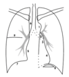
The following image shows the pathways of various central venous catheters - where does each line originate from?

A

Yellow - peripheral central catheter (cephalic, basilic, brachial)

Purple - Right subclavian vein

Light blue - Right jugular vein

Dotted blue - Left jugular vein

21
Q

What point in the heart is highlighted by the red circle in this image?

A

Cavoatrial Junction

The tip of a venous catheter should sit at the cavoatrial junction (SVC meets and melds with superior wall of right atrium)

22
Q

What abnormality is visible on this CXR?

A

Pneumoperitoneum

(perforation of hollow viscus resulting in gas within peritoneal cavity)

23
Q

What artery is affected in this ischaemic stroke?

A

Left PCA (posterior cerebral artery)

24
Q

Which artery is affected in this ischaemic stroke?

A

Right ACA (anterior cerebral artery)

25
Which artery is affected in this ischaemic stroke?
Right MCA (middle cerebral artery)
26
What is visible in this CT and at what stage would you expect to see this during the evolution of a stroke?
= Hyperdense segment of a vessel Direct visualisation of intravascular thrombus/embolus Often EARLIEST visible sign on CT
27
What is visible on this CT and at what point in a stroke evolution would you expect to see this?
* = Loss of grey-white differentiation, and hypoattenuation of deep nuclei * Cortical hypodensity with associated parenchymal swelling (with resultant gyral effacement) * Typically seen within first few hours of presentation
28
What is visible on this CT and at what stage in a stroke evolution would you expect to see this?
* This is a later sign in a stroke presentation * With time, the hypoattenuation and swelling becomes more profound * Results in significant mass effect
29
True or False: Gliosis is an early finding on CT in stroke?
FALSE ## Footnote Gliosis develops over time, eventually appearning as a region of low density with volume loss
30
What type of cerebral haemorrhage is this?
Intra-axial haemorrhage/Intra-cerebral haemorrhage Bleeding into brain parenchyma
31
What type of cerebral haemorrhage is this?
Extra-dural haemorrhage
32
What type of cerebral haemorrhage is this?
Sub-dural Haemorrhage
33
What type of cerebral haemorrhage is this?
Sub-dural haemorrhage
34
What type of cerebral haemorrhage is this?
Subarachnoid Haemorrhage
35
What are the names of these different types of herniation?
A - Subfalcine B - Central C - Uncal D - Tonsillar
36
What pathology is imaged here?
Hydrocephalus
37
What is the correct descriptions for this fracture types?
A - Transverse B - Oblique C - Spiral D - Comminuted
38
What is the correct descriptions for these fracture types?
A - Avulsion B - Impacted C - Torus D - Greenstick
39
What fracture is shown here?
Humerus surgical neck fracture
40
What abnormality in this X-ray indicates there could be a possible pathology?
The anterior fat pad (and posterior slightly) is more visible than it would usually be This indicates perfusion within that joint (blood or fluid)
41
Which fractures (adults + children) will most commonly show abnormal fat pad?
Adults - radial head fracture Children - supracondylar fracture (anterior sail sign)
42
What is a Colles fractures and who is at most risk?
Fracture of distal forearm in which broken end of radius moves dorsally Common in elderly, fall onto outstretched hand
43
What is a Smith's fracture?
Fracture of distal radius With associated volar angulation of distal fracture fragments (aka Reverse Colles fracture)
44
True or False: Intracapsular fracture of the femur have better healing than extracapsular fractures
FALSE Intracapsular - lose blood supply to femoral head = AVN Extracapsular - blood supply remains so improved healing
45
What type of femur fracture is shown here?
Intracapsular fracture of femur
46
What type of femur fracture is shown here?
Intertrochanteric fracture of femur
47
What are some advantages and disadvantages of CT?
A - quick, accurate, allows better planning of surgery or intervention D - radiation exposure, renal impairment,
48
What pathology is seen on this CT?
Acute Appendicitis
49
What pathology is this most likely to be?
Acute Cholecystitis
50
What type of test is this?
MRCP MR Cholangiopancreatography (Can be used to show stones in common bile duct or GB causing obstruction)
51
What pathology is shown here and who is most likely to get it?
Emphysematous Cholecystitis = acute infection of GB wall caused by gas-forming organisms (Air in gallbladder wall visible) Seen in Diabetics
52
What are some common causes and symptoms of Small Bowel Obstruction?
Causes - adhesions, cancer, herniae, gallstone ileus Symptoms - vomiting, pain, distension Signs - increased bowel sounds, tenderness, palpable loops
53
What are some common causes of large bowel obstruction?
Colorectal cancer (60%) Volvulus (15%) Diverticulitis (10%)
54
What are some potential causes of perforation?
Common: * Perforated ulcer (decreasing incidence as treatment improved) * Diverticular perforations (1-2% generalised, most localised) Less common: * Secondary to cancer * Secondary to ischaemia
55
What is the normal blood flow to the GI tract and at what level does ischaemia develops?
Normal GI blood flow - 20% cardiac output If \<20% ischaemia develops
56
What are some symptoms + signs of bowel ischaemia?
Severe abdominal pain Vomiting Diarrhoea Distension inconsistent Borderline amylase Raised WCC Acidotic
57
What pathology is seen on this scan?
Right Ureteric Calculus
58
What pathology is seen on this scan?
Leaking AAA