Week 2 Flashcards
5 sets of 2 (34 cards)
The second week of development is characterized by what?
What are they?
(5) Sets of two
- (2) Trophoblasts
- (2) Embryoblasts
- (2) Cavities
- (2) Yolk Sacs
- (2) Layers of Extraembryonic Mesoderm
What causes the Trophoblast to differentiate?
What are the resulting cells called?
- During Implantation, the trophoblast comes in direct contact with the endometrium, causing proliferation and invasion into the endometrium, thereby imbedding it self and giving rise to a multi-layered (stratified) mass
- The Trophoblast in direct contact with (and invading) the Endometrium is now called Syncytiotrophoblasts, while the trophoblasts not in direct contact with the endometrium are now referred to as Cytotrophoblasts

What is the roll of the Syncytiotrophoblast and Cytotrophoblast?
Syncytiotrophoblast; Invades the endometrium, allowing the embryo to embed itself into the uterine lining
Cytotrophoblasts; remain as a single layer of cells, continuing to divide and produce new cells to be assimilated into the growing syncytiotrophoblast

Once fully embedded into the uterine lining, what temporarily marks the site of implantation?
The Coagulation Plug temporarily marks the site of implantation until the epithelial cells reseal

The Embryoblast reorganizes into what (2) Distinct layers?
What is this process called?
Bilaminar Germ Disc formation;
Epiblast (primary ectoderm): The layer of cells in direct contact with the cytotrophoblast
Hypoblast (Primary Endoderm): The layer of cells closer to the abembryonic pole
What are the (2) Cavities?
Amniotic Cavity & Chorionic cavity (EE Coelom)

How does the Amniotic cavity form?
Fluid accumulates in the center of the Epiblast tissue, following Bilaminar Disc Formation, splitting the Epiblast into (2) sections;
Epiblast cells in contact with the cytotrophoblasts become Amnioblasts, while cells in contact with the hypoblast retain their name and function.

How does the Chorionic Cavity form?
Fluid accumulation in the Extraembryonic Mesoderm layer, splitting it (near completely) in two sections, with the exception of the Connecting Stalk near the Amnioblasts

What are the (2) Yolk sacs that form?
Primary Yolk sac (exocoelomic cavity) & Secondary Yolk Sac (Definitive Yolk Sac)
How does the Primary Yolk Sac form?
Hypoblast Cells lining the Blastocyst cavity begin migrating along the inside of the Cytotrophoblast, forming Heusers Membrane.
Once migration is complete, Hypoblast cells completely encircles the Blastocyst Cavity, now called the Primary Yolk Sac

How does the Secondary (Definitive) Yolk Sac form?
Following the formation of the Chorionic Cavity, a 2nd migration of Hypoblast cells pushes a portion of the Primary Yolk Sac across the Chorionic cavity, pinched off, and becomes buried within the Extraembryonic Mesoderm.
The Yolk Sac remaining associated with the Bilaminar Germ Disc is considered the Definitive Yolk Sac

What are the roles of the Definitive Yolk Sac?
- Major site of Hematopoeisis
- Thought to be involved in Nutrient Metabolism
- Generation of Primordial Germ cells (PGC)
What process gives rise to (2) layers of Extraembryonic Mesoderm?
Chorionic Cavity formation leads to (technically speaking) (3) layers of extraembryonic Mesoderm.
EEM between the Chorionic cavity & EE Endoderm contributes to the Primary Yolk Sac
EEM between the Chorionic Cavity and Cytotrophoblast contributes to the Placenta
EEM between the Amnioblasts & Cytotrophoblast forms the Connecting Stalk (and future umbilical cord)
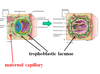
What begins to form on Day 9 within the Syncytiotrophoblast, to facilitate Waste/Nutrient exchange with the mother?
What simultaneously occurs in the mother?
How are they physically connected?
- Trophoblastic Lacunae (Vacuoles)
- Maternal Uterine Capillaries expand, forming Maternal Sinusoids
- Sinusoids connect to the Lacunae via Anastomoses, connecting bloodflow

When approximately does Primary Stem Villi form?
How do they form?
- Day 11-13
- EE Mesoderm induces the Cytotrophoblast to proliferate and migrate into the overlying Syncytiotrophoblast, forming Primary Stem Villi
What must occur to the Primary Stem villi to convert them into Secondary Stem Villi?
Day 16: EE Mesoderm penetrates the core of the Primary Stem Villi
What must occur to give rise to Tertiary Stem Villi?
Blood vessel formation within the EE Mesenchyme.

What is a Hydatidiform Mole?
A mass of vesicular tissue (resembling a bunch of grapes) that results from the build up of fluid and swelling of placental villi, due to failed embryo development (which normally absorbs the fluid).
* The trophoblast will stil form a placenta (and placental villi) regardless of failed embryo development.
What are the (2) types of Hydatidiform moles and how do they differ?
- Complete & Partial Hyditidiform Moles
- Complete H. Moles contain no Embryo at all, while Partial H. Moles contain an embryo which is underdeveloped.
How do Complete Hyditidiform Moles occur?
These occur when the Ovulated egg lacks it’s own Pronucleus & receives (2) Paternal Genomes through either
Dispermic Fertilization
Monospermic Fertilization and subsequent mitosis of the Male Pronucleus w/o Cleavage

How do Partial Hyditidiform Moles occur?
Occurs when a Normal egg is fertilized by either
Two Sperm (Dispermic)
One abnormal Diploid sperm (2N)

While Hydatidiform Moles can be aborted spontaneously, a __&__ (surgical procedure) may be necessary to prevent what disease?
- D&C procedure: Dilation of the Cervix, followed by suction and curettage
- Gestational Trophoblastic Disease
Persistent Trophoblastic Disease will occur if what occurs?
What are they two outcomes?
- Persistent Trophoblastic Disease will occur if any residual trophoblastic tissue remains following a D&C
- The remnants can grow into a benign tumor (Non-Life-threatening) or into Invasive Moles or Metastatic Choriocarcinoma (May be Life-threatening)
What are the symptoms experienced by women with Gestational Trophoblastic Diseases?
- Vaginal Bleeding
- Severe Morning sickness (due to high levels of hCG secreted by trophoblastic tissues)


