Week 3 Flashcards

(85 cards)

1
Q

Steps in reading X-Ray

A

Demopgrahics

Evaluate for adequacy RIIP

Evaluate heart and mediastinum

Evaluate lungs

Evaluate bones and soft tissues

2
Q

Adequacy of Chest Radiograph

A

Rotation - spinous processes equidistant from clavicle ends (if spinous closer to right then rotated to left)

Inspiration - 9-10 posterior ribs seen on inspiration

Anatomy - 1st ribs, costophrenic angle and lateral edges

Penetration - see thoracic vertebral body underneath heart

3
Q

Mistake of diagnosing if X-Ray

A

Underpenetrated - Pleural disease

Overpenetrated - underiagnosed pleural disease

4
Q

What is left hear border is indistinct? Right?

A

Left lingular consolidation

Right middle lobe consolidation

5
Q

Widened mediastinum (+8cm) causes

A

Thoracic aneurysm

Ruptured aorta

Aortic dissection

Mediastinal lymphadenopathy

6
Q

What is the obliteration of silhouette sign diagnostic of?

A

pus, blood or fluid

7
Q

Structures in contact with lungs on X-Ray
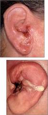
A
8
Q

Otitis external

Another name?

Predisposing factors

Pathogens

Severe form

Treatment

A

Swimmer’s ear

Trauma (removing cerumen), High temp, Derm disease, insertion of foreign objects

Pseudomonas aeruginosa, Staphylococcus auerus

Malignant otitiss externa

Acetic acid-hydrocortisone eardrops; cirpo-hydrocortisone

9
Q

Otitis externa presentation

Malignant otitis externa presentation

A

>38.3C, severe pain, purulent, otorrhea, necrosis can spread to mastoid, diabetes association, possibly fatal

10
Q

Otitis media

Population

Most frequent diagnosis if febrile children

Who has reccurent otitis media

Who should be inspected for otitis media

Pathogen

In

Preceeded by

Treatment

A

Children

Otitis media

People with immune deficiencies

Children w/ purulent conjunctivitis or rhinosinusitis

Streptococcus pneumoniae, H. influenza (nontypeable), Moraxella catarrhalis (also S. aureus and S. pyogenes)

Gram negative bacilli

Viral infection

Amoxicillin

11
Q

Otitis media presentation

A

Blocked eustachian tube, serous effusion, pain fever, tympahnic membrne bulge,

12
Q

Hordeola

What is it?

Pathogen

Complication of

Treatment

What is Chalazia?

A

Acute purulent papules that occur at the lid margin

S aureus

blepharitis (blockage and infection of Zeiss or Moll sebaceous glands or Meibomian glands in the tarsal plate

Lancing (external), dicloxacillin (internal), good hygiene

granulomatous lesions that are not painful

13
Q

Preseptal and orbital celulits

Infection of what

Preseptal cellulitis (PC) vs. Orbital cellulitis (OC)

Cause for orbital cellulitis

Complication

Pathogens

A

eyelid and periorbital soft tissues

OC much more serious

Ethmoid sinutitis

Cavernous venous thrombosis

S. pneumoniae, S. aureus
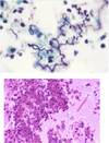
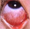
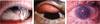
14
Q

Conjunctivitis

Common name

What is it?

Other complication

Most common Pathogens

Purulen conjuctivitis

Hyperpurulent conjunctivitis

Follicular (inclusion) conjunctivitis

Disease caused by N. gonorrhoeae and C. trachomatis

A

Pink eye

Inflammation of palpebral and bulbar conjuctiva

Keratoconjunctivitis

Adenoviruses, HSV1/2 (less common but serious)

S. aureus; S. pneumonia; Moraxella; H. ifluenzae

Neisseria gonorrhoeae

Chlamydia trachomatis -> trachoma (blindness) -> conjuctival scarring & hypertrophy

Opthalamia neonatorum -> invasive -> rapid perforation

15
Q

Viral conjunctivitis presentation

A

Injection (blood vessel dilatation)

Burning / Grit sensation of foreign body

Vision not impaired

16
Q

Ophthalmia neonatorum presetnation

treatment

A

Karatoconjucitivtis -> progressing to perforation of cornea

Ceftriaxone

17
Q

Trachoma presentation

Stages

End stage?

Treatment

A

Active trachoma, follicular response, trichiasis (Scarring)

Blindness

Azithromycin

18
Q

What is the inflammation of cornea?

Cornea and conjuctiva?

Complication?

Risk factor?

Pathogens

A

keratitis

keratoconjunctivitis

vision-threatening

lenses

HSV12, S. aureus, fungi, Acanthamoeaba

19
Q

HSV keratitis

treatment

A

trifluridine (often) and acyclovir

corticosteriods to prevent scarring

20
Q

Uveitis

Causes

Pathogens

A

Inflammation of uvea – pigmented middle layer

Autoimmune, infections, trauma, idiopathic

Herpetic infections and toxoplasmosis

21
Q

Uveitis types

pathogen/structure/description:

Anterior uveitis

Posterior uveiti

Panuveitis

Endopthalmitis

A

Anterior uveitis (iritis) == eye pain, desced vision, ciliary flush, cells in anterior == herpes simplex

Posterior uveiti (choroiditis/retinitis) == painless loss of vision, many cells in viterous == Toxoplasma gondi

Panuveitis (all) == Treponema pallidum

Endopthalmitis == fungal inf of viterous or aqueous humor or obth == Staph aureus, Strep, Gram(-)

22
Q

Pathogenesis of common cold

Cause

Mecahnism

Progression

A

Rhinoviruses

Infect ciliated columnar epithelium cells

Host cells kiled inflammation

Can progress to paranasal sinusitits or otitis media or bronchitis

23
Q

Acute rhinosinusitits

What is it

Cause

Pathogens

Complication

Pathogens of complication

Immuncomporomised pathogens

A

Inflammation or infection of nasal passage mucosa and at least one of the paranasal sinuses <4wks

Respiratory viruses (common cold, dentral etraction)

Rhinovirus, parainfluenza virus, adenovirus, respiratory syncytial virus

Acute bacterial rhinosinusitits

Strep pneu, Heam Inf, Moraxe catarrhalis

Mucor, Rhizopus, Asperigillus

24
Q

Pharyngitis

Most common cause

Seriousness

Pathogens

A

Viruses ; S pyo (children)

Except diptheira it is mild

Rhinoviruses, adenoviruses, S. pyo, Cornybacterium, Candida

25
Viral pathogensis of pharyngitis? Bacterial pathogensis of pharyngitis?
Viruses gain access to mucosal cells lining the nasopharynx and replicate and damage them . Pyogenes attaches to the mucosal epithelial cells using M protein. Production of protease and hyaluronidase to assist invasion.
26
Importance of rapid strep test?
If rapid Strep A test is positive, antibiotics should be prescribed; negative test means wait for cultures to start antibiotics
27
Pharyngitis to candida Pain? Oral symptoms Treatmnent
No Dysphagia Nystatin or clortrimazole
28
Diptheria presentation
Pseudomembrane Bull neck mycarditts Antitoxin
29
Croup What is it? Symptoms
laryngotracheobronchitis dysphonia (hoarseness), odynophonia (pain when speaking), and dysphagia (difficulty swallowing), inspiratory stridor, barking cough
30
What is most serious complication of croup, laryngitis, or epiglottitis?
Airway obstruction
31
Most common cause of epiglottitis
H. influ
32
Thumbprint sign
Epiglossitis
33
Emphensyma types and causes
Centraacinar - smoker (upper lung) Panacinar - a-tripsin (lower lung) Panseptal - pneumothorax
34
Bronchitis definition
3mo of cough productive for 2 consequctive years
35
Acute Bronchitis vs. Emphensyma
36
Chronic Bronchitis histological changes
Increase in goblet cells Thickness of submucosa Squamous metaplasia Loss of cillia
37
Chronic Bronchitis cardiomyopathy mechanism
Hypoxia Vasoconstriction Afterload Cor pulmonale
38
Pulmonary values
39
Obstructive Pulmonary Disease values
low FEV low FEV/FCV low FEF normal-high TLC high RV
40
Obstructive Pulmonary Disease examples
Emphysema (COPD) Chronic Bronchitis(COPD) Asthma Bronchiectasis
41
Emphysema definition
Anatomic definition ## Footnote Irreversible enlargement of the airspaces distal to the terminal bronchioles
42
Emphysema vs. Chronic Bronchitis Clinical Features
E: dyspnea, wieght loss, barrel-chest, prologed expiratory, sitting forward, pursed lipids CB: persisten cought; mild cyanosis, hypercapnea
43
Persistent asthma symptoms What is it
Status asthmaticus Persistent symptoms lasting for days to weeks
44
Asthma histology
Curschmann spirals–Whorls of shed epithelium forming spiral shaped mucous plugs Charcot-Leyden crystals
45
Asthma types
Atopic - Hyper Type I Non-Atopic - hyperirritability of the bronchial tree
46
Asthma (non-atopic) ASA Triad (Samter's Triad) Difference
aspirin-exacerbated respiratory disease (AERD) More severe, adult onset
47
Asthma histological changes
smooth muscle hyperrophy mucous hypertophy basemembrane fibrosis increase vasculatorure eosinophilic inflitrate
48
Asthma treatment
Methacholine
49
Definitions Bronchitis Bronchiolitis
Bronchitis – Inflammation of the large and mid-sized airways; primarily viruses Bronchiolitis - inflammation of the bronchioles, the smallest air passages of the lungs; primarily viruses, e.g., RSV (50-90%)
50
Definitions Acute pneumonia Atypical pneumonia Chronic pneumonia
Acute pneumonia - Inflammation of lungs caused by microbial infection of the alveoli and surrounding lung; present for days Atypical pneumonia – Characterized by moderate amounts of sputum, absence of physical findings of consolidation, only moderate elevation of WBC, and lack of alveolar exudates Chronic pneumonia – Inflammation of lungs caused by microbial infection of the alveoli and surrounding lung or non-infecious causes; present for weeks to months
51
Pulmonary host defenses
**Nasopharynx** Nasal hair Anatomy of upper airways Mucocilliary apparatus **Oropharynx** Saliva Cough Bacterial inferference **Trachea and bronchi** Cough, epiglottal reflexes = alcohol&aspiration Mucocilliary apparatus = cigarette smoking, bacteria destorying Airway surface liquid (lysozyme, lactoferrin) **Terminal airways and alveoli** Alveolar lining fluid (surfactant, fibronectin, iron-binding proteins) Alveolar macrophages = cigarette Neutrophil recruitment
52
Acute pneumonia children adults
RSV, Para, Ortho Ortho, RSV, metapneumovirus, adenovirus
53
Atypical pneumonia Characteristics
Nonlobar, patchy, or interstitial pattern on chest radiography Failure to identify organism on gram stain Slower, and absence of crisis (like in pneumococcal pneumonia)
54
Atypical pneumonia Cause
Mycoplasma Viruses (adenovirus, RSV, cytomegalovirus, SARS) Legionella Pneumocystis jirovecii
55
Empirical treatment of adult patients with community-acquired acute pneumonia
56
Empirical treatment of adult patients with health care-acquired acute pneumonia
57
Klebsiella pneumoniae characteristics
Gram - Capsule Lactose Fermenting Rod Enterobacteria Uusualy in immune suppressed / impaired respiratory defenses Hightly resistance e.g Carbapenem-resistant Klebsiella pneumonaie (CRKP)
58
Legionnaries vs. Pontaic fever Severity Exposure Mortality
Severe actute pneumona / Mild (no pneumonia) aerosol / aerosol \<1% / 15-20%
59
Legionnaries pathogenesis
Inhale aerosol Infect alveoalar macrophages, monocytes, and alveolar epithlial Inhibits phagolysosomal fusion (no H2O2) Intracellular proliferation (primary virulence) Inflammatory response Cell-mediated immunity
60
Diagnosis of Legionnaire disease
Culture X-Ray Urinary antigen test (LPS serogroup)
61
Diagnosis of Legionnaire disease What is host in water?
Free-living amoebas
62
Mycobacterium pneumonia pathogenesis
Adheres to respiratory epithelium via an attachment organelle P1 protein is primary adhesin Destroyes, cillia, then epithelial cells Causes persistent cough
63
Mycobacterium pneumonia clinical diseases
Tracheobronchitits - low-grade fever, malaise, headache, non-productive cough Primary atypical pneumonia - less acute than pneumonia r influenza (walking pneumonia)
64
Mycobacterium pneumonia diagnosis
Poor microscopy, culture, NAA Serology: complement fixation (no sens/spec), cold agglutinin (no sens/spec)
65
Histoplasma
66
Morphology of yeasts
histoplasmosis: (M) tuberculate macroconidia (Y) small, oval, budding yeast
67
Acute / Chronic pulmonary histoplasmosis Progressive disseminated histoplasmosis African histoplasmosis symptoms
fever, headach, nonproductive, chills, chest pain fever, productive cough, chest pain, cavitary lesions multiple organs, risk +55yo or immunosupp H. capsulatum var. duboisii; skin and skeletal
68
Blastomycosis diseases
Acute pulmonary blastomycosis (often asymptomatic) but might progress to myalgias, arthralgias, chills, and fever Disseminated disease -- skin most often but also bones and joints
69
Blastomycosis Histology
70
Histoplasmosis histology
71
Carbapenem-resistant Klebsiella pneumoniae (CRKP) mechanism
Hydrolyze all known beta lactam antibiotics Resistant to beta lactamase inhibitors
72
Coccidioides diseases
**Primary pulmonary coccidioidomycosis:** symptoms 7-21 after exposure, cough chest pain ,SOB, fever, fatigue **Pulmonary nodules and cavities:** pleuritic pain, cough, hemoptysis **Extrapulmonary dissemination:** immunosuppression (meningitis most serious form)
73
coccidioidomycosis histology
74
Cord Factor - M. tuberculosis Molecule What does it do
Trehalose dimycolate - glycolipid, subset of mycolic acids Binds to macrophage surface receptor called mincle Blocks macrophage activation by INF-g, induced secretion of TNFa Causes cord formation
75
76
Primary tuberculosis pathogenesis
Phagocytosis of bacteria Survival in phagosome Secretion of IL-12 and TNF-a T differentiate into TH1 secretion of INF-g (DHT) DTH may lead to granuloma (lymphocytes+macrophage+epitheiod+fibroblasts+giant cells) Resolution if killed or dormant
77
Tuberculin skin test what is it read for false positive false negative
Intradermal injection of 5 tuberculin units (TU) of PPD 48-72h read for induration infection with non-Mtb immunosuppression
78
IFN-γ-release assay what is it advantages
Whole blood incubated measures release of INF-g no reaction if received BCG, no booster effect
79
Diagnosing M tubercluosis
Smear acid-aclohol decolorized counterstained with methylene blue Fluoresence dyes most sensitive Nucleic acid testits (can also asses resistance to rifampin) Culture (gold standard) in Lowenstein Jensen -3-8wks
80
M tuberculossi antibiotics
Isoniazid (INH) – inhibits synthesis of mycolic acid Rifampin (RMP) – inhibits DNA-dependent RNA polymerase Ethambutol (EMB) – inhibits cell wall synthesis Pyrazinamide (PZA) – mechanism not well understood
81
Resistance of Multidrug-resistant Mtb (MDR TB) Extensively drug-resistant Mtb (XDR TB)
isoniazid and rifampin isoniazid + rifampin + quinolones + at least one second-line drug
82
BCG vaccine What is it
less virulent strain of M. bovis (live vaccine)
83
Mycobacterium avium Complex (MAC) specifc disease
M. avium and M. intracellulare Prinary infection (no reactivation) Chronic localized pulonary - patient have intact immunity, smoking/COPD firsk factor Disseminated CD4\<100 Cervial lymphadentitis
84
Mycobacterium avium Complex (MAC) Indetification Source Treatment Prophylaxis
Culture / blood (for dissemtinated) Env sources (no human-human spread) Very difficult (high fail rate) Clarithromycin or azithromycin + ethambutol and rifabutin Azithromycin prophylaxis if CD4 \< 50 cells/mm2
85
Virulence of Nocardia
Related to ability to avoid intracellular killing Catalase and superoxide dismutase inactivate toxic metabolites **Cord factor** Prevents intracellular killing Prevents phagosome-lysosome fusion