Week 3: Skull, Brainstem and Cranial Nerve Flashcards

(89 cards)

1
Q

outline hthe bones of the vsicerl cranium

A

Aka the facial skeleton-- Nasal bones; make up the bridge of the nose

The paired maxillary bones make up the upper jaw, floor of the orbit and parts of nose and hard palate

Mandible forms lower jaw and houses the lower teeth

Paired zygomatic bone; lateral aspects of orbit and bony processes of the cheek (cheek bones)

The zygomatic also joins the temporal bone to form the zygomatic arch.

2
Q

what bone of the skull is associated with the mastoid process

A

temporal

3
Q

what is the largest fontanelle in children

A

The largest fontanelle is the anterior fontanelle, which is between the frontal and parietal bones, and closes between 18-24 mos of age.

The coronal suture runs between the frontal and parietal bones.

The sagittal suture runs between the two parietal bones, and meets with the two lateral lambdoid sutures, which separates the parietal bones from the occipital bone of the skull.

The external occipital protuberance is a key landmark and can be felt posteriorly on the skull. It’s the attachment site for a number of neck muscle.

4
Q

suture line between occipital and parietal

A

lamboid

5
Q

suture line between the two aspects of the parietal bones in a fetus

A

the saggital suture

6
Q

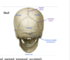
how can fracture of the pterion cause a hematoma? what type of hematoma?

A

Pterion– worse consequences, formed by the joining of the frontal, parietal, sphenoid and temporal bones. The middle meningeal artery runs between the skull bones and the dura mater at the point of the pterion. A fracture in this region can sever this artery and lead to a life threatening epidural hematoma.

7
Q
A
8
Q

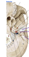
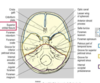
which fossa contains the sella turcica and pituitary gland

A

middle cranial fossa; hosues the tmpeoral lobe, forms the sella turcica, and contains the pituitary glan

9
Q

the spinal cord exit hole

A

foramen magnum

10
Q
A
11
Q

Cribriform plate: has many holes were the extensions of the ___ __ pass through into the nasal cavity.

A

Cribriform plate: has many holes were the extensions of the olfactory nerve pass through into the nasal cavity.

12
Q

Optic canal: houses the __ __ as well as some blood supply to the eye.

A

Optic canal: houses the optic nerve as well as some blood supply to the eye.

13
Q

what nerves run through the superior orbital fissure?

A

3, 4, 6, and V1(opthalmic of trigeminal nerve)

14
Q

what CN runs through the foramen rotundum

A

the V2 (maxillary nerve)

15
Q

what CN runs throguh the foramen ovale

A

V3 (mandibular nerve of the trigeminal nerve)

16
Q

the foramen spinosum contains the __ ___ artery

A

middle meningeal artery

17
Q

what moves through the carotid canal?

A

internal carotid artery, which tontributes to the circle of willis

18
Q

what moves throuhg the internal acoustic meatus?

A

cranial nerve 7 and 8; facial and vestibulocochlear

19
Q

what nerves move throuhg the jugular foramen?

A

in addition to the jugular vein, CN 9, 10, 11 also run through the jugular foramen

20
Q

what moves through the foramen magnum?

A

vertebral artery travels superiorly to the foramen magnum. lateral to the foramen magnum is the occipital condyle which artculates with the C1 vertebrae. there is a mandibular fossa where the TMJ os lovated.

21
Q

exit of the facial nerve

A

the facial nerve (7) runs through the internal acoustic meatus, and also exits out of the stylomastroid foramen.

22
Q

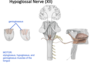
what CN exits the hypoglossal canal?

A

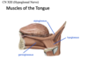
hypoglossal nerve, which provides motor output for the tongue

23
Q
A
24
Q
A
25
The cerebral peduncles are large bundles of white matter that communicate with other regions of the brain that are located more superiorly. These cerebral peduncles often contain bundles of white matter that are part of the motor ___ \_\_.
## Footnote The cerebral peduncles are large bundles of white matter that communicate with other regions of the brain that are located more superiorly. These cerebral peduncles often contain bundles of white matter that are part of the motor **corticospinal tract**
26
CN 1 ## Footnote There are little fibers that extend FROM the **olfactory** __ and PAST the cribriform plate which are called olfactory \_\_. These olfactory __ synapse onto secondary neurons and form the olfactory \_\_, which contain and project back to the olfactory cortex of the brain in the uncus.
There are little **fibers** that extend FROM the olfactory **bulb** and PAST the cribriform plate which are called olfactory **nerves**. These olfactory nerves synapse onto secondary neurons and form the olfactory **tracts**, which contain and project back to the olfactory cortex of the brain in the uncus.
27
28
Lesioning CNII in one side will result in: lesioning one optic tract will result in: lesioning of an optic radiation will result in: lesioning the optic chiasm will result in:
Lesioning one of the cranial nerve will result in the loss of vision of the eye (monocular blindness) Lesioning one of the tracts (behind the optic chiasm) will result in a specific visual field being lost (either right or left), called homonymous hemianopsia Lesioning of the optic radiations will result in random gaps in the visual fields. Lesioning of the optic chiasm will result in “tunnel vision” called bitemporal hemianopia This is seenin pituitary adenomas-- due to its close relationship, pituitary adenomas can result in disruption of the cross over of visual information.
29
what causes this
Lesioning one of the cranial nerve will result in the loss of vision of the eye (monocular blindness)
30
not being able to see an entire visual field is called:
homonoymous hemianopsia. due to lesioning of the optic tract. Lesioning one of the tracts (behind the optic chiasm) will result in a specific visual field being lost (either right or left), called homonymous hemianopsia
31
what common presentation results in this:
pituitary adenoma can cause the person to have bitemporal heminaopia because the chiasm is cut
32
what muscles is CN III responsible for innervating?
CNIII: medial, superior, inferior rectus muscle, and inferior oblique muscles
33
what parasympathetic function does CNIII have
sphincter pupillae and ciliary muscle filation
34
what muscle is innervated by the trochlear nerve
the superior oblique muscle. with an injury to the usperior oblique muscle, the eye will drift upwards and laterally, resulting in doulb eviison,
35
what muscle is innervated by the abducens nerve (CN6)
Lateral rectus muscle to abduct the eyeball.
36
3 divisions of the opthalmic nerve
**Ophthalmic nerve (V1):** travels through the superior orbital fissure, towards the forehead, and exits the superior aspect of the orbit through the supra-orbital notch or foramen **Maxillary nerve V2**; enters skull through foramen rotundum, and exits via foramen below the eye (infraorbital foramen), and innervates the area over the maxilla, as well as upper teeth and palate **Mandibular nerve branch (v3**); enters skull through foramen ovale and exits traveling down onto the deep side of mandible, through mandible, and out the mental foramen. Provides sensory info from mandible, lower teeth and external ear. **Near the entrance of the mandible, it gives off a branch called the lingual nerve, which goes onto the tongue and carries somatic sensation from anterior ⅔ of the tongue.** The mandibular division of the trigeminal nerve also carries motor to the muscles of mastication, including the temporalis, masseter, and tergoid muscles?
37
does the mandibular branch provide taste sensation to the tongue?
no. the mandibular branch gives off a branch called the **lingual nerve,** which carries out comatic anterior2/3 tongue movement. - they provide motor movement to masseter muscles
38
explain how trigeminal cross talk can cause brainfreeze
The nerve branches can suffer from “cross talk”-- in the same way that we think about referred pain, the brain interprets info from the cross talk. Pain from the palate, which are sensed by V2, can be relayed back to **the brainstem and can excite the V1 branch, producing a forehead brain (brain freeze)**
39
The facial nerve exits skull through the ___ \_\_\_ and divides into 5 branches near the parotid gland, which innervate the entire face and the back of the ear.
The facial nerve exits skull through the stylomastoid foramen and divides into 5 branches near the parotid gland, which innervate the entire face and the back of the ear.
40
which nerve innervates the anterior 2/3 of tongue to give a taste sensation
facial nerve.
41
outline the parasympathetic function of the facial nerve
**Parasympathetic motor to the salivary and lacrimal glands**
42
outline how lesioning LMN vs UMN pathway involving the facial nerve can result in a complete vs partial facial droop
Lesion of the LMN of the facial nerve results in ipsilateral total loss of motor to the muscles of facial expression, producing characteristic facial drooping and dry eye, as the face is paralyzed to that side In contrast to **UMN due to cortical lesion,** there is some compensation that can occur in the upper ⅓ of the face due to contralateral overal of the facial nerve. **If the forehead contraction is preserved, it is most likely an UMN in the cortex.**
43
which structures does the CN8 associated with for balance and hearing
balance; semicircular canals hearing; chochlea
44
45
special sensory to posterior ⅓ of tongue, and somato-sensory to ⅓ tongue, and posterior pharynx is CN\_\_\_ responsibility
CN9 ## Footnote Carries visceral sensory info from the carotid artery and sinus. Measures blood quality as it enters brain Also has special sensory to posterior ⅓ of tongue, and somato-sensory to ⅓ tongue, and posterior pharynx. Involved in the gag reflex, along with the vagus nerve. Info sensed to the glossopharyngeal nerve and the motor efferent reflex is produced by the vagus nerve, cranial nerve 10. This nerve carries parasympathetic innervation to the parotid salivary gland.
46
parasympathetic innervation of the CN9
parotid gland
47
Taste to the anterior ⅔ of the tongue is conducted by the __ nerve, and taste to the posterior ⅓ of the tongue is conducted by the ___ nerve.
Taste to the anterior ⅔ of the tongue is conducted by the **facial** nerve, and taste to the posterior ⅓ of the tongue is conducted by the **glossopharyngeal** nerve.
48
Epiglottis taste is provided by the __ nerve.
Epiglottis taste is provided by the vagus nerve.
49
For the somatic sensation, on the right, the anterior ⅔ is provided by the ____ division of the ___ nerve, where the posterior ⅓ is also provided by the ____ nerve. Some somatic sensation of the epiglottis is provided by the vagus nerve.
For the somatic sensation, on the right, the anterior ⅔ is provided by the mandibular division of the trigeminal nerve, where the posterior ⅓ is also provided by the glossopharyngeal nerve. Some somatic sensation of the epiglottis is provided by the vagus nerve.
50
Vagus nerve provides somatic motor innervation to muscles of larynx and superior esophagus. Recall that the ___ \_\_ ___ goes to the vocal chords and folds. All vocal muscles are supplied by the vagus nerve, except for the cricho-thyroid muscle this nerve also provides ____ motor to cardiac and pulmonary plexus, adn follows to esophagus to foregut and midgut to provide parasympathetic innervation Visceral sensory fibers allow for visceral sensation of the thorax and abdomen to travel with the nerve.
Vagus nerve provides somatic motor innervation to muscles of larynx and superior esophagus. Recall that the recurrent laryngeal nerve goes to the vocal chords and folds. All vocal muscles are supplied by the vagus nerve, except for the cricho-thyroid muscle this nerve also provides parasympathetic motor to cardiac and pulmonary plexus, adn follows to esophagus to foregut and midgut to provide parasympathetic innervation Visceral sensory fibers allow for visceral sensation of the thorax and abdomen to travel with the nerve.
51
two key muscles that the spinal accessory nerve innervates
SCM and trapezius muscle
52
T/F the nuclei of the spinal nerve are functioning in the brainstem
false. ## Footnote Actually a debated cranial nerve because it actually originates off of the **upper spinal nerve segments**, and those fibers travel up through the **foramen magnum and pass out through the jugular foramen, a**longside the cranial nerve 9 and 10.
53
54
The hypoglossal nerve will exit the skull via the ___ \_\_\_, and go on to innervate the muscles of the tongue Lesion of the HG nerve (ex in LMN), then the tongue cannot \_\_\_\_\_, and the tongue will ___ TO THE ____ OF THE LESION.
The hypoglossal nerve will exit the skull via the hypoglossal canal, and go on to innervate the muscles of the tongue Lesion of the HG nerve (ex in LMN), then the tongue cannot pull it out on that side, and the tongue will POINT TO THE IPSILATERAL SIDE OF THE LESION.
55
\_\_\_\_ nerve goes to the tongue, and is a branch of the mandibular division, provides general sensation to ⅔ anterior of tongue
Lingual nerve goes to the tongue, and is a branch of the mandibular division, provides general sensation to ⅔ anterior of tongue
56
Travels through the internal acoustic meatus.This travels through inner ear, through the petrous portion, and exits inferior through the stylo-mastoid foramen.
the facial nerve
57
Vertebral arteries travel up through ___ \_\_\_ and merge together to form the __ artery which supplies postieorr circulation to brain
Vertebral arteries travel up through foramen magnum and merge together to form the basilar artery which supplies postieorr circulation to brain
58
Internal carotid arteries supply the anterior neural tissue, and travels through the ___ canal. Has a medial trajectory
Internal carotid arteries supply the anterior neural tissue, and travels through the carotid canal. Has a medial trajectory
59
\_\_\_ \_\_: where the middle meningeal artery travels. Lateral to the foramen ovale (where the maxillary V3 branch of the trigeminal nerve moves)
Foramen spinosum: where the middle meningeal artery travels. Lateral to the foramen ovale (where the maxillary V3 branch of the trigeminal nerve moves)
60
A 19-year old male presents with a bleeding and externally deformed nose following a bar fight. Further examination reveals clear liquid leaking from his nostrils, and the physician immediately orders a head CT. Why?
This individual likely has a nasal fracture that involves the cribriform plate. Cerebrospinal fluid is leaking into the nasal cavities.
61
Externally, two thin nasal bones form the bridge; **the ethmoid bone, which forms the superior and middle nasal concha,** as well as the **nasal septum.** Ethmoid bone is highlighted in pink Anteriorly, the septum is cartilaginous, and posteriorly, the V**omer and perpendicular plate of ethmoid bone** fuse to form the posterior septum. Cribiform Plate; olfactory nerve fibers pass through to transmit olfactory sensory information to the brain.
62
review the location and pattern of CSF circulation
Produced by choroid plexus, found in lateral, third and fourth ventricles From lateral ventricles, CSF fills both ventricles, then makes its way to the third vent via the foramen of monro. The cerebral aqueduct then leads to the 4th vent, which is located between the pons anteriorly and cerebellum posteriorly. The CSF then escapes to subarachnoid space via medial foramen of magendie and the two lateral foramen of luschka. The CSF enters the dural sinus(between pia and arachnoid mater) from the subarachnoid space via the arachnoid granulations. It mixes with venous blood and returns to the heart.
63
This artery supplies dura mater and periosteum of the inner cranial bones, and is a branch of the maxillary artery
the middle meningeal artery ## Footnote In addition, just deep to the portion on the interior aspect of the skull is the middle meningeal artery. This is a branch of the maxillary artery, a branch of the external carotid artery. **This artery supplies dura mater and periosteum of the inner cranial bones (see right image)** **The MMA is sandwiched between skull bones laterally, and dura mater medially.**
64
site of primary and secondary neruon synapse for the olfacotry nerve tract
Numerous small neurons projecting through the cribriform plate to the olfactory bulb. From the olfactory bulb, they synapse with secondary neurons, which move as an olfactory tract to go to the cortex.
65
Info from right visual field is projected to left primary visual cortex. specifically from the right INFERIOR visual field (seen below) is projected to the left primary visual field cortex ____ the calcarine sulcus.
Info from right visual field is projected to left primary visual cortex, specifically from the right INFERIOR visual field (seen below) is projected to the left primary visual field cortex **ABOVE the calcarine sulcus.** CN2: Optic Nerve. Light info from the retina is carried by the optic nerve. At the chiasm, fibers carrying info from the left hand side cross over and move to the right side of the brain, and fibers carrying info from the right hand side cross over and move to the left side of the brain. Because of the crossing at the chiasm, visual info from the left side of both retinas, which carries info from the right visual field, is carried by the left optic tract to the lateral geniculate nucleus of the left thalamus. After the thalamus, the optic radiations project to the primary visual cortex, and the info projects retinotopically on either side of the calcarine sulcus. ex/ Info from right visual field is projected to left primary visual cortex, specifically from the right INFERIOR visual field (seen below) is projected to the left primary visual field cortex ABOVE the calcarine sulcus.
66
67
68
69
70
71
4. A young person presents with pain behind her left ear, drooping left face, and sensitivity to loud sounds in the left ear. Examination reveals pronounced left facial weakness including the forehead. What might be producing these symptoms? Specifically, what neuron int he circuit?
This individual is likely suffering from Bell’s palsy, a dysfunction of the left facial nerve affecting the lower motor neuron. Note that a lesion affecting the upper motor neuron **will spare the forehead, due to bilateral innervation of the portion of the motor nucleus for the facial nerve that supplies the muscles of facial expression.**
72
73
74
parasympathetic innervation to lacrimal gland, and sublingual and submandibular salivary gland, and carries a special sensation, Taste, to anterior ⅔ of tongue. What nerve is this?
facial nerve
75
T/F facial nerve innervates the sublingual and parotid salivary gland
false. it innervates the sublingual and submandibular salivary gland
76
77
Outline the differences in symptoms between if the red star was lesion compared to if the blue star area was lesioned
Upper motor neuron; innervates upper ⅓ of face Receives bilateral input from cortico-bulbar fibers If there is an upper MN lesion, (ex/ on the left hand side like in the diagram below), the patient will have a facial droop on the lower ⅔ of the face on the right hand side, but the pt will still be able to wrinkle the forehead because it receives input from the ipsilateral upper motor neuron. Lower neuron; innervates lower ⅔ of face. Lower facial nerve receives input from contralateral cortico-bulbar fibers from the primary motor cortex If there is a lower facial nerve lesion, (ex on RS) they will experience facial droop. The entire ipsilateral face will droop.
78
Why do things seem louder if you have bells palsy
Mechanism; there is an eardrum, hammer, anvil, stapes. Stapes is the smallest bone in the human body, controlled by the stapedius muscle. **CN7 also innervates the stapedius muscle, which contracts and tethers/reining in the stirrup, which dampens down noises.** When the stapedius muscle isn’t working because of CN7 Bells’ Palsy, sounds seem louder because you can’t dampen down the sounds.
79
80
General sensation of this region. Sensory branch of gag reflex Also caries chemo and baroreceptors from carotid bodies Taste sensation posterior ⅓ Parotid salivary gland innervation
glossopharyngeal nerve
81
Completes gag reflex by providing motor for larynx and esophagus Parasympathetic motor to cardiopulmonary, foregut and midgut structures.
Completes gag reflex by providing motor for larynx and esophagus Parasympathetic motor to cardiopulmonary, foregut and midgut structures. VAGUS NERVE
82
83
84
ventral vs tegmentum region of the pons
Pons= bridge **Ventral pons= responsible for coordination of movement with the cerebellum** **Transversely oriented fibers between pons and cerebellum give striated appearance of the pons.** **Tegmentum region= responsible for arousal and attentiveness. Also houses cranial nerve nuclei (trigeminal, abducens and facial)** Medulla oblongata is also near the pons.
85
Taste Anterior ⅔ of tongue: Light touch ⅔ of tongue: Posterior ⅓ of tongue taste and light touch Posterior oropharynx taste and sensation
Taste Anterior ⅔ of tongue: taste is carried cranial nerve 7 by chorda tympani branch of the facial nerve Light touch ⅔ of tongue: CN5 (maxilla via lingual nerve) Posterior ⅓ of tongue taste and light touch-- both carried by CN9 Posterior oropharynx taste and sensation-- vagus nerve CN10
86
A middle-aged woman presents with difficulty swallowing, decreased left palate movement, hoarseness, and left vocal cord paralysis, left trapezius and sternocleidomastoid weakness, left tongue deviation and atrophy, decreased hearing in the left ear, alteration in taste and left-sided headache. Where is the most likely location of the lesion?
This individual likely has a large lesion outside the medulla, near the left internal auditory meatus, jugular canal, and hypoglossal foramen.
87
88
89
The hypoglossal nerve exits the cranium via the ___ canal. At the base of the skull, the canal opens anterior to the __ \_\_.
The hypoglossal nerve exits the cranium via the **hypoglossal** canal. At the base of the skull, the canal opens anterior to the **occipital** **condyle**.