Week 4: Brain and Spinal Cord Circulation Flashcards
(26 cards)
The brain has a high demand for oxygen, receiving___% of cardiac output.
The brain has a high demand for oxygen, receiving 15% of cardiac output.


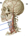
explain how the anatomy of the common carotid arteries differ on either side
The brachiocephalic trunk on the right hand side gives way to the right common carotid artery, while on the left side, the left common carotid artery arises straight from the aortic arch.
outline the difference in supply between the external and internal carotid arteries

Vertebral Arteries;
Arise from ___ arteries, ascending up the neck through the ___ ____ of the ___ vertebrae
Superiorly, the left and right vertebral artery to form the single midline vessel, __ artery.
Vertebral Arteries;
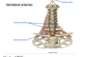
Arise from subclavian arteries, ascending up the neck through the transverse foramina of the cervical vertebrae
Superiorly, the left and right vertebral artery to form the single midline vessel, basilar artery.
differentiate between epidural, subdural and subarachnoid hemorrhage
Epidural hemorrhage; produced by tearing of a branch of the middle meningeal artery, which is located between the calvarium and the periosteal dura.
Subdural hemorrhage: produced by tearing of a superior cerebral vein, and is located between the dura and arachnoid mater
Subarachnoid hemorrhage; often due to the rupture of a saccular aneurysm, and is located in the subarachnoid space.


There are three key branches of the internal carotid arteries:
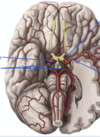
- posterior communicating artery
- middle cerebral arteries
- anterior cerebral arteries
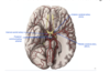
__ cerebral artery supplies medial surface of hemisphere, and covers supply to the lower limbs.
__ cerebral artery covers a lot of territory– face, arm, speech centers. In most people.
Anterior cerebral artery supplies medial surface of hemisphere, and covers supply to the lower limbs.
Middle cerebral artery covers a lot of territory– face, arm, speech centers. In most people.


Ophthalmic artery; supplies structures of the orbits, and anastomosis of the external carotid artery that are supplying the face


Outline the order that the vertebral arteires give rise to in ascending order
- PICa
- AICA
- pontine arteries
- superior cerebral arteries, supplying bloos to the superior cerebellar cortex and the superio cerebellar peduncles


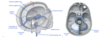
General flow of venous drainage through sinuses
Superior and inferior sagittal sinuses are at the superior and inferior margin of the falx cerebri.
Both drain into the ___ sinus, which meet with the superior sagittal sinus and transverse sinus at the ___ of sinuses/___ at the occipital point.
The confluence of sinuses drains through the ___ sinus to the ___ sinus and to the ____ ___ vein
The internal jugular vein exits the skull through the jugular foramina, along with CN 9, 10, and 11.
Most of the venous blood flows in this pathway.
Superior and inferior sagittal sinuses are at the superior and inferior margin of the falx cerebri.
Both drain into the straight sinus, which meet with the superior sagittal sinus and transverse sinus at the confluence of sinuses/torcula at the occipital point.
The confluence of sinuses drains through the transverse sinus to the sigmoid sinus and to the internal jugular vein
The internal jugular vein exits the skull through the jugular foramina, along with CN 9, 10, and 11.
Most of the venous blood flows in this pathway.
Inferiorly, the internal jugular vein drains into its respective brachiocephalic vein, which then joins to form the superior vena cava, and the blood is returned to the heart.
There are anteriorly located sinuses that drain through some additional pathways:
Cavernous sinus; associated with internal carotid artery, and CN 3, 4, 6, branches V1 and V2 of trigeminal nerve
Due to connections of the facial veins, facial infections can go into the CNS via conduction through the cavernous sinus
Central region of face = danger triangle.

Transverse sinus is in the posterior margin of ___ __, that overlays the cerebellum, separating it from the occipital cortex.
Transverse sinus is in the posterior margin of tentorium cerebelli, that overlays the cerebellum, separating it from the occipital cortex.
outline the exit of CSF

Necessary to prevent build up of pressure.
Escapes the subarachnoid or ventricular system by dumping into the dural sinuses from the sbuarachnoid space via the
Necessary to prevent build up of pressure.
Escapes the subarachnoid or ventricular system by dumping into the dural sinuses from the sbuarachnoid space via the arachnoid granulations; protruding knots of tissue in the dural venous sinuses
Once in the sinus, it mixes with venous blood and returns to heart.
Once in the sinus, it mixes with venous blood and returns to heart.


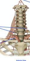
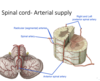
outline the arterial supply of the spinal cord
The spinal cord is supplied longitudinally by the anterior spinal artery, a branch of the vertebral artery. It runs inferiorly in the anterior median fissure of the spinal cord, giving off branches as it descends.
The anterior spinal artery supplies anterior ⅔ of spinal cord
The posterior ⅓ spinal cord is supplied by the paired posterior spinal arteries, which run in the posterior-lateral sulcus of the spinal cord. These arteries arise from the vertebral arteries.
The anterior and posterior spinal arteries are fed by the radicular segmental arteries that arise from spinal arteries off the vertebral, intercostal, lumbar or sacral arteries depending on the level
There are several anastomoses to give some redundancy.

venous drainage of the spinal cord

One __ and one ___ ___ vein, along with a series of __ segmental vein that all drain into the vertebral venous plexus and subsequent ___ system to return to the heart.
One anterior and one posterior spinal vein, along with a series of radicular segmental vein that all drain into the vertebral venous plexus and subsequent azygous system to return to the heart.

outline the general sensory cranial nerve pathway
Ex/ Sensory cranial nerve pathway: sensory to tongue, the mandibular branch of the trigeminal nerve.
Sensory info comes into the brainstem, synapses and decussates at the brainstem. Second order goes up to the thalamus, where it synapses with the third order neuron. The third order neuron goes to the primary sensory cortex.
Lesion of the trigeminal nerve mandibular branch will result in ipsilateral sensory loss. If you have a lesion in the secondary or tertiary neuron of the trigeminal sensory nerve, it will affect the sensory of the contralateral side since it is already decussated.
outline the general motor cranial nerve pathway
Ex/ Motor cranial nerve pathway– corticobulbar pathway involving the hypoglossal nerve
There is an upper and motor neuron. The UMN goes down from the cortex to the brain stem. In the brain stem, there is a decussation and synapse with the LMN, which becomes the hypoglossal nerve, which innervates the tongue
If you damage the right hypoglossal nerve, the right hand side of the tongue will atrophy (ipsilateral deficit)
If you damage the UMN, you will have a contralateral sign because it has not yet been decussated.
note:
Therefore, generally speaking, if you damage the ACTUAL cranial nerve, you will have deficits on the IPSILATERAL side. If you damage the upper neuron or secondary order neuron, you will have damage on the CONTRALATERAL side because it already has crossed over and decussated at the brain stem.
