Week 6-9 quiz questions Flashcards

1
Q

Immunosurveillance is defined by three events:

  1. B cell response, T cell response, macrophage response
  2. escape, imbalance, elimination
  3. escape, equilibrium, elimination
  4. B cell response, T cell response, macrophage response
A

escape, equilibrium, elimination

2
Q

Immunosurveillance is defined by three events:

  1. B cell response, T cell response, macrophage response
  2. escape, imbalance, elimination
  3. escape, equilibrium, elimination
  4. B cell response, T cell response, NK response
A

escape, equilibrium, elimination

3
Q

Immunoscore is based on

  1. the number of leukocytes and T helper cells infiltrating a tumor
  2. the number of leukocytes and B cells infiltrating a tumor
  3. the number and the phenotype of all leukocytes in the tumor
  4. the number of T cells and NK cells infiltrating a tumor
A

the number and the phenotype of all leukocytes in the tumor

4
Q

In regard to tumor immunology

  1. adaptive immunity is involved
  2. innate immunity is involved
  3. B cell are involved
  4. adaptive and innate immunity are involved
A

adaptive and innate immunity are involved

5
Q

Macrophages

  1. are always deleterious for tumor growth
  2. are helping tumor growth if they are M1 type
  3. are deleterious for tumor growth if they are M2 type
  4. are favouring tumor growth if they are M2 type
A

are favouring tumor growth if they are M2 type

  • M1 macrophages can kill tumour cells by mechanisms that they also use to kill infectious organisms. Prominent among these is production of nitric oxide (NO), which has been shown to kill tumours in vitro and in mouse models in vivo.
  • There is evidence that some macrophages in tumours contribute to tumour progression and have an M2 phenotype. These cells secrete vascular endothelial growth factor (VEGF), transforming growth factor-β (TGF-β), and other soluble factors that promote tumor angiogenesis.
6
Q

NKG2D

  1. is an activating receptor on NK cells
  2. is an inhibitory receptor on NK cells
  3. is an inhibitory receptor on the tumor cell surface
  4. is an activating receptor on the tumor cell surface
A

is an activating receptor on NK cells

  • NK cells kill many types of tumor cells, especially cells that have reduced class I MHC expression and express ligands for NK cell–activating receptors. In vitro, NK cells can kill virally infected cells and certain tumor cell lines, especially hematopoietic tumors.
  • NK cells also respond to the absence of class I MHC molecules because the recognition of class I MHC molecules delivers inhibitory signals to NK cells. Some tumors lose expression of class I MHC molecules, perhaps as a result of selection against class I MHC–expressing cells by CTLs.
  • This loss of class I MHC molecules makes the tumors particularly good targets for NK cells. Some tumors also express MIC-A, MIC-B, and ULB, which are ligands for the NKG2D activating receptor on NK cells.
  • In addition, NK cells can be targeted to IgG antibody–coated tumor cells by Fc receptors (FcγRIII or CD16). The tumoricidal capacity of NK cells is increased by cytokines, including interferon-γ (IFN-γ), IL-15, and IL-12, and the anti-tumor effects of these cytokines are partly attributable to stimulation of NK cell activity.
  • IL-2–activated NK cells, called lymphokine-activated killer (LAK) cells, are derived by culture of peripheral blood cells or tumor-infiltrating lymphocytes from tumor patients with high doses of IL-2. These cells are more potent killers of tumors than are unactivated NK cells.
7
Q

A cancer with substantial defect in DNA repair

  1. is poorly immunogenic
  2. is able to elicit a weak immune response
  3. is able to produce few neoantigens
  4. is able to elicit a strong immune response
A

is able to elicit a strong immune response

8
Q

Myeloid derived supressor cells are deleterious for an immune response vs. tumors because

  1. Increase cancer cell stemness and inhibit T cells
  2. Decrease cancer cell stemness and inhibit NK cells
  3. Inhibit NK and T cells and decrease M2 macrophages
  4. Inhibit the whole immune response and increase M2 macrophages
A

Inhibit the whole immune response and increase M2 macrophages

9
Q

CTLA4 and PD1

  1. are expressed only on tumor cells
  2. are expressed on leukocytes and stromal cells
  3. are activatory proteins of the immune response
  4. are inhibitory proteins of the immune response
A

are inhibitory proteins of the immune response

  • Tumors may engage inhibitory mechanisms that suppress immune responses. There is strong experimental and clinical evidence that T cell responses to some tumors are inhibited by the involvement of CTLA-4 or PD-1, two of the best-defined inhibitory pathways in T cells.
  • A possible reason for this role of CTLA-4 is that tumor antigens are presented by APCs in the absence of strong innate immunity and thus with low levels of B7 costimulators. These low levels may be enough to engage the high-affinity receptor CTLA-4.
  • PD-L1, a B7 family protein that is a ligand for the T cell inhibitory receptor PD-1 is expressed on many human tumors, and animal studies indicate that anti-tumor T cell responses are compromised by PD-L1 expression. PD-L1 on APCs may also be involved in inhibiting the activation of tumor-specific T cells.
  • As we will discuss later, blockade of the CTLA-4 and PD-L1/PD-1 pathways is now being used in the clinic to enhance tumor immunity.
10
Q

NK cells

  1. kill target expressing MHC class I molecules
  2. kill target cell not expressing MHC class I molecules
  3. kill targets cells only when virus-infected
  4. kill only epithelial malignant cells
A

kill target cell not expressing MHC class I molecules

11
Q

Cytotoxic CD8 positive cells

  1. are T cells
  2. are T helper cells
  3. are B cells
  4. are NK cells
A

are T cells

12
Q

M1 macrophages

  1. help establish an angiogenic milieu
  2. kill neighbouring T cells
  3. kill tumor cells
  4. protect tumor cells from apoptosis
A

kill tumor cells

  • M1 macrophages can kill tumour cells by mechanisms that they also use to kill infectious organisms. Prominent among these is production of nitric oxide (NO), which has been shown to kill tumours in vitro and in mouse models in vivo.
  • There is evidence that some macrophages in tumours contribute to tumour progression and have an M2 phenotype. These cells secrete vascular endothelial growth factor (VEGF), transforming growth factor-β (TGF-β), and other soluble factors that promote tumor angiogenesis.
13
Q

Cells involved in chronic inflammation are typically

  1. neutrophils, lymphocytes and plasma cells
  2. eosinophils, neutrophils and plasma cells
  3. macrophages, lymphocytes and plasma cells
  4. macrophages, lymphocytes and neutrophils
A

macrophages, lymphocytes and plasma cells

14
Q

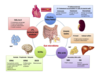
Most common cancer metastasis

  1. lungs, liver, bone, lymph nodes
  2. lungs, liver, spleen, bone
  3. lungs, liver, brain, bone
  4. lungs, liver, heart, kidneys
A

lungs, liver, bone, lymph nodes

15
Q

Coagulative necrosis

  1. is typical of brain tissue
  2. is typical of cardiac tissue
  3. is related to a bacterial infection
  4. is never related to hypoxia
A

is typical of cardiac tissue

  • When many cells undergo necrosis at once, then definable patterns of necrosis are produced, depending upon the nature of the injury, the type of tissue, and the length of time.
  • This is an example of coagulative necrosis.
  • This is the typical pattern with ischemia and infarction (loss of blood supply and resultant tissue anoxia).
16
Q

Chronic inflammation is defined by

  1. platelets and neutrophils in the lesion
  2. the cellular components that are the main driver of the lesion
  3. lymphocytes, red cells and neutrophils in the lesion
  4. the vascular extravasation of plasma
A

the cellular components that are the main driver of the lesion

17
Q

Hyperplasia

  1. is synonym with hypertrophy
  2. means an increased number of cells
  3. means an increased size of cells
  4. is the same of eutrophy
A

means an increased number of cells

18
Q

Abscopal effect

  1. is caused by an immune response
  2. elicits an immune response
  3. does not use an intact immune system
  4. happens only when Nk cells are available
A

elicits an immune response

19
Q

Humans

  1. acquire their first microbiome in utero
  2. acquire their first microbiome at birth
  3. acquire their first microbiome few days after birth
  4. acquire their first microbiome few years after birth
A

acquire their first microbiome at birth

20
Q

Fibrosis of the liver

  1. takes stage before cirrhosis
  2. precedes liver tumor
  3. precedes hepatitis B or C
  4. takes stage after cirrhosis
A

takes stage before cirrhosis

21
Q

Prognosis of a myocardial infarction

  1. is worse with right coronary dominance
  2. is better with right coronary dominance
  3. is better with left coronary dominance
  4. is the same irrespective of coronary dominance
A

is better with right coronary dominance

22
Q

Maximal blood perfusion is achieved in the heart wall

  1. during systole
  2. at the end of systole
  3. at the end of diastole
  4. during diastole
A

at the end of diastole

23
Q

Damage due to hypoxia in the myocardium

  1. stars always from the epicardial layers
  2. starts always in the endocardial layers
  3. starts always in the middle layers
  4. start often in the endocardial layers
A

starts always in the endocardial layers

24
Q

Oxygen extraction from blood in the coronaries

  1. is not maximal
  2. is maximal and cannot be modified by endogenous molecule
  3. is maximal and can be modified by endogenous molecules
  4. depends on NO secretion
A

is maximal and cannot be modified by endogenous molecule

25
Q

Oxygen supply in the heart wall

  1. depends solely on coronary vessel diameter
  2. depends also form oxygen extraction
  3. depends from both
  4. depends essentially form cardiac output
A

depends solely on coronary vessel diameter

26
Q

Nutmeg liver

  1. occurs mostly in right Heart Failure (HF)
  2. occurs only in left HF
  3. occurs in both cases equally
  4. occurs acutely
A

occurs mostly in right Heart Failure (HF)

27
Q

Right chronic heart failure

  1. typically does not show a systemic venous congestion
  2. shows only a neck venous congestion
  3. typically does have an early pulmonary congestion
  4. typically shows a systemic venous congestion
A

typically shows a systemic venous congestion

28
Q

Stroke volume in heart failure is by definition

  1. Increased
  2. Decreased
  3. Normal
  4. decreased only in the pulmonary circulation
A

Decreased

29
Q

Most common cause of heart failure

  1. ascending aortic aneurysm
  2. aortic valve stenosis
  3. aortic valve stenosis
  4. ischaemic heart disease
A

ischaemic heart disease

30
Q

Left chronic heart failure

  1. is determined by the impossibility to “move” venous blood returning form the periphery
  2. is determined by the impossibility to oxygenate venous blood in the lungs
  3. is determined by the impossibility to push blood in the pulmonary circulation
  4. is determined by the impossibility to “move” blood from lungs
A

is determined by the impossibility to “move” blood from lungs

31
Q

Myocardial oxygen extraction from blood at rest is

  1. is very low (20-30%)
  2. near maximal (60-80%)
  3. quite low (40-50%)
  4. maximal (100%)
A

near maximal (60-80%)

32
Q

Determinants of myocardial oxygen consumption

  1. heart rate and systolic pressure
  2. systolic pressure and left ventricular contractility
  3. heart rate, systolic pressure and diastolic pressure
  4. systolic pressure, heart rate, left ventricular contractility
A

systolic pressure, heart rate, left ventricular contractility

33
Q

Shortening diastole

  1. dampens coronary perfusion
  2. decreases contractility
  3. always increases cardiac output
  4. favours coronary perfusion
A

dampens coronary perfusion

34
Q

Sub-endocardial flow starts to decrease

  1. above a mean coronary pressure of 40 mm Hg
  2. below a mean coronary pressure of 50 mm Hg
  3. below a mean coronary pressure of 40 mm Hg
  4. below a coronary pressure of 20 mmHg
A

below a mean coronary pressure of 40 mm Hg

• Subendocardial flow occurs primarily in diastole and begins to decrease below a mean coronary pressure of 40 mm Hg. By contrast, sub-epicardial flow occurs throughout the cardiac cycle and is maintained until coronary pressure falls below 25 mm Hg.

35
Q

Nitric oxide (NO)

  1. is released by pericytes near the vascular wall
  2. is released by smooth muscle cells near the vascular wall
  3. is released by macrophages neat the vascular wall
  4. is released by endothelial cells of the vascular wall
A

is released by endothelial cells of the vascular wall

  • Nitric Oxide (Endothelium-Derived Relaxing Factor). NO is produced in endothelial cells by the enzymatic conversion of l-arginine to citrulline via type III NO synthase (NOS). Endothelial NO diffuses abluminally into vascular smooth muscle, where it binds to guanylyl cyclase, increasing cGMP production and causing relaxation through a reduction in intracellular calcium.
  • NO-mediated vasodilation is enhanced by cyclic or pulsatile changes in coronary shear stress. Chronic upregulation of NOS occurs in response to episodic increases in coronary flow, such as during exercise training, which also potentiates the relaxation to various endothelium-dependent vasodilators.
  • NO-mediated vasodilation is impaired in many disease states and in patients with one or more risk factors for coronary artery disease (CAD). This occurs via inactivation of NO by superoxide anion generated in response to oxidative stress.
  • Such inactivation is the hallmark of impaired NO-mediated vasodilation in atherosclerosis, hypertension, and diabetes.
36
Q

Sub-epicardial flow starts to decrease

  1. below a mean coronary pressure of 35 mm Hg
  2. below a mean coronary pressure of 45 mm Hg
  3. below a mean coronary pressure of 25 mm Hg
  4. no, it occurs throughout the cardiac cycle irrespective of mean coronary pressure
A

below a mean coronary pressure of 25 mm Hg

37
Q

Stenosis of a coronary tract becomes symptomatic

  1. when is more than 60% of the section area
  2. when is less than 40% of the section area
  3. when is more than 40% of the section area
  4. when is more than 95% of the section area
A

when is more than 60% of the section area

38
Q

Impaired NO-mediated vasodilation occurs

  1. only in high blood pressure subjects
  2. only during a heart attack
  3. only in diabetic patients in peripheral arteries and later on even in coronaries
  4. in several diseases such as arteriosclerosis, diabetes and high blood pressure
A

in several diseases such as arteriosclerosis, diabetes and high blood pressure

39
Q

Irreversible injury begins in the myocardium

  1. after 20 minutes from hypoxia
  2. after 5 minutes from hypoxia
  3. after 15 minutes from hypoxia
  4. after 2 hours from hypoxia
A

after 20 minutes from hypoxia

40
Q

Renal release of renin during heart failure

  1. is mainly stimulated by reduced renal perfusion pressure
  2. is mainly stimulated by Na and liquid retention
  3. is mainly stimulated by the sympathetic nervous system
  4. is mainly stimulated by diuretic therapy
A

is mainly stimulated by reduced renal perfusion pressure

mechanoreceptors detect under-filling (left ventricle, aortic arch, carotid sinus, renal afferent arterioles)

afferent impulses via vagus & glossopharyngeal nerves to the cardio-regulatory centres (medulla) causing cerebral response via

sympathetic motor nerves resulting in:
– tachycardia, myocardial contractility, vasoconstriction
(arterial & venous)
– release of renin & angiotensin = renal vasoconstriction
– direct (non-osmotic) release of vasopressin from hypothalamus

Net effect: increased total body sodium, increased total body H2O (up to 25%)

41
Q

Left ventricular remodeling in heart failure

  1. does not change the shape of the heart
  2. change shape and function of the left ventricle
  3. is a compensatory event
  4. is not an adaptive event
A

change shape and function of the left ventricle

42
Q

Distributing arteries

  1. are the major target of arteriosclerosis
  2. they have a minimal layer of smooth muscle cells
  3. they have the biggest layer for smooth muscle cells
  4. they have a thin wall
A

they have the biggest layer for smooth muscle cells

43
Q

Conducting arteries

  1. are elastic
  2. no smooth muscle cells layer is present
  3. they are not a target of arteriosclerosis
  4. they have a very thick wall
A

are elastic

44
Q

In any given moment in the body most of the blood volume

  1. is contained in the arteries of the upper body
  2. is in the conducting arteries
  3. is in the distributing arteries of the lower part of the body
  4. is in the veins
A

is in the veins

45
Q

Two pivotal hormones that directly increase blood pressure

  1. Vasopression and testosterone
  2. Angiotensin II and epinephrine
  3. Angiotensin II and calcitonin
  4. Vasopressin and osteocalcin
A

Angiotensin II and epinephrine

46
Q

Foamy (or foam) cells

  1. are macrophages filled with fatty acids
  2. are lymphocytes filled with fatty acids
  3. are platelets filled with fatty acids
  4. are fibroblasts filled with fatty acids
A

are macrophages filled with fatty acids

47
Q

Oxidation of Low Density Lipoproteins (LDLs) is the major cause of

  1. endothelial wall damage
  2. foamy cells formation
  3. smooth muscle cells damage
  4. platelet activation
A

foamy cells formation

48
Q

Inflammatory mediators of arteriosclerosis include

  1. Interleukins 4 and 5
  2. Interleukin 2
  3. Interleukin 1beta
  4. Interleukin 3
A

Interleukin 1beta (followed by IL-6)

“By blocking IL-1β — the main circulating form of IL-1 — canakinumab blocks this pivotal mediator of inflammation, which is produced when cholesterol crystals activate the NLRP3 inflammasome as well as when oxidized LDL are produced locally, likely with the contribution of other pathological mechanisms.

IL-1β then induces expression cascade of several pro-inflammatory molecules, such as IL-6, a cytokine that triggers production of CRP and that has been implicated directly in atherosclerotic development and plaque rupture.”

“ …We have to highlight and acknowledge the definitive proof of the inflammatory hypothesis in atherosclerosis pathogenesis and its natural history in a clinical setting: this knowledge potentially opens to a plethora of a new clinical research era that can modify further the outcome of these still deadly diseases.”

49
Q

TMAO (trimethylamine N-oxide)

  1. increases macrophage infiltration in the atherosclerotic wall
  2. increases the levels of oxidized LDLs in the blood
  3. increase smooth cells replication in the vessel wall
  4. increase platelet reactivity and thrombosis potential
A

increase platelet reactivity and thrombosis potential

Trimethylamine N-oxide (TMAO) is a gut microbiota-derived metabolite that enhances both platelet responsiveness and in vivo thrombosis potential in animal models, and TMAO plasma levels predict incident atherothrombotic event risks in human clinical studies. TMAO is formed by gut microbe-dependent metabolism of trimethylamine (TMA) moiety-containing nutrients, which are abundant in a Western diet. Here, using a mechanism-based inhibitor approach targeting a major microbial TMA-generating enzyme pair, CutC and CutD (CutC/D), we developed inhibitors that are potent, time- dependent, and irreversible and that do not affect commensal viability. In animal models, a single oral dose of a CutC/D inhibitor significantly reduced plasma TMAO levels for up to 3 d and rescued diet-induced enhanced platelet responsiveness and thrombus formation, without observable toxicity or increased bleeding risk. The inhibitor selectively accumulated within intestinal microbes to millimolar levels, a concentration over 1-million-fold higher than needed for a therapeutic effect. These studies reveal that mechanism-based inhibition of gut microbial TMA and TMAO production reduces thrombosis potential, a critical adverse complication in heart disease.

50
Q

TMAO (trimethylamine N-oxide)

  1. is produced by gut microbiota
  2. is in the dairy foods
  3. is not depending on the type of diet of the subject
  4. is linked to sugar-rich drinks
A

is produced by gut microbiota

51
Q

Pathophysiology

  1. studies the causes of disease and related functional changes
  2. studies the mechanisms of disease and related functional changes
  3. studies the therapy of diseases and related functional changes
  4. studies the prognosis of diseases and related functional changes
A

studies the mechanisms of disease and related functional changes

52
Q

Chronic bronchitis

  1. is an example of obstructive lung disease
  2. is an example of restrictive lung disease
  3. is an example of restricted and obstructive lung disease
  4. is not leading usually to COPD
A

is an example of obstructive lung disease

53
Q

An example of restrictive lung disease phenotype not dependent from pulmonary disease

  1. pulmonary fibrosis
  2. silicosis
  3. obesity
  4. pleurisy (pleuritis)
A

obesity

54
Q

Allergic asthma

  1. is an example of restrictive lung disease
  2. is an example of obstructive lung disease
  3. is caused only by smoking
  4. is a rare condition
A

is an example of obstructive lung disease

55
Q

FEV1s/FVC ratio

  1. is always 1 in obstructive lung disease i
  2. s always less than 0.4 in obstructive lung disease
  3. is always less than 0.8 in obstructive lung disease
  4. is always less then 0.5 in restrictive lung disease
A

is always less than 0.8 in obstructive lung disease

56
Q

An acute cause of restrictive lung disease is

  1. acute bronchitis
  2. asthma
  3. CO inhalation
  4. Acute respiratory distress syndrome (ARDS)
A

Acute respiratory distress syndrome (ARDS)