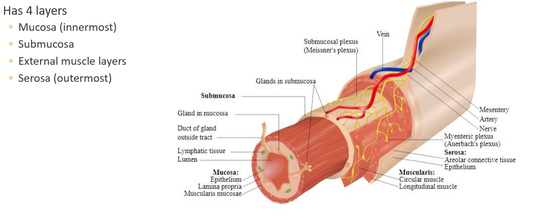
State the four layers of the GI tract innermost to outermost
State 4 roles of the epithelial layer of the mucosa
It is a selectively permeable barrier which:
- Facilitates transport and digestion of food
- Promotes absorption
- Produces hormones
- Produes mucus
State the three layers of the mucosa innermost to outermost
- Epithelium
- Lamina propria
- Muscularis muosae
State the main role(s) of the lamina propria in the mucosa
Has lots of lymphoid tissue and macrophages to produce antibodies to protect against bacterial or viral infection
What antibodies are mainly produced by lamina propria of mucosa and why?
IgA as reistant to proteases
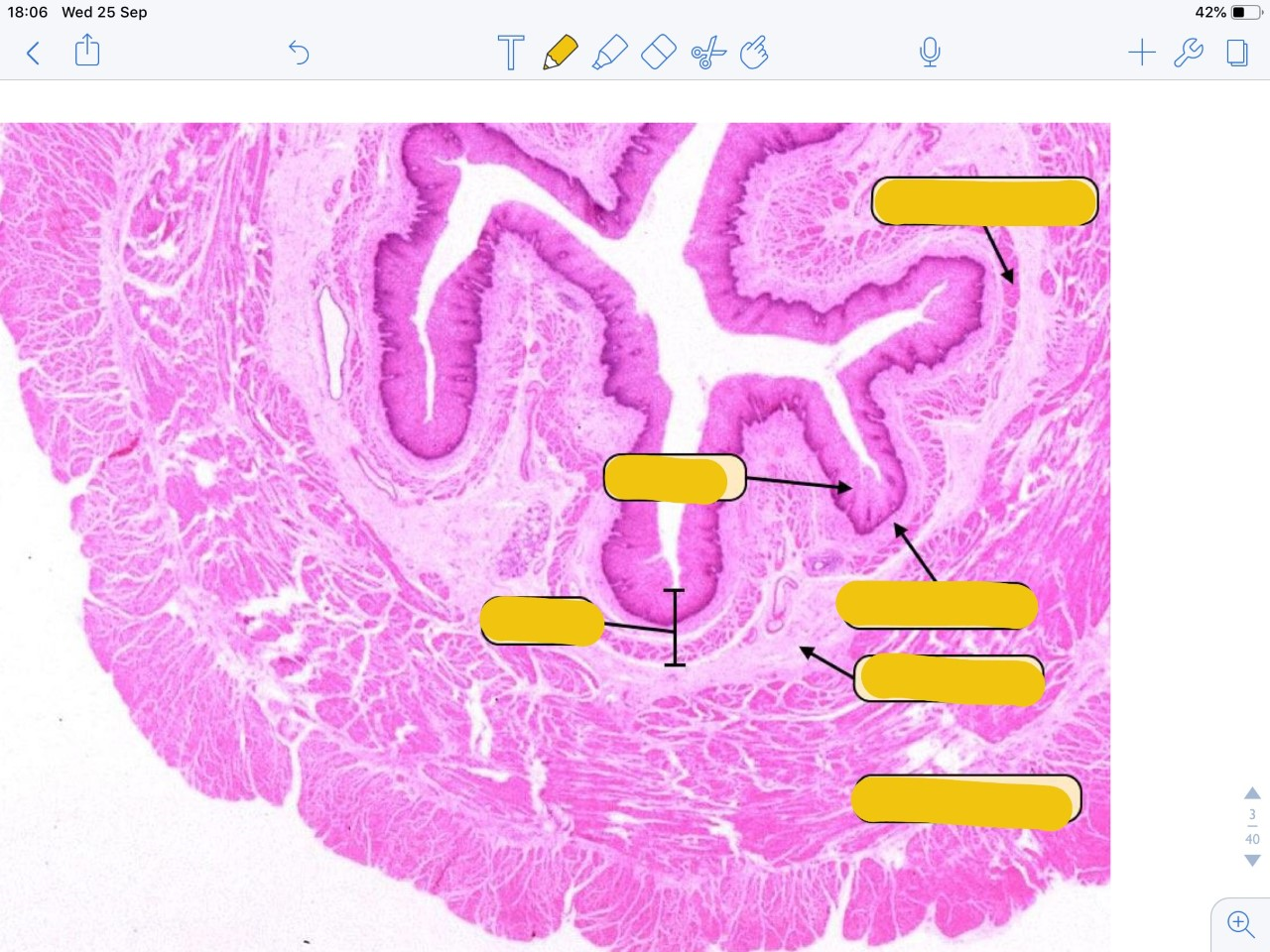
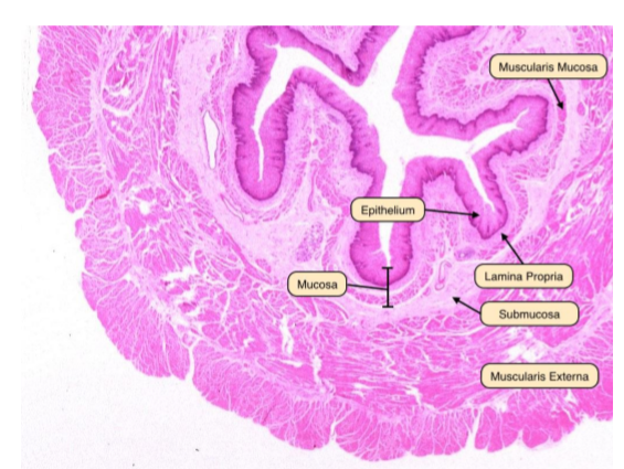
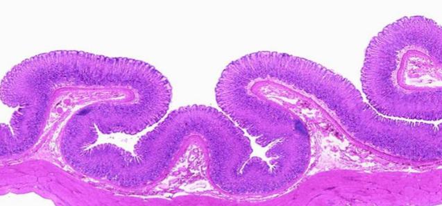
Label the following image
State 2 roles of the muscularis mucosa layer of the mucosa
Layers of smooth muscle orientated in different directions which:
- Keep epithelium in contact with gut contents
- Help keepy crypt contents dynamic
State the contents of the submucosa
- Dense connective tissue
- Blood vessels
- Glands
- Lymphoid tissue
- Submucosal (Meissner’s) plexus
Describe the arrangement of muscle fibres in the muscularis propria and state what is found betwen the two layers
- Outer longitudinal muscle (shorten gut)
- Myenteric (Auerbach’s plexus)
- Inner circular muscle (peristalsis)
State 3 contents of the serosa
- Blood vessels
- Lymph vessels
- Adipose tissue
The serosa is continuous with mesenteries; true or false
True
Give a brief overall description of layers, and sublayers, of the GI tract

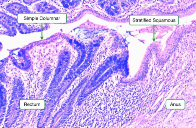
What epithelia is found in the alimentary canal? CLUE: does it change as you go down the alimentary canal
- Stratified squamous in oesophagus and distal anus
- Everything inbetween is simple columnar
What type of epithelial cell is an enterocyte and what does it do?
Simple columnar epithelial cell that absorbs
Why must blood vessels and lymphatics lie immediately below enterocyte in the lamina propria?
Nutrients must be transported through both apical and basolateral membrane to be absorbed
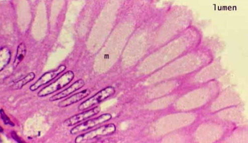
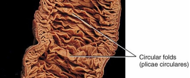
State 3 anatomical features of small intestine to increase surfacea area
- Plicae circulares (permanent folds)
- Villi
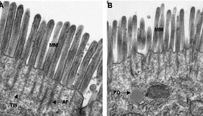
- Microvilli
What is meant by the brush border?
The microvilli (increases surface area but also contains digestive enzymes)
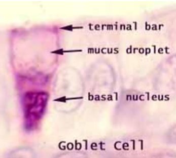
Where do you find goblet cells in GI tract?
Scattered between enterocytes increasing in number from duodenum to colon
Why is nucleus of a goblet cell at the base?
Mucus compresses nucleus
Mucus protects epithelia from what three things?
- Friction (it is a lubricant)
- Chemical damage (environment can be acidic)
- Bacterial inflammation (physical barier)
Where are foveolar cells found and what is their role?
Foveolar cells lien gastric mucosa and secrete mucus, which contains HCO3, which acts as a barrier against stomach acid
What are the permanent folds in the small intestine called?
Plicae circulares
What are rugae and why are they needed?
Rugae are temporary folds in the stomach which allow it to expand
What are haustra and where are they found?
Haustra are small pouches/sacculations which gives colon segmented appearance. As a result of contraction of longitudinal muscle


-
1a.) Purpose of Gut37
-
1b.) Basic Anatomy60
-
2a.) Hernias40
-
2b & 3a.) Embryology75
-
3b.) Salivation & Swallowing36
-
4a.) Intro to Stomach43
-
4b.) Pathology of Stomach40
-
5a.) Chyme, Liver & Pancreas56
-
5b.) The Intestines55
-
6a.) Liver & Biliary System62
-
6b.) Jaundice & LFTs30
-
7a.) Large Intestine/Inflammatory Bowel Disease44
-
7b.) Distal GI Tract Pathology73
-
8a.) GI Malignancy74
-
9b.) GI Emergencies63
-
9a.) GI Infections59
-
10a.) Imaging of GI Tract55
-
11a.) Endoscopic Tour of GI Tract8
