Heart Flashcards
Briefly explain the portal circulation
- venous blood from unpaired abdominal organs (stomach, intestine, pancreas, spleen) absorbed by intestine
- carried to v. portae
- collected in capillary bed in liver
- collected by vv. hepaticae
- conveyed to v. cava inferior
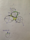
1 - 5

1) apex cordis
2) basis cordis
3) sulcus coronarius
4) sulcus interventricularis anterior
5) sulcus interventricularis posterior
6 - 10
How is #8 called inside?

6) truncus pulmonalis
7) sinus trunci pulmonalis
8) conus arteriosus → inside: infundibulum cordis
9) atrium dextrum
10) ventriculus dexter
11 - 14

11) aorta
12) v. cava superior
13) auricula sinistra
14) auricula dextra
1 - 5

1) aorta ascendens
2) arcus aorticus
3) truncus pulmonalis
4) a. pulmonalis sinistra
5) truncus brachiocephalicus
6 - 10

6) a. carotis communis dextra
7) a. subclavia dextra
8) a. carotis communis sinistra
9) a. subclavia sinistra
10) lig. arteriosum
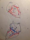
1 - 5

1) arcus aorticus
2) aorta descendens
3) aorta ascendens
4) truncus brachiocephalicus
5) a. carotis communis sinistra
6 - 10

6) a. subclavia sinistra
7) v. cava superior
8) a. pulmonalis dextra
9) a. pulmonalis sinistra
10) v. cava inferior
11 - 15

11) vv. pulmonales dextrae
12) vv. pulmonales sinistrae
13) atrium dextrum
14) atrium sinistrum
15) sulcus coronarius
16 - 20

16) sinus coronarius
17) sulcus terminalis
18) auricula sinistra
19) sinus transverus pericardii
20) ventriculus sinister
21 - 25

21) ventriculus dexter
22) sulcus interventricularis posterior
23) apex cordis
24) basis cordis
25) truncus pulmonalis
1 - 5

1) v. cava superior
2) v. cava inferior
3) sinus coronarius
4) vv. pulmonales dextrea
5) vv. pulmonales sinistrae
6 - 10
What forms #6?

6) crista terminalis → outside: sulcus terminalis
7) mm. pectinati
8) sinus venarum cavarum
9) valvula Eustachii
10) valvula Thebesii
11 - 15
Give another name for #11.

11) valva artrioventricularis dextra → tricuspid valve
12) chordae tendinae
13) m. pappilaris anterior
14) m. pappilaris septalis
15) m. pappilaris posterior
16 - 20
Give another name for #17.

16) trabeculae carnae
17) trabecula septomarginalis → moderator band
18) septum interventriculare pars muscularis
19) septum interventriculare pars membranacea
20) septum artrioventriculare
22 - 25
Give another name for #25.

22) septum interartriale
23) limbus fossae ovalis
24) fossa ovalis
25) valva artrioventricularis sinistra → bicuspid valve
26 - 28

26) m. pappilaris anterior
27) m. pappilaris posterior
28) thicker wall of right ventricle
Describe the structure of a semilunar valve
Give another name for the smallest part
- pars densa: lower part
- pars flaccida: upper part
- 2 lunuli: thickened edges
- 1 nodulus (=corpus Arantii): closes cusps
The inflow and outflow tract of the right ventricle are divided by.. ?
- crista supraventricularis
- trabecula septomarginalis
The inflow and outflow tract of the left ventricle is divided by.. ?
How is the structure called?
anterior cusp of bicuspid valve
→ vestibulum aortae
Which vessel leaves the left ventricle?
aorta
What are the 3 layers that make up the heart wall?
- endocardium
- myocardium
- epicardium
Which structures make up the myocardium?
- atrial muscle
- ventricular muscle
Differentiate btw atrial muscle layers
- deep layer: surrounding each atrium
- superficial layer: covers both atria
Differentiate btw ventricle muscle layers
- stratum subendocardiale: forms trabeculae carnae, mm. papillares, sulcus coronarius, sulcus interventricularis ant./post
- stratum musculare: only septum interventr., wall of left ventricle
- stratum subepicardiale: forms vortex cordis
Describe the structure of the endocardium
= continuation of inner layer of wall vessels, therefore:
- endothelium
- conn. tissue
Describe the structure and function of the epicardium
- mesothelium
- conn. tissue
- adipose tissue → smoothes unevenness of heart surface
What is the function of the cardiac skeleton?
- electrical insulation of atria/ventricles
- attachment site for:
- myocardium
- leaflets and cusps of valves
- keeps orifices of valves stable
1 - 5

1) valva atrioventricularis dextra
2) valva atrioventricularis sinistra
3) valva aortae
4) valva pulmonalis
5) cuspis anterior
6 - 10

6) cuspis posterior
7) cuspis commisuralis
8) cuspis anterior
9) cuspis posterior
10) cuspis septalis
11 - 15

11) cuspis semilunaris sinistra
12) cuspis semilunaris dextra
13) cuspis semilunaris posterior
14) cuspis semilunaris dextra
15) cuspis semilunaris anterior
16 - 22

16) cuspis semilunaris sinistra
17) sinus trunci
18 - 20) sinus aorticus
21) a. coronaria dextra
22) a. coronaria sinistra
23 - 27
Another name for #27?
23) anulus fibrosus dexter
24) anulus fibrosus sinister
25) tendo coni arteriosi
26) trigonum fibrosum dextrum
27) fasciculi atrioventricularis → bundle of HIS
28 - 30

28) trigonum fibrosum sinistrum
29) lunulus valvulae semilunaris
30) nodulus valvulae semilunaris
1 - 5

1) v. cava superior
2) truncus pulmonalis
3) aorta
4) v. cava inferior
5) apex cordis
6 - 11

6) apex cordis
8) a. coronaria sinistra
9) a. coronaria dextra
10) r. interventricularis anterior
11) r. interventricularis posterior
12 - 16

12) r. circumflexus
13) r. marginalis dexter
14) r. marginalis sinister
15) rr. atriales
16) r. atrialis anastomicus
17 - 21
Another name for #21?
17) rr. ventriculares
18) r. lateralis
19) rr. interventriculares septales
20) rr. atrioventriculares
21) v. obliqua atrii sinistra (= Marshall vein)
22 - 25

22) v. cordis magna
23) sinus coronarius
24) v. cordis parva
25) v. cordis magna
1 - 5
Another name for #1 - 3

1) nodus sinuatrialis (= SA-node)
2) nodus atrioventricularis (=AV-node)
3) fasciculus atrioventricularis (=bundle of HIS)
4) crus dextrum
5) crus sinistrum
6 - 8
Another name for #6

6) trabecula septomarginalis (= moderator-band)
7) Purkinje fibers
8) septum interventriculare pars muscularis
1 - 5
Give another name for #4

1) pericardium serosum → lamina parietalis/visceralis
2) pericardium fibrosum
3) porta arteriosa
4) porta venosa (= Sappey’s T)
5) truncus pulmonalis
6 - 10

6) aorta
7) vv. pulmonales dextrae
8) vv. pulmonales sinistrae
9) v. cava superior
10) v. cava inferior
11 - 15
What is the function of #13?
Which additional nerve not shown on the drawing innervates the pericardium?

11) sinus obliquus pericardii
12) n. phrenicus
13) a. + v. pericardiophrenica → blood supply of pericardium
14) n. vagus
15) lig. arteriosum
→ truncus sympathicus
16 - 18

16) centrum tendineum (caudal border)
17) cavitas pleuralis (lateral borders)
18) sinus transversus pericardii
1 - 5

1) facies sternocostalis
2) facies diaphragmatica
3) facies posterior cordis → mainly: left atrium
4) facies pulmonales
5) mediastinum posterius
6 - 10

6) mediastinum medium
7) mediastinum anterius
8) pericardium
9) heart
10) esophagus
12 - 14

12) lung
13) pleura / cavitas pleuralis
14) radix pulmonis
1 - 5

1) intercostal space
2) sulcus coronarius, left edge
3) apex cordis
4) sulcus coronarius, right edge / v. cava inferior, right margin
5) v. cava superior, right margin
6 - 10

6) 2 cm
7) midclavicular line
8) 2 cm
9) 2 cm
10) right atrium
11 - 15
Give another name for #12

11) v. cava superior
12) arcus aorticus, aortic knuckle
13) truncus pulmonalis
14) auricula sinistra
15) left ventricle
16 - 20
Give another name for #18
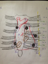
16) auscultation site: valva aortae
17) auscultation site: valva pulmonis
18) auscultation site: punctum quintum (= Erb’s point)
19) auscultation site: bicuspid valve
20) auscultation site: tricuspid valve
21 - 23

21) valva trunci pulmonis/aortae
22) bicuspid/tricuspid valve
23) sulcus coronarius