GI system embryology Flashcards
Which arteries supply the different parts of the gut?
unpaired branches of abdominal aorta
- truncus coeliacus → foregut
- a. mesenterica sup. → midgut
- a. mesenterica inf. → hindgut
What are the derivatives of the foregut?
pharynx → ampulla pancreatica
- pharynx
- lower resp. system
- esophagus + stomach
- duodenum proximal to opening of bile duct
- liver + biliary apparatus + pancreas
How does the esophagus develop?
Where do its structures originate from?
- seperation from trachea by treacheoesophageal septum (cf. flashcards for resp. system)
- obliteration of lumen + recanalization
orgin:
- endoderm → epithelium + glands
- 4th + 6th pharyngeal arch → striated m.
- splanchnic mesenchyme → smooth m.
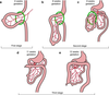
How does the stomach develop?
slight dilation in caudal part of foregut in median plane
- dorsal border grows faster than ventral
→ curvatura gastrica major - rotation 90° clockwise around longitudinal axis
Explain the innervation of the stomach w/r/t its rotation
since:
- left side → ventral surface
→ ant. wall innervated by truncus vagalis sin. - right side → dorsal surface
→ post. wall → innervated by truncus vagalis dex.

How do you call the structure attaching to the stomach?
Where else does it attach?
What does it eventually form?
dorsal mesogastrium
- attaches to: post. abdominal wall
- contains: spleen, celiac artery
- forms: omental bursa
ventral mesogastrium
-
attaches to: ant. abdominal wall
- duodenum to liver
How is the omental bursa formed?
Which structures does it form?
clefts forming in dorsal mesogastrium coalesce and form foramen omentale, demarcates opening to omental bursa (sac formed during rotation of stomach)
forms:
- superior recess persists dorsal to right lung
- inferior recess → disappears as layers of omentum majus fuse
How does the duodenum develop?
section around junction btw foregut and midgut
- forms C-shaped loop (= ansa duodenalis)
- pushed into retroperitoneal position by developing stomach (not pars superior!) → sec. retroperitoneal
- lumen obliterates → recanalization
How do the liver and biliary apparatus develop?
outgrowth from distal part of foregut (= hepatic diverticulum) extends into septum transversum
- enlarges and divides into 2 parts btw ventral mesogastrium
- cranial part: liver primordium
* - caudal part:* gall bladder primordium
- stalk of caudal part: ductus cysticus
- stalk connecting both parts: common bile duct - future common bile duct moves to dorsal aspect of duodenum as duodenum grows
How does the “interior” of the liver develop?
endodermal cells → cords of hepatocytes + endothelial lining of intrahepatic part of biliary apparatus
→ hepatic cords anastomose around hepatic sinusoids
When does hematopoiesis in the liver begin?
6th week → bright reddish color to the liver
When does bile formation begin?
How do you call the dark green intestinal contents?
12th week
meconium
The ventral mesentery gives rise to which structures?
- omentum minus (= lig. hepatogastricum, lig. hepatoduodenale, lig. hepatoesophageale)
- lig. falciforme
- lig. coronarium
How does the pancreas develop?
endodermal cells from caudal end of foregut form pancreatic buds btw the layers of the mesentery
- ventral pancreatic bud (near entry of bile duct)
- dorsal pancreatic bud (larger + more cranially)
- duodenal rotation carries ventral pancreatic bud dorsally → posterior to dorsal pancreatic bud
- pancreatic buds fuse (ventral pancreatic bud → proc. uncinatus)
- pancreatic ducts fuse
Which pancreatic bud is responsible for which pancreatic duct?
ventral pancreatic bud:
- ductus pancreaticus
dorsal pancreatic bud:
- proximal part → ductus pancreaticus acc.
- distal part → ductus pancreaticus
How does the first part of the intestines develop?
- midgut enlarges
- physiological umbilical hernia
- rotation of midgut loop
- retraction of intestinal loops
- fixation of intestines
meanwhile: cecum and appendix develop