Uro.... system Flashcards
Describe the general structure of the kidney.
What is its peritoneal relation?
Size and shape.?
retroperitoneal organ
- 120 - 200g
- 10 - 12cm long, 5-6cm wide, 4cm thick
2 different types of structures:
- urine formation: nephrons + uriniferous tubules
- urine collection: renal calyces + renal pelvis
What are the functions of the kidney?
-
homeostasis of bodily fluids (amount, osmolarity, pH, ion concentration)
∽ 180l primary urine filtered out of the blood/day -
excretion of metabolic end products (urine)
1.5 - 2l final urine formed/day - endocrine function (renin-angiotensin, vit D3, erythropoietin)
1 - 5

1) extremitas superior
2) extremitas inferior
3) hilum renale
4) sinus renalis
5) pyramidis renalis
6 - 10
Another name for #8.

6) capsula fibrosa
7) capsula adiposa
8) fascia renalis (= GEROTA)
9) medulla renalis
10) cortex renalis
11 - 13

11) papilla renalis
12) columna renalis
13) lobus renalis
What is a floating kidney?
What are other names?
loss of capsula adiposa → hypermobility of the kidney which descends into the pelvis
-
other names:
nephroptosis, nephroptosia, renal ptosis, renal descensus, renal prolapse
Which structures can be found in the cortex?
radii medullares (= medullary rays) = continuation of medullary substance
cortex corticis = contains radii medullares
labyrinthus corticis = area btw radii medullares
What are the projections of the kidney?
in fossa lumbalis
- right: Th12 → L3
- left: Th11 → L3
- hilum: L2
⇒ left kidney moves (2-3 cm) during deep inspiration
What are the boundaries of the kidney?
- laterally - medially
- cranially
- dorsally
laterally:
- 6) m. transversus abdominis
medially:
- 8) m. psoas major
cranially:
- diaphragm
- suprarenal gll.
dorsally:
- 7) m. quadratus lumborum
- n. subcostalis
- n. iliohypogastricus
- n. ilioinguinalis

What are the ventral boundaries of the kidney?
Differentiate btw right/left.
right:
- 1) right lobe of the liver
- 2) pars descendens duodeni
- 3) flexura coli dextra
- 4) loops of small intestine
left:
- 6) stomach
- 7) spleen
- 9) pancreas
- 10) flexura coli sinistra
- 11) radix mesocolica
- 12) loops of small intestine

What are the dorsal relations of the kidney clinically relevant?
contact to n. ilioinguinalis + n. iliohypogastricus explains why pain can spread up to the inguinal region in case of renal diseases
Explain the segmentation of the kidney.
5 segments
- segm. superius
- segm. anterius superius
- segm. anterius inferius
- segm. inferius
- segm. posterius
⇒ correspond to the arterial supply via a. renalis
What is the microscopic unit of the kidney?
Describe it.
nephron
- renal corpuscle
- glomerulus
forms together with renal tubule the uriniferous tubule
List some genetic abnormalities of the kidney.
- additional kidneys
- horseshoe kidney = fused kidneys
- renal aplasia = one kidney is missing
- renal hypoplasia = underdevelopment of one kidney
Explain the vasculature of the kidney
arterial supply:
- a. renalis dex./sin.
-
type I r. principalis ant./post.
* *type II** r. principalis ant./post./inf. - aa. interlobulares
- aa. arcuatae
- aa. corticales radiatae (= aa. interlobulares)
- arteriolae afferentes
drainage: beginning at glomerulus
- arteriolae efferentes
- vasa recta/peritubular cap.
- vv. corticales radiatae
- vv. arcuatae interlobulares
- vv. renales
- IVC
What are clinically important abberations of the renal vasculature?
- accessory renal aa.: persisting branches of aorta that didn’t redevelop during fetal dev., esp. important in case of surgeries
- abberant renal aa.: aa. don’t enter through hilum, but through sup./inf. pole
What is important abt v. renalis sin.?
Why is it clinically important?
3 tributaries:
- v. suprarenalis
- v. testicularis/ovarica
- v. phrenica inf.
⇒ cancer in the left renal v. can cause reflux into v. testicularis → dilation of scrotum (= varicocele)
What innervates the kidneys?
sympathetic innervation via plexus renalis
What are calices renalis?
Differentiate.
drain urine from papilla renalis into pelvis renalis
- major
- minor
What are the 2 shapes of the pelvis renalis?
type depends on calices renalis
volume: 3 - 8ml
- branching: minor calices open consistently into major which eventually open into pelvis renalis
- ampullary: minor and major calices open into pelvis renalis
What are the 2 ways for X-ray examinations to investigate the pelvis renalis?
- urogram: iodid containing contrast material injected intravenously, eventually excreted by kidney
- retrograde pyelogram: contrast material injected into ureters via a catheter
Which structures are connected by the ureter?
What are its 3 points of constriction?
pelvis renalis → urinary bladder
- exit from pelvis renalis
- crossing of a. iliaca communis/externa when entering the lesser pelvis
- wall of urinary bladder
Why are the points of constriction of the ureter clinically relevant?
⇒ renal colic = type of abdominal pain commonly caused by kidney stones, often wavelike in phases
What are the parts of the ureter?
- pars abdominalis in retroperitoneal space
- pars pelvica in lesser pelvis
- pars intramuralis in wall of urinary bladder
Explain the crossings of the ureter.
over - under - over - under
- over n. genitofemoralis
- under a./v. testicularis/ovarica
- over a. iliaca communis (left)/externa (right)
- under ductus deferens (male)/a. uterina (female)

What is important during a uterectomy?
a. uterina is close to ureter → injury/ligation of ureter can lead to loss of kidney
Which vessels supply and drain the ureter?
supply:
- pars abdominalis: a. renalis, aorta abdominalis, a. testicularis/ovarica, a. iliaca communis
- pars pelvica: a. iliaca interna, a. vesicalis inf. (in females also often a. uterina)
drainage:
→ v. testicularis → v. iliaca int. → plexus venosus vesicalis
Which structures surround the urinary bladder?
- paravesicular adipose tissue
- anteriorly spatium retropubicum (RETZIUS)
- posteriorly in males excavatio rectovesicalis
- in female*s excavatio vesicouterina
additionally posteriorly located in males from medial to lateral (cf. picture):
- ductus deferens
- gl. vesiculosa
- ureter
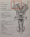
1 - 3
Origin, insertion, innervation.

m. levator ani
-
ORIGIN:
- m. pubococcygeus/-rectalis: ramus sup.
- m. iliococcygeus: arcus tendineus m. levatorius (fascia of m. obturator int.)
-
INSERTION:**
- m. pubo-/iliococcygeus: sacrum, coccyx
- m. puborectalis forms sling around rectum
-
INNERVATION:
- plexus sacralis (S3/4)
- m. puborectalis also by n. pudendus
1 - 5

1) pubic bone
2) ischium
3) membrana obturatoria
4) m. obturator int.
5) membrana perinei
6 - 10

6) vesica urinaria
7) prostate
8) urethra - pars prostatica
9) urethra - pars intramuralis
10) urethra - pars membranacea
11 - 15

11) urethra - pars spongiosa
12) m. sphincter urethrae ext.
13) m. levator ani
14) fossa ischioanalis
15) peritoneum
16 - 20
What is formed by #17?
Another 2 names for #20.

16) corpus spongiosum
17) corpus cavernosum → crura of penis
18) m. ischiocavernosus
19) m. bulbospongiosus
20) fascia perinei profunda (= GALLAUDET), investing layer of perineal fascia
21 - 25
Another 2 names for #22. What is it continuous with?
What does #25 partially form?

21) skin
22) fascia perinei superficialis (= COLLES), membraneous layer of perineal fascia → continuous w/ fascia penis sup., tunica DARTOS
23) spatium superficiale perinei
24) spatium profundum perinei
25) m. transversus profundus (part of urogential diaphragm)
26 - 29

26) trigonum vesicae
27) ostium uteris
28) saccus subcutaneus perinei
29) paracystium
1 - 5

1) ischium
2) pubis
3) membrana obturatoria
4) m. obturator int.
5) membrana perinei
6 - 10
Another name for #10.

6) vagina
7) cervix
8) fornix lat.
9) vestibulum vaginae
10) ostium uteri (= ext. os)
11 - 15
Another 2 names for #15

11) m. levator ani
12) m. sphincter urethrae ext.
13) m. compressor urethrae
14) peritoneum
15) lig. cardinale (MACKENRODT), lig. cervicale lat.
16 - 20
Which structure is formed by #18?

16) labia min.
17) labia maj.
18) corpus spongiosum (both form bulbus vestibuli)
19) m. bulbospongiosus
20) corpus cavernosum clitoridis
21 - 26
Another 2 names for #22, 23.

21) m. ischiocavernosus
22) fascia perinei profunda, investing layer of per. fascia (= GALLAUDET)
23) fascia perinei superficialis, membraneous layer of per. fascia (= COLLES)
24) lig. teres uteri (= remnant of gubernaculum)
25) spatium perinei profundum
26) spatium perinei superficiale