...genital sytem Flashcards
Differentiate btw male genetalia.
internal genetalia:
- testes + epidydimis
- ductus deferens + funiculus spermaticus
- accessory sex glands: prostate + gl. vesiculosa + gl. bulbourethralis (COWPER)
testes + epidydimus belong to int. genetalia bc they originate from abd. cavity, descended w/ peritoneal covering (cavitas serosa scroti) into scrotum
external genetalia:
- penis
- urethra
- scrotum
Briefly explain the function of the male genetalia.
- testes produce spermatozoa (∽ 74d)
- transported to epididymis → maturation (∽ 8-17d)
- pass through ductus deferens to urethra, sperms mixed with secretions of accessory sex glands
- leave body cavity through urethra
Describe the macroscopic structure of the testes.
- sup./inf. pole
-
epididymis
- 3 parts: caput, corpus, cauda
- fixed to sup. pole by lig. epididymis sup./inf.
- becomes ductus deferens at inf. pole
- covered by capsule = tunica albuginea
Briefly describe the microscopic structure of the testes.
What is their function?
seperated by septula testis into lobuli testes
→ contain seminiferous tubules
⇒ spermatozoa are produced in wall of seminiferous tubules
- in interstitium btw seminiferous tubules: LEYDIG cells → produce testosterone
Which remnants of embryological structures can be found in mature testes?
appendix testis:
- 3-4 mm wide at sup. pole
- remnant of MÜLLERIAN duct
appendix epididymis:
- at sup. pole of head of epidiymis
- remnant of WOLFFIAN duct
Which vessels supply/drain the testes?
Innervation?
supply:
-
a. testicularis (from aorta pars abdominalis)
orginate from lumbar region, follow during descencus
drainage:
-
plexus pampiniformis → unite in canalis inguinalis
→ v. testicularis dextra → v. cava inf.
→ v. testicularis sin. → v. renalis sin.
innervation:
- symph: plexus testicularis (rr. from plexus intermesentericus/renalis)
What is a varicocele and what might be the cause?
e. g. kidney tumors can grow into v. renalis → cause constriction of left v. testicularis →
* *dilation of plexus pampiniforis/vv. testicularis** → changes in blood circulation → reduced spermatic production
List the coats of the testes/scrotum from the innermost to the outermost layer.
-
tunica vaginalis = mesorchium
- epiorchium
- periorchium
- fascia spermatica int.
- m. cremaster
- fascia spermatica ext.
- scrotum - tunica DARTOS
- scrotum - skin
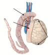
1 - 5
Which structures are formed by #2
No #4.
Another name for #3 and #5.

1) testes + epididymis
2) peritoneum → proc. vaginalis + deep ing. ring
3) epiorchium (visceral layer of tunica vaginalis)
5) periorchium (parietal layer of tunica vaginalis)
6 - 10

6) fascia transversalis abdominis
7) m. transv. abd.
8) m. obliq. int.
9) m. obliq. ext.
10) m. cremaster (prod. by #7 + #8)
11 - 16

11) fascia spermatica ext.
12) scrotum
13) tunica DARTOS
14) skin
15) cavitas serosa scroti
16) fascia spermatica int.
What is the function of tunica dartos?
movement of scrotal skin → temperature regulation (optimally 2 °C below body temperature)
Which structure can be found on the dorsal aspect in the middle of the scrotum?
raphe scroti = continuation of raphe perinei
What might be the reason for an innate inguinal hernia?
no obliteration of proc. vaginalis peritonei that forms tunica vaginalis testis
What is a hydrocele testis?
accumulation of fluid in cavitas serosa scroti → balloon-like enlargement
What can cause a testicular torsion and what are possible consequences?
thin mesorchium → testicular torsion → strangulation of blood vessels → irreversible damage to testes
What causes the cremaster reflex?
petting of inner surface of thigh → r. femoralis of n. genitofemoralis + cutaneous rr. of n. obturatorius → reflectory contraction of m. cremaster
What is cryptorchidism?
non-descent of testis into scrotum → stay in abd. cavity → high body temperature → damage to parenchyme of testes → no spermatic production
Which vessels supply/drain the scrotum?
Innervation?
supply:
- coats of testes: a. cremasterica (a. epigastrica inf.)
- scrotum: a. pudenda int.
drainage:
- v. pudenda ext. → v. saphena magna
- v. pudenda int. → v. iliaca int.
innervation:
- rr. scrotales of n. ilioinguinalis/n. pudendus
Which structures are connected by vas deferens?
How long is it, how thick?
Relate the structure of its wall to its function.
connects epididymis + urethra
- 35 - 40cm long
- 3mm thick
thick muscular layer → emission of sperms
What are the parts of vas deferens?
- pars epididymica ductus deferentis in inner aspect of epididymis
- pars funiculi spermatici in spermatic cord
- pars inguinalis in canalis inguinalis
- pars pelvica in lesser pelvis
then:
- ampulla ductus deferentis before entering prostate
- ductus ejaculatorius in prostate
- opens into colliculus seminalis of urethra
Which vessels supply/drain vas deferens?
Innervation?
supply:
- a. ductus deferentis (from a. umbilicalis)
drainage:
- plexus pampiniformis (cf. scrotal supply/innervation)
- *innervation:
- symph: plexus hypogastricus inf.
What are the layers of funiculus spermaticus?
from innermost to outermost
- fascia spermatica int
- m. cremaster
- fascia spermatica ext.
What are the contents of funiculus spermaticus?
-
vas deferens
- a. ductus deferentis
- plexus pampiniformis
- 2 aa. testiculares
- r. genitalis of n. genitofemoralis
- parasymph. fibers of plexus testicularis
- lymph vessels
<strong></strong><em>cf. histology flashcards</em>
Which structures accompany funiculus spermaticus?
- n. ilioinguinalis
- a. cremasterica
What are the 3 accessory sex glands in males?
What is their common function?
- paired gl. vesiculosa
- paired gl. bulbourethralis (COWPER)
- prostate
⇒ produce chief constituent of ejaculate
Where is gl. vesiculosa (= seminal vesical) located?
How big is it?
How much of the ejaculate is produced by it?
behind bladder, lateral to ampulla ductus deferentis
- 5cm long
- 1cm wide
- 1cm thick
⇒ produce 50-80% of ejaculate
How is gl. vesiculosa examined?
palpable via rectum
Which vessels supply/drain gl. vesiculosa?
supply:
- a. vesicalis inf.
- a. rectalis med.
- a. ductus deferentis
drainage:
- plexus venosus vesicalis/prostaticus
innervation:
- plexus hypogastricus inf.
<u>only difference btw supply/drainage innervation of gl. vesiculosa/prostate:</u><br></br>- <em>gl. vesiculosa:</em> a. ductus deferentis (since more cran.)<br></br>- prostate: a. pudenda int. (since more caud.)
What are the boundaries of the prostate?
How big is it, weight?
How much of the ejaculate is produced by it?
⇒ produces 15 - 30% of the ejaculate containing e.g. acid phosphatase
- 3cm long, 4cm wide, 2cm thick
- 20g
boundaries:
- ant: lig. puboprostaticum to pubic bone
- post: fascia rectoprostatica (DENONVILLIER) to rectum
- cran: base attaches to bladder
- caud: apex is sitting on pelvic diaphragm
Divide the prostate into zones.
- periurethral zone around urethra
- anteromedial zone anterior part of prostate (no glands)
- central zone encloses ductus ejaculatorii
- peripheral zone main mass laterally
- transitional zone btw central/peripheral zone

Why are the prostatic zones clinically relevant?
- benign prostate hyperplasia develops mainly in central/transitional zone (occurs in 50% of all men over 50)
- prostate carcinomas develop mainly in peripheral zone
⇒ impairs micturition due to occlusion of urethra
Which vessels supply/drain the prostate?
Innervation?
supply:
- a. rectalis med.
- a. vesicalis inf.
- a. pudenda int.
drainage:
- plexus vensosus vesicalis/prostaticus
innervation:
- plexus hypogastricus inf.
<u>only difference btw supply/drainage innervation of gl. vesiculosa/prostate:</u><br></br>- gl. vesiculosa: a. ductus deferentis (since more cran.)<br></br>- prostate: a. pudenda int. (since more caud.)
Where are gll. bulbourethrales located?
Another name?
COWPER glands on urogenital diaphragm
⇒ lubricate pars spongiosa of urethra
Which 2 structures attach the penis to the body?
- lig. fundiforme penis hooks around penis
- lig. suspensorium penis attaches at dorsum
⇒ both attach it to abd. wall / symphysis pubica
MNEMONIC: fundiforme = forms a fundus, “holds” penis
1 - 5

1) urethra pars intramuralis
2) urethra pars prostatica
3) urethra pars membranacea
4) urethra pars spongiosa
5) ostium ureteris
6 - 10
Another name for #10.
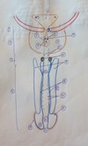
6) trigonum vesicae
7) colliculus seminalis
8) prostate
9) ductuli prostatici
10) gll. bulbourethrales (COWPER)
11 - 15

11) crura of penis
12) ductus gll. bulbourothrales
13) corpus cavernosum
14) glans penis
15) preputium
16 - 20

16) fossa navicularis
17) ostium urethrae ext.
18) corpus bulbospongiosum
19) corona glandis
20) crista urethralis
21 - 23
Another name for #23.

21) openings of ductuli ejacultorii
22) ostium urethrae int.
23) openings of gll. urethrales (LITTRE)
1 - 5

1) corpus cavernosum
2) corpus spongiosum
3) urethra
4) septum penis
5) tunica albuginea
6 - 10
Another name for #6.

6) fascia penis (BUCK)
7) subcutis
8) cutis
9) a. prof. penis
10) v. dorsalis prof. penis
11 - 13

11) v. dorsalis sup. penis
12) a. dorsalis penis
13) a. bulbi penis
1 - 5
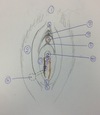
1) mons pubis
2) labium majus pudendi
3) labium minus pudendi
4) commissura labiorum ant.
5) commissura labiorum post.
6 - 10

6) frenulum labiorum pudendi
7) carunculae hymenales
8) rugae vaginales
9) crista urethralis vaginae
10) ostium urethrae ext.
11 - 14
Another name for #14.

11) frenulum clitoridis
12) glans clitoridis
13) preputium clitoridis
14) projection of gl. vestibularis maj. (BARTHOLIN)
1 - 5
Another name for #2.
What is the difference btw #4 and #5 besides their location?

1) excavatio vesicouterina
2) excavatio rectouterina (DOUGLAS pouch)
3) corpus uteri
4) cervix uteri - portio supravaginalis → attached to parametrium = paracervix
5) cervix uteri - portio vaginalis
6 - 10
Another name for #10.

6) endometrium
7) myometrium
8) perimetrium
9) fornix post. vaginae
10) ostium anatomicum uteri int. (= internal os)
11 - 15
Another name for #11, 13, 15.

11) ostium uteri (= external os)
12) cavitas uteri
13) spatium retropubicum (RETZIUS)
14) isthmus uteri
15) facies intestinalis/post.
16 - β
Another name for #16.
⍺ and β are pointing at the angles.

16) facies vesicalis/ant.
⍺) angle of anteflexio
β) angle of anteversio
1 - 5

1) lig. ovarii proprium
2) lig. teres uteri
3) mesometrium
4) mesovarium
5) mesosalpinx
6 - 10

6) lig. latum
7) lig. suspensorium ovarii
8) lig. cardinale
9) cervix portio supravaginalis
10) cervix portio vaginalis
11 - 15
Another name for #11.

11) pars uterina tubae (= intramural part)
12) isthumus tubae uterinae
13) ampulla tubae uterinae
14) infundibulum tubae uterinae
15) fimbriae
16 - 20

16) ureter
17) a./v. iliaca int.
18) a./v. uterina
19) r. vaginalis a. ut.
20) r. helicanus a. ut.
21 - 24

21) r. tubarius a. ut.
22) r. ovarius a. ut.
23) a./v. ovarica
24) plexus ovaricus