Lecture - Lower Limb Vessels and Nerves Flashcards
Explain this diagram:

Bascially, gray matter is H and the thinner ends of H is the posterior side.
Spinal nerve sits in intervetebral foramen and it’s called spinal nerve bc has sensory and motor axons. It will split into 2 things - posterior and anterior ramus (or dorsal and ventral ramus respectively).
The ventral or anterior ramus will supply all muscles except intrinsic and back muscles whereas the posterior or dorsal will only supply the intrinsic back muscles
What is nerve plexus?
Anterior rami of spinal nerves divide and recombine in nerve plexuses (plexi).
No thorax plexus. Plexus is a network/meshwork. But in thorax, the anterior ramus goes and forms the intercostial nerves in between ribs - formed from T1-T12 anterior ramus

What plexus is the lower limb supplied by?
It’s supplied by the lumbosacral plexus (which is L1-S4) and that is joined by lumbosacral trunck (L4,5).
Lumbar plexus (L1-L4) embedded in the psoas major whereas the sacral plexus formed on the piriformus muscle

What are the roots of the femoral nerve and what does it supply (skin and muscles)? How is it different to the obturator nerve?
Femoral nerve is L2, 3 and 4. These are the posterior divisions of the anterior rami whereas with the obturator nerve, it’s still got L2, 3 and 4 but they are the anterior divsions of anterior rami.
Femoral nerve is main nerve for anterior compartment of the thigh (illiopsoas, pectineus, sartorius, quads). It also supplies the anterior, medial thigh. and medial side of leg (because only one branch of femoral nerve goes below knee - saphenous nerve)

Obturator nerve: what roots, supplies what part of skin and muscles?
L2, 3 and 4 but they’re the anterior divisions of anterior rami.
Supplies medial thigh skin and it is the nerve to medial compartmennt (aka the adductors: obturator externus, adductor magnus, breven and lonugs plus gracillis)

Lateral femoral cutaneous nerve: what fibres does it have and what are its roots? Where does it go to?
It only has sensory fibres and roots are L2 and L3.
It is deep to inguinal ligament and supplies the skin of lateral aspect of thigh. It picks up the sensory information - sometimes the inguinal ligamment can become fibrotic and can squash this nerve so get funny sensation on lateral aspect of thigh lol

What is the black line, a and the bottom red line?

Black = lumbosacral trunk (L4,5)
A = sacral plexus (anterior rami S1-4) but like, lumbosacaral trunk is part of it sorta
red line = siatic nerve (L4,5 S1,2,3). It’s formed from sacral plexus and has 2 divisions:
- common fibular: goes to leg - anterior and lateral compartemnts
- tibial nerve: goes to posterior compartment of thigh and leg and foot (plantar muscles)
From the beginning, they are separte nerves but in one sheath = called sciatic nerve but then will split into two branches in popletial fossa area
Where do the gluteal nerves come from and what do they innervate?
They come from sacral plexus and they innervate gluteal muscles. They’re named in relation to the piriformis muscle (superior gluteal nerve is above the piriformis and inferior gluteal nerve is below the piriformis)

Now moving a little down, what does the siatic nerve supply? Which muscles and skin?
So remember that is has two divisions: common fibular and tibial nerve.
Common fibular: anterior and lateral leg
Tibial: posterior thigh, leg and foot
So overall: all muscles in leg and foot but only posterior thigh muscles
Skin: on gluteal region, posterior side of lower limb and. on lateral side of leg and foot and sole (femoral does anterior+medial skin on thigh and femoral’s saphenous does medial side of leg)

What is a dermatome and why is it useful?
What is a mytome?
Dermatome:
- area of skin supplied by a single spinal nerve
- useful for testing nerve root function
Mytome:
- Region or group of muscles innervated by efferent fibres from a single spinal cord level
- A particular myotome is mainly supplied by one or two) spinal nerves. It is not usually exclusively supplied by that spinal level

Memorise this.

In lumbar area - only 5 spinal nerves so stop at 5 and start at S1.
Now name them
What does this slide say?

Well that ligamanent is the sacrotuberous and it (plus the sacrospinous) forms the greater siatic formaen which the siatic nerve passes through.
When have intramuscular injection, want to be as far away from siatic nerve as possible and what you do is: Place index finger on ASIS and middle finger along iliac crest and inject in the triangle – green area
What is sciatica?
Bc siatic nerve has so many origin points, it can get compressed by disc prolapse. Discs are catrilaginous in between the vertebral bodies and the outer part is fibours and inner is nucelus so the inner can come out? And usually they protrude posterior-laterally so squash the nerves leqving the intervertbral foramen area.
Usually compresses bottom: so if protrusion in between L4 and L5 then it will squash L5 (the bottom one is squahed) and you get shooting pain

Again, what are the two branches/components of the siatic nerve and what do they supply?
The common fibular nerve - supplies two compartments - it’s divded after popliteal fossa (post part of knee) and goes past neck of fibular and divides into deep and superficial nerve. Deep goes to anterior aspect and supplies extensor muscles. Superficial goes to lateral where the fibularis longus and brevis are - everters are here

What if there is a fracture near neck of fibula (aka the common fibular nerve is damaged)
Causes foot drop due to:
- loss of dorsiflexsion (ant leg muscles)
- loss of eversion of foot (lateral leg muscles)

Arteries! Name these

- Abdominal aorta splits into common illiac arteries
- Internal illiac (split from common illiac) - supplies strucutes inside pelvis and gives a branch called obturatory artery - comes througn the obturator canal
- Obturator arterior - branch of internal illiac
- Deep artery of thigh: goes and supplies all compartment of thigh (ant, medial, post). But medial compartment gets help from obturator artery
- Femoral artery: Changes name after goes past inguinal ligament (from external illiac artery)

What are the labels?

Femoral artery: femoral artery conintues down deep to the sartorius muscle and tries to go towards posterior part of knee. Lies in area called subsartorial canal and then it also has a number of perforating branches and anastomoses with smaller branches
Adductor hiatus: Little hole in adductor magnus and it goes through this and into posterior part of knee joint and when it reaches apex of region (popliteal fossa), it becomes the popliteal artery (change name)

Label
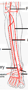
- Popliteal artery: At bottom of fossa, the artery divides into 2. So above interosseous membrane, there is a hole so the anterior tibial artery will go through to anterior of interossecous membrane. The posterior will continue behind the interessoeous membrane (supplies posterior compartment strucutres of leg)
- Fibular artery: Posterior gives off fibular artert Supplies strucutre in lateral compartmet (near fibula)
- Dorsalis pedis artery: Anterior changes name to thsi in front of ankle. Can palpate pulse here
- Medial and lateral plantar arteries: Once the post tibial arteru enters, it dives into two and one goes to medial plantar surface and other goes to other side

What does the popliteal fossa look like?
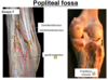
Label


Label the red arrows

- Superficial inguinal nodes: About 10 of these and they drain lymph from thigh etc and they lie on medial aspect of femoral vein and they can also drain into external illiac lymph nodes
- Lymphatics start from capilliaries bed and eventually they drain back into veinous system

What is the base, medial border, lateral border, roof, floor, apex and contents of femoral triangle?
Adductor canal:

In this, you have femoal artery, vein and a few nerves like saphenous nerve but not inside sheath
This canal goes through adductuor hiatus and conects to posterior part of knee joint/popliteal fossa
Read this slide and stuff
- Hernia can occur in femoal canal/ring of femoral sheath
- In female, hernia common bc wider pelvis so broader femoral ring so more prone to femoral hernias more
- Femoral hernias occur down there below the vein and artery
