Anatomic Variants and Pitfalls Flashcards
(55 cards)
What is the purpose of the Eustachian valve?
The eustachian valve (also known as the “valve of the inferior vena cava”) is a ridge of variable thickness in the inferior right atrium. It is a remnant of a fetal structure that directed incoming oxygenated blood to the foramen ovale and away from the right atrium.

Highly oxygenation fetal blood in the IVC goes directly to the coronaries via the Eusatchian valve thorough the fossa ovalis
Chiari networks can result in what?
Thromboembolic phenomenon

What is the Crista Terminalis thought to promote?
Atrial Tachyarrythmias
high density of adrenergic nerve fibers

Other than thromboembolic events, what are chiari networks also associated with? (Name 3)
- PFO
- Aneurysmal intraatriaal septum
- Atrial Arrythmias
What is the Thebesian Valve?
Valve of the Coronary Sinus

What is the problem with a prominent thebesian valve?
Problems with retrograde cardioplegia due to insertion of the cannula into the coronary sinus
What is another name for the Moderator Band?
septomarginal trabecula
What is another name for the Coumadin Ridge?
A coumadin ridge, also called warfarin ridge or left lateral ridge, is a band-like embryological remnant in the left atrium between the left superior pulmonary vein and the left atrial appendage. The ridge contains the ligament of Marshall, autonomic nerve bundle, and small atrial or sinoatrial node artery.
What is the primary problem with Cor Triatriatum Sinister from a physiological standpoint?
LV inlet obstruction
“Mitral stenosis like physiology”
What is the primary problem with Cor Triatriatum Sinister from a surgical standpoint?
Open-chamber procedure
What is the primary etiology with Cor Triatriatum Sinister?
Failure of resorption of common pulmonary veins during embryogenesis
Which Cor triatriatum is more common:
Sinister or Dexter?
Sinister > Dexter
Label 1
1 Eustachian Valve
Label 2

2 Septum Primum
Label 3

3 Septum Secundum
What can be associated with lipomatous hypertrophy?
Atrial Arrythmias
Where is the location of the fat that develops within the lipomatous hypertrophy?
Superior Interatrial Groove
AKA
Waterston’s Groove

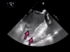
Identify the green structure
It’s “serpentine” and hypermobile on the echo clip

Chiari Network
Where does the Chiari network originate from?
Eustachian Valve
Are the chiari networks clinically significant?
No
but can be associated with PFO, Aneurysmal intraatrial septums or thromboembolic disease
What is the major echo differences from chiari network vs. eustachian valve
Chiari Network = Serpentine and Mobile
Eustachian valve = Thicker less mobile
What is the Eustachian valve an embryological remnaant of?
Sinus Venosus
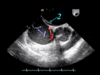
Identify 1

1 Crista Terminalis
Identify 2

2 Eustachian Valve