Cardiology Flashcards
(37 cards)
What are the signs of severity of aortic stenosis?
Vital signs:
- Narrow pulse pressure (and not usually hypertensive)
Palpation:
- Slow rising, low volume pulse (pulsus parvus et tardus)
- Palpable systolic / aortic thrill
- Heaving / pressure loaded character to apex beat which is not displaced (or displaced just a little)
Auscultation:
- Soft S2, or reverse splitting of S2
- A long, late-peaking ESM (not the volume of the murmur)
- The later the peak, the more severe (see below)
- Fourth heart sound (won’t get if in AF)
Other:
- Signs of cardiac failure

What are the echocardiographic feeatures of severe AS?
Aortic area < 1cm2
Maximum velocity > 4 m/s
Mean pressure gradient > 40mmHg

What is the management of aortic stenosis?
Four main questions:
- Is it severe? (and if it’s not, is it a low flow/low gradient)
- Are they symptomatic?
- Is the EF < 50%
- Are the undergoing cardiac surgery anyway?
If yes to 1. AND any of 2. / 3. / 4. then there is indication for SAVR (TAVI if they are frail)

What are the signs of AS (not associated with severity)
- ESM loudest at the base, radiating to carotids
- The murmur is usually graded as 3 OR 4/6 (unless stroke volume is low when the murmur may then be soft).
- The murmur can radiate to the apex, this is known as the Gallavardin phenomenon (changes quality from harsh at the base, to musical at the apex)
- To differentiate from MR, it should still be ejection systolic (rather than holosystolic)
- Sinus rhythm (AF is unusual)
What are the causes of aortic stenosis and regiritation?
Causes of aortic stenosis
- Degeneration / calcification of tricuspid (normal) aortic valve
- Degeneration / calcification of bicuspitd aortic valve
- Rheumatic heart disease (but rarely in isolation)
Causes of aortic regurgitation
3/4 of patients with AR are male!
- Valvular
- Congenital (in association with VSD)
- Rheumatic (usually not isolated)
- IE
- Aortic root
- Aortic root dissection
- Ankolysing spondylitis
- Syphillitic aortitis
- Marfan’s syndrome
What are the signs of aortic regurgitation (all of the signs, not of severity)?
Vital signs
- Widened pulse pressure (but not in severe AR with high LVEDP)
Peripheral exam
- Collapsing, water-hammer pulse
- Quinke’s sign (nail bed pulsation)
- de Musset sign (head bobbing in time with heart rate)
- Corrigan’s sign (‘dancing carotids’, visible pulsatations at the neck)
- Traubes sign: ‘pistol shot’ sounds during both systole and diastole over the femoral arteries
Praecordiam exam
- Diastolic thrill over LLSE
- Volume loaded, displaced apex beat
- Decresndo diastlic mumur, loudest at end expiration at the LLSE
- Austin-Flint Murmur: diastolic murmur at apex (sounds like MS) - regurgitatnt jet is directed at free wall
- Soft A2
- S3
What are the signs of severity of AR?
Vital signs
- Widened pulse pressure (except very severe AR with high LVEDP)
Peripheral
- Collapsing, water-hammer pulse
- Diastolic thrill over LLSE
- Displaced, volume loaded apex beat
- Length of the decrescendo, diastolic murmur
- Presence of Austin flint murmur
- Soft A2
- S3
NB: An ejection systolic murmur may be present in patients with aortic regurgitation and often indicates a large stroke volume rather than coexistent aortic stenosis.
What is the management of AR?
Questions to ask:
- Is it caused by the aortic root or the valve
- If the root, then for aortic root surgery if exceeds x diameter
- If the valve….
- Is it SEVERE on TTE?
- Are they symptomatic?
- Is the EF < 50%
- Is their TTE evidence of LV dilation?
- Are they undergoing cardiac surgery anyway?
If answers YES to 2, and YES to any of 3-6 - indication for AVR

What are the indications for surgery in mitral regirgitation?
Questions to ask
- Is its primary to functional MR (if the latter - no surgery)
- Is it severe?
- Are they symptomatic?
- Is their EF 30-60?
- Do they need a simultaneous CABG?
If the answer is yes to 2, and yes to 3, 4 or 5 then there is an indication for surgical intervention.

What are the signs of severity of mitral valve regurgitation?
Peripheral exam
Small pulse volume in very severe mitral regurgitation
Praecordial examination
- Volume loaded, displaced axped beat
- Apical thrill
- Soft S1
- S3
Signs of pulmonary hypertension and LVF
What is the murmur of a mitral valve prolapse?
Mid-systolic click, followed by mid-late systolic murmur, best heard at apex
Dynamic manoeuvres:
- Valsalva: click earlier & longer murmur [think: like HOCM in which there is a problem with the movement of the MV]
- Handgrip (increases after load) & squatting (increases preload): later click & shorter murmur

Dynamic manoeuvers for HOCM / AS / MVP?
HOCM = louder on valsalva
AS = louder on squatting
MVP = valsalava causes ejection click earlier and makes mid-late systolic murmur longer, whereas squatting/clenched fists make it later/shorter (think: also to do with MV leaflets like HOCM)
Mechanisms:
Valsalva decreases preload
Squatting increases preload

Which murmurs increase on inspirtaion / expiration?
Right sided murmurs increase on inspiration
Left sided murmurs increase on expiration

How do *most* murmurs change on valsalva and squatting?
Think of valsalva and squatting as the opposite of each other
Valsalva decreases preload - most murmurs are quieter
Squatting increases preload - most murmurs are louder

What murmurs increase with clenched fists?
Clenched fists increases afterload
- Mitral stenosis murmur is increased
- Murmurs of valve regurgitation also increase
What are the signs (and signs of severity) of mitral stenosis?
- Narrow pulse pressure (like in AS)
- Prominent a wave (forceful RA contraction against a non-compliant RV like in HOCM)
- Tapping apex beat, palpable S1
- Loud S1 (softer with severity)
- Apical diastolic thrill
- Opening snap
- The closer S2 and OS the more severe
- Diastolic murmur loudest at the apex
- The longer the murmur the more severe
- Signs of pulmonary hypertension and left heart failure
What are the signs of MS plus MR?
mid diastolic + holosytolic murmur
- If S1 / OS is soft or absent think of MS + MR
- If S3 after OS think of MS + MR
What is the murmur of aortic regurgitation?
There are two or three murmurs:
- Decrescendo diastolic murmur, heard best sitting forward, at the end of expiration, over the left LSE
- Austin Flint murmur: mid-diastolic murmur heard at the apex, sounds like MS (regurgitant jet is directed against free wall)
- NB: An ejection systolic murmur may be present in patients with aortic regurgitation and often indicates a large stroke volume rather than coexistent aortic stenosis.
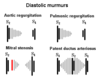
How to differentiate between diastolic murmurs?
AR: early diastolic decrescendo murmur
MS: late diastolic murmur after an opening snap
Signs of pulmonary HT
- Right ventricular heave
- Palpable thrill over pulmonary area
- Loud P2, which may be split
- TR
- R heart failure
What are the common causes of MS and MR?
MITRAL STENOSIS
- 2/3 female
- Almost always rheumatic heart disease
MITRAL REGURGITATION
- Primary valvular aetiologies
- Degeretion, myxomatous mitral valve
- Mitral valve propapse
- Chordae tendinae rupture / ischaemia to papillary muscles
- IE
- Congenital heart disease
- Secondary / functional MR
- Dilated cardiomyopathy
What are the signs of tricuspid regurgitation?
*most readily clinically diagnosed on the basis of peripheral signs
- Observation:
- JVP - large V waves, rapid Y descent
- Palpation:
- RV heave
- Auscultation:
- panysystolic / holosystolic murmur loudest at LSE
- loudest on inspiration
- Right ventricular S3
- Other:
- pulsatile liver
- may also develop portal hypertension / ascites (cardiac cirrhosis)
- Commonly accompanies pulmonary hypertension
- Right ventricular heave
- Palpable thrill over pulmonary area
- Loud P2, which may be split
- TR
- R heart failure
- Commonly accompanies MR
- Which causes pulmonary hypertension and therefore TR
What are the causes of tricuspid regurgitation?
- Functional - due to RV dilation
- Right IE (secondary to IVC / IVDU)
- Complication of PPM insertion
- Complication of frequent trans jugular biopsies
- Commonly accompanies MR & pulmonary hypertension (and with associated features)
- Congenital: Ebstein’s anomaly (tricuspid valve not formed properly, box shaped heart on XR)
Clinical findings of HOCM
Pulse
- Jerky, bifid pulse (rapid ejection by hypertrophied ventricle —> obstruction)
JVP:
- Prominent a wave (forceful RA contraction against a non-compliant RV)
Palpation:
- Double or triple impulse apex (presystolic expansion of the ventricle)
- LV heave
Auscultation
- Late systolic murmur LSE (LVOT obstruction), loudest at the LSE, does not radiate to carotids
- Pan systolic murmur at the apex (MR, due to systolic anterior motion)
- S4
Dynamic
- Increases with Valsalva (strain; reduces preload —> small LV size —> more obstruction —> louder)
- Decreases with Squatting and Handgrip


