E2: Stomach and GDV Flashcards

1
Q

What are the gastric layers? Which is strongest?

A

From outside to inside: Serosa - Muscle - Submucosa - Mucosa

Submucosa is the strongest (holdng layer)

2
Q

Pica predisposes animals to gastric foreign bodies. Name some conditions that predispose an animal to pica.

A

Iron deficiency

Hepatic encephalopathy

Pancreatic exocrine insufficiency

3
Q

Which of these is not a clinical sign associated with a gastric foreign body?

Vomiting

Fever

Lethargy

Abdominal pain

Anorexia

A

Fever

4
Q

Which of these are possible lab findings for an animal with a gastric foreign body?

Anemia

Leukocytosis

Leukopenia

Neutropenia

Renal azotemia

Pre-renal azotemia

Metabolic alkalosis

Metabolic acidosis

Hyperkalemia

Hypokalemia

Hyperchloremia

Hypochloremia

A

Anemia

Leukocytosis

Pre-renal azotemia

Metabolic alkalosis

Metabolic acidosis

Hypokalemia

Hypochloremia

5
Q

If you decide to medically manage a foreign body, what do you need to include in your therapy?

If you needed to induce vomiting, what would you use?

A

Fluid therapy: rehydrate, correct electrolyte imbalances

Monitor using serial rads

Induce vomiting in dog- Apomorphine

Cat- Xylazine

6
Q

T/F: Many gastric foreign bodies can be removed endoscopically.

A

True

7
Q

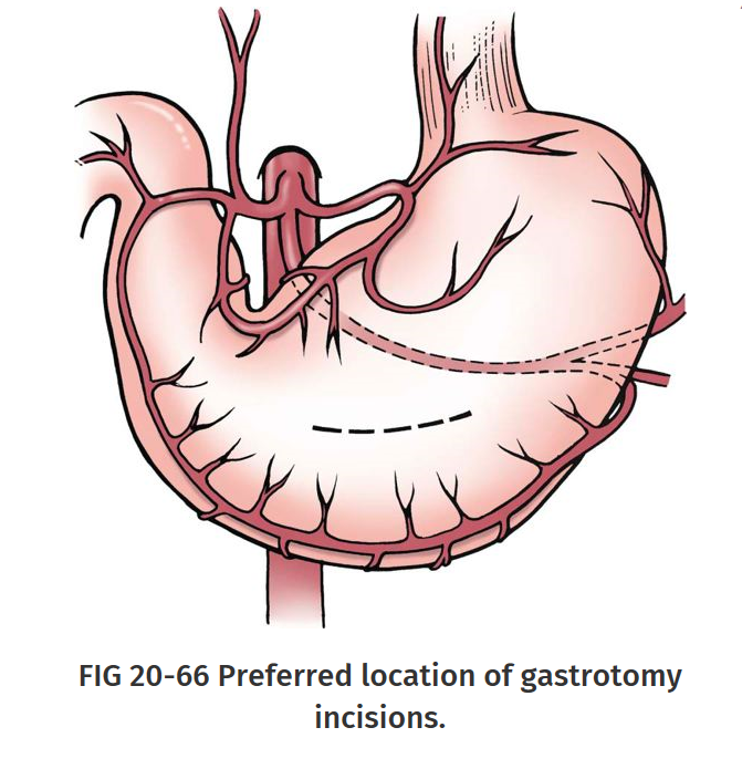
Where should you make your surgical approach for gastric foreign body removal?

A

Ventral midline celiotomy (from xiphoid to pubis)

Incision in hypovascular aspect of stomach between greater and lesser curvature

8
Q

To reduce contamination during a gastrotomy, isolate the stomach from the remaining abdominal contents with moistened _________. Place _________ to assist in manipulation of the stomach and help prevent spillage of gastric contents.

A

Laparotomy sponges

Stay sutures

9
Q

How do you close a gastrotomy incision- what pattern(s) could you use in the first layer and what tissue(s) will this layer incorporate? Second layer?

What alternative method could you use to reduce post-op bleeding?

A

First layer: Cushing or simple continuous pattern

Tissues: Serosa, muscularis, and submucosa

Second layer: Lembert/Cushing pattern

Tissues: Serosa, muscularis and submucosa

Alternative: Close mucosa in simple continuous pattern as separate layer followed by inverting pattern including all other tissue layers

(Cushing then Cushing/Lembert = Double layer inverting

Simple then another simple followed by Cushing/Lembert= Double layer appositional then inverting)

10
Q

What clinical signs are associated with gastric outflow obstruction?

A

Chronic intermittent vomiting of partially digested food hours after feeding

(If congenital signs seen at weaning)

11
Q

What congenital abnormality causes benign gastric outflow obstruction?

A

Pyloric stenosis

12
Q

What artery supplies the lesser curvature of the stomach? What is/are the parent artery/arteries?

Greater curvature? What is/are the parent artery/arteries?

A

Lesser- Gastric arteries (left and right)- parent- Celiac artery

Greater- Gastroepiploic arteries (left and right)- parent = Celiac A

Short gastric arteries - parent= Splenic A

13
Q

What breeds are predisposed to congenital pyloric stenosis?

A

Brachiocephalic dog breeds (Boxers, bulldogs)

Siamese cats

14
Q

What is the suspected etiology for congenital pyloric stenosis?

A

Excess gastrin production

(trophic for gastric smooth muscle and mucosa)

Also possible cause of Chronic Hypertrophic Pyloric Gastropathy

15
Q

What are the diagnostic modalities for determining pyloric stenosis?

A

Radiographs- to look for gastric distenssion or delayed gastric emptying (evidenced by a not-empty stomach after 8 or more hours of fasting)

Contrast radiography- to look for ‘beak’ or ‘apple core’ sign

Ultrasonography-

16
Q

What surgeries can you perform to correct congenital pyloric stenosis?

A

Pyloromyotomy (Fredet-Ramstedt procedure)

Transverse pyloroplasty (Heineke-Mikulicz procedure)

17
Q

Aquired hypertrophy of which layer or layers of the pyloris causes CHPG (chronic hypertrophic pyloric gastropathy)? Which breeds are predisposed? Is there a sex or age prediliction?

A

Mucosa and or muscular hypertrophy

Small breed dogs (<10kg) such as Shih-tse, Lhasa apso, Maltese

Males predisposed

Middle aged to older

18
Q

What diagnostic tool can be used to evaluate the middle and pyloric wall thickness to diagnose CHPG? What alernative modality can you use if you also need to take biopsies?

A

Ultrasound

Endoscopy

19
Q

In addition to a Heineke-Mikulicz pyloroplasty, which 2 other surgical techniques can you use to manage CHPG? What are advantages and disadvantages of each?

A

Y- U Pyroplasty

_Advantages:_Increase diameter of pylorus, access to excise hypertrophied mucosa

Disadvantages: Potential flap tip necrosis, possible side effect of rapid gastric emptying

Pylorectomy w/Gastroduedenostomy (Bilroth I)

Advantages: All diseased tissue can be removed

Disadvantages: technically more demanding, increased risk of “dumping” syndrome and reflux gastritis (due to direct connection between duodenum and stomach)

20
Q

_________ tumors are commonly found near the cardia.

_________ tumors are commonly found in the pyloric antrum or the lesser cuvature of the stomach.

A

Leiomyoma, LSA

Adenocarcinoma

21
Q

What are the sigalment, physical finding, treatment options and prognosis for gastric adenocarcinoma?

A

Signalment: Male, 8-10yo, Staffies, Belgian shepherds

Physical findings: plaque-like mucosal lesions with ulcers, raised sessile or polypoid lesions, diffuse infiltration (linitis plastic, scirrhous stomach wall), on US see mural thickeing and loss of normal gastric wall

Treatment options: aggresive sx exision via gastrectomy, 5cm margins

Pallitative tx: By-pass procedure (Bilroth I or II), chemotherapy?

Prognosis: Guarded to poor, no sx= 2-4mo, aggressive therapy= 10mo

22
Q

What are the sigalment, physical finding, treatment options and prognosis for gastric leiomyosarcoma?

A

Signalment: 7-8 yo

Physical findings: ulceration, mass near cardia protruding into the gastric lumen

Treatment options: submucosal resection, Partial gastrectomy (if extensive or ulcerated)

Prognosis: median survival 21 mo, good to guarded, recurrence possible

23
Q

What are the sigalment, physical finding, treatment options and prognosis for gastric leiomyoma?

A

Signalment:older dogs, INCIDENTAL finding

Physical findings: mass near cardia protruding into the gastric lumen

Treatment options: submucosal resection, Partial gastrectomy (if extensive)

Prognosis: Good to guarded

24
Q

How would you describe this stomach wall that has been affected by a gastric adenocarcinoma?

A

Scirrhous

25
Q

What are the sigalment, physical finding, treatment options and prognosis for Pythiosis?

A

Signalment: Young dogs, Labrador Retrievers, Cavalier King Charles Spaniels and German Shepherd Dogs predisposed (highest incidence in Gulf Coast States in fall or early winter and usually after a summer of flooding and large amounts of precipitation)

Physical findings: severe inflammation and infiltration (submucosa and muscularis affected), intense fibrotic reaction, transmural thickening (esp gastric outflow area), vomiting, inappetence, weight loss, diarrhea

Treatment options: Wide surgical excision (antifungals INEFFECTIVE)

Prognosis:Guarded to poor, MST 26.5 days

26
Q

What type of suture should be avoided in gastric surgery?

A

Chromic gut

27
Q

What is a Bilroth II and what are the indications to perform it?

What are some disadvantages of using this procedure?

A

Gastroenterostomy- partial gastectomy followed by gastroenterostomy

Indications: Extensive gastric resection making gastroduodenostomy impossible

Disadvantages: A LOT of complications - Alkaline gastritis (bile and pancreatic secretions flow into stomach), “Blind loop” syndrome (gastric contens move orally and putrefy), Marginal ulceration (of jejunal mucosa due to acid)

28
Q

What is a Bilroth I and what are the indications to perform it?

What are some advantages and disadvantages of using this procedure?

A

Pylorectomy Gastroduodenostomy

Indications: neoplasia, outflow obstruction caused by muscular hypertrophy, ulceration of gastric outflowtract

Advantage: All dieased tissue can be removed

Disadvantages: difficulty, risk for “dumping” syndrome and reflux gastritis (must use extreme care when incising in pyloric area to avoid damaging COMMON BILE DUCT where it traverses lesser omentum)

29
Q

How does a Bilroth I differ from a Bilroth II?

A

In Bilroth II the distal stomach and proximal duodenum are closed after pylorectomy, and the jejunum is attached with a side-to-side anastomosis to the diaphragmatic surface of the stomach

In Bilroth I stomach and duodenum are connected where the pylorus has been removed

30
Q

_________ is the distension of the stomach with fluid, food, and/or gas. Treatment is _______ (medical/surgical).

_________ is the enlargement of the stomach associated with rotation on its medenteric axis. Treatment is _______ (medical/surgical).

A

Dilation/ dilatation - Medical

Dilation-Volvulus - Surgical

31
Q

What risk factors for developing GDV have been identified?

A

Exercise: following large meals? - largely disproven

Diet: highly processed food?, lots of water before exercising? large volume once daily, eating rapidly, feeding from raised feed bowl, dry kibble, fats/oils in 1st 4 ingredients (fish or egg supplements decreased risk)

Age: older=higher risk

Weight: UNDERweight

Body confirmation: Deep, narrow thorax

History: Having a 1st degree relative with hx of GDV, being stressed, vomiting, fearfulness, anxiety, spending 5 hours/day with owner

Genetics: Irish Setters, Great Danes, GSD, males

32
Q

Gastric enlargement is thought to be associated with ___________ obstruction.

A

Gastric outflow

(mechanical or functional)

33
Q

Once the stomach dilates, normal physiologic means of ______________ are hindered because the esophageal and pyloric portals are obstructed.

A

Removing air

34
Q

Generally, with GDV the stomach rotates in a ______direction when viewed from the surgeon’s perspective (with the dog on its back and the clinician standing at the dog’s side, facing cranially)

A

Clockwise

(usually 220-270 deg, but may be 90-360)

35
Q

In what direction/where are the duodenum, pylorus, and spleen displaced with GDV?

A

Duodenum and pylorus move ventrally, left of midline (end up between stomach and esophagus)

Spleen is usually displaced to right ventral side of abdomen

36
Q

Why does myocardial ischemia occur following GDV?

A

Caudal vena cava and portal vein are compressed by distended stomach leading to decreaed venous return and decreased cardiac output

37
Q

T/F: Arrhythmias occur in many dogs with GDV, particularly those with gastric necrosis.

A

True

38
Q

Fill in the blanks

A

Blue: Gas accumulation

Red: Abnormal gastroesophageal function and Delayed gastric emptying

39
Q

What contributes to the gas which collects in the stomach during GDV?

A

Aerophagia

Bacterial fermentation of carbs

Diffusion from bloodstream

Metabolic reactions

40
Q

What has been implicated as causing much of the tissue damage that ultimately results in death after correction of GDV?

A

Reperfusion injury

41
Q

The term “torsion” implies a ______ (clockwise/counter-clockise) rotation of less than ____ degrees while the term volvulus implies a ______ (clockwise/counter-clockise) rotation of more than ____ degrees.

The term “displacement” implies a _______(clockwise/counter-clockise) rotation of less than _____degrees.

A

Torsion: (less than) 180 clockwise

Volvulus: (more than) 180 clockise

Displacement: (less than) 90 counter-clockwise

42
Q

What covers the stomach when it is displaced clockwise but not when it is displaced counter-clockwise?

A

Omentum

43
Q

What are the typical clinical signs associated with GDV? Which is most common?

A

Most common: Nonproductive retching

Restlessness

Hypersalivation

“Praying” position

Vomiting

Weakness

Collapse

(note- acute)

44
Q

What diagnostic modality do you use to differentiate dilation from dilation + volvulus? What views are warrented and why?

A

Radiographs

Right lateral + DV: to facilitate filling the abnormally displaced pylorus with air so it can be easily identified

(Rlat w/GDV pylorus lies cranial from body and separate from rest of stomach by soft tissue = Reverse C Sign/Double bubble

DV w/GDV pylorus is gas-filled structure to left of midline)

45
Q

You take rads of a dog with suspected GDV and in addition to noting the classically reverse-C sign, you also see free abdominal air and air within the wall of the stomach. What does this suggest?

A

Free air -Gastric rupture

Air in stomach wall- Necrosis

SURGICAL EMERGENCY (both or either)

46
Q

Which type of GDV is most commonly associated with a history of chronic GI signs?

A

Counterclockwise displacement

(Partial GDV)

47
Q

T/F: You can differentiate GDV from gastric dilatation without volvulus if you are able to pass a stomach tube.

A

False, stomach tubes canf requently be passed in dogs with twisted stomachs

48
Q

What are the key components of pre-op stabilization of GDV patients and in what order do you want to perform these treatments?

A
  1. Fluids, pain meds and monitoring
    Cystalloid slns or hypertonic saline + colloids
    Full mu
    EKG/BP
  2. Decompression via tube (1st choice) or trocharization then tube
  3. Radiographs and CBC/Chem: Correct and A/B or significant e-lyte disturbances, especially LACTATE
  4. Antimicrobials and Free radical scavengers
  5. Pre-oxygenate, rapid induction (Etomidate, Alfaxalone, Propofol)
49
Q

What are the options for correcting the hypovolemic shock often associated with GDV?

A

Isotonic fluids (90 ml/k/hr)

Hypertonic (7%) saline (4-5 ml/kg q5-15min)

Hetastarch (5-10ml/kg q10-15min)

7.5% Saline + Hetastarch (7.5% sln) 4 ml/kg q5min

50
Q

What methods can be used to decompress the stomach? Describe them as well, including precautions that must be taken.

A

Preferred method: Orogastric intubation- use bite block, measure and mark tube length using xiphoid as landmark, advance tube slowly while rotating. After removing air, lavage with warm water.

Trocharization- use large bore needle/catheter at most tympanic site which has been clipped and cleaned, followed with tube

51
Q

What are the potential complications associated with trocharization?

A

Hitting the spleen causing hemorrhage

Leakage of gastric contents into abdominal cavity resulting in infalmmatory response/damage

52
Q

What are some examples of free radical scavengers and why are they potentially beneficial in treating GDV patients?

A

Acetylcysteine

Vitamine C and E

Selenium

Desferoxamine (iron chelator)

Lidocaine

Purpose: prevent damage caused by reperfusion which releases ROS into circulation

53
Q

What are the advantages of early surgical correction in the management of GDV?

A

Improves bloodflow (prevent gastric necrosis)

Prevent arrhythmias

54
Q

Why must the stomach be lavaged following decompression?

A

Otherwise the stomach will redilate after the tube is withdrawn

55
Q

What area of the stomach is most commonly affected by vascular compromse?

A

Greater curvature near short gastric arteries (junction between fundus and body)

56
Q

How is viability of the stomach typically assessed? How can palpation of the stomach wall be helpful in assessing viability?

A

Color

Pulsation of blood vessels

Bleeding from cut surface

Peristalsis (should resume almost immediatly after respositioning)

Surface oximetry

Palpation is helpful becasue a thinned wall indicates vascular compromise and potential necrosis (now or in the future)

57
Q

What abnormalities in the spleen may occur with GDV and how are they managed?

A

Venous congestion- is self-limiting and tends to resolve when stomach is derotated

Vessel thrombosis- Splenectomy

Splenic torsion- Splenectomy

58
Q

What is a disadvantage of using a double inverting pattern to close a lesion? Where would you never want to use an inverting pattern?

A

It decreases the lumen

Close to the pylorus (or cardia)

59
Q

What layer(s) of the stomach are affected by congenital pyloric stenosis?

A

Only the muscularis

60
Q

What layers can be affected with aquired pyloric stenosis?

A

Muscularis

Mucosa

61
Q

What pathologic classification would you give a dog with CHPG if muscular and mucosal hypertrophy are present?

A

Grade II

62
Q

T/F: If only muscular hyperplasia is present with CHPG, its pathologic classification is Grade I.

A

FALSE, muscular HYPERTROPHY

63
Q

What are the 2 pathologies that indicate you have a grade III CHPG?

A

HyperPLASIA of the mucosa

Inflammation of the mucosa and submucosa

64
Q

What is the difference between hypertrophy and hyperplasia?

A

Hypertrophy- increase in cell SIZE

Hyperplasia- increase in cell NUMBER

65
Q

The higher the grade of CHPG, the more invasive the procedure to correct it needs to be. Name 2 surgeries you could do for a low classification and 1 you could do for a higher classification.

A

Low: Heineke-Mikulicz Pyroplasy or Y-U pyroplasty

High: Bilroth I gastroduodenostomy

66
Q

What type of suture pattern would you use to close an incision close to the pylorus?

A

Single layer appositional

67
Q

What are the goals of a gastropexy?

A

Inspect the stomach and spleen to identify and remove damaged tissues

Decompres the stomach and correct any malpositioning

Adhere stomach to body wall to prevent subsequent malpositioning (doesn’t prevent dilation)

68
Q

To create a permanent adhesion, the gastric muscle must be in contact with the muscle of the body wall. Why?

A

Intact gastric serosa does not form permanent adhesions to an intact peritoneal surface.

69
Q

Describe the technique, advantages and disadvantages of an incisional/muscular flap gastropexy.

A

Technique:

  1. Make incision in seromuscular layer of gastric antrum
  2. Make another incision in right ventrolateral abdominal wall through peritineum and internal facia of rectus abdominis or trasnverse abdominis muscles
  3. Suture edges of incision in simple continuous pattern (2.0 absorbable or non-abs)
  4. Ensure contact between muscularis layer and abdominal wall muscle
  5. Suture cranial margin first, then caudal margin (or can raise flaps in stomach and body wall to increase tissue contact)

Advantages: Easier than circumcostal, quicker, gastric lumen not opened

Disadvantages: less strong than circumcostal, no direct access to gastric lumen if need post-op decompression

70
Q

Describe the technique, advantages and disadvantages of a Belt Loop gastropexy.

A

Technique:

  1. Elevate a seromuscular flap in gastric antrum and make 2 transverse incision in ventrolateral abdominal wall through peritoneum and abdominal musculature (2.5-4cm appart 3-5cm long)
  2. Create a tunel under abdominal musculature with forceps
  3. Place stay sutures in edge of antral flap and use them to pass flap from cranial to caudal under the muscular flap
  4. Suture flap to original gastric margin in simple continuous pattern (2.0 abs or non-abs) or use skin staples

Advantages: Easier than circumcostal, quicker, gastric lumen not opened

Disadvantages: less strong than circumcostal, no direct access to gastric lumen if need post-op decompression

71
Q

Describe the technique, advantages and disadvantages of a circumcostal gastropexy.

A

Technique:

  1. Make either 1 or 2-layer hinged flap by incising through seromuscular layer of pyloric antrum (dont incise gastic mucosa)
  2. Elevate flap by dissecting under muscularis (if 1-hinged flap place hinge toward lesser curvature)
  3. make 5-6cm incision over 11th or 12th rib at costochondral junction (do not penetrate diaphragmatic attachments to body wall)
  4. Form tunnel under rub using Carmalt/hemostat
  5. Place stay sutures in flap (if 2-flap technique on flap closest to lesser curvature)
  6. Pass gastric antral flap craniodorsal under rib and suture to original gastric margin

Advantages: stronger adgesion than most other techniques, dimished risk of gastric leakage and abdominal contamination (gastric lumen not opened)

Disadvantages: technically challenging, complications include pneumothorax and rib fracture, no direct access to gastric lumen if need post-op decompression

72
Q

Describe the technique, advantages and disadvantages of a tube gastropexy.

A

Technique:

  1. Stab incision into R abdominal wall caudal to last rib, 4-10cm from midline
  2. Foley catheter through incision
  3. Place purse-string sutures (2.0 absorbable) so catheter/balloon is in hypovascular region of seromuscular layer of ventral surface of pyloric antrum (where it won’t obstruct gastric outflow)
  4. Inflate balloon with SALINE and secure purse-string sutures
  5. Preplace 3-4 absorbable sutures between pyloric antrum and body wall where tube exits
  6. Draw stomach to body wall and tie preplaced sutures
  7. Secure tube to skin using Roman sandle suture pattern (DO NOT PENETRATE TUBE)
  8. Leave tube in place 7-10days
  9. To remove delfate baloon, leave skin incision open to facilitate drainage

Advantages: Quick, simple, allows post-op gastric decompression and placement of meds directly into stomach

Disadvantages: Longer hospitalization (must allow adhesions to form), peritonitis if improperly placed

73
Q

Describe the technique, advantages and disadvantages of an incorporating/ laproscopic-assisted gastropexy.

A

Technique:

  1. Place first cannula just caudal to umbilicus
  2. Place second cannula jusr right of midline, 2-4cm behind last rib (incision parallel to rib)
  3. Pull stomach through incision
  4. Place stay sutures between stomach adn body wall
  5. Perform muscular flap/incisional pexy

Advantages: Pyloric antrum accurately visualized, don’t need special instruments

Disadvantages: Expertise in gastrscopy required, potential organ trauma

74
Q

What is the most critical time period for post-op GDV patients?

A

The first 4 days after surgery

75
Q

What types of arrhythmias are most common with GDV? When do these usually occur?

A

Ventricular arrhythmias:

V-tac

VPCs

Paroxysmal runs

Idioventricular rhythm (slow v-tach)

Mutifocal VPCs

Begin 12-36hrs after surgery (usually abate 24-72 hrs post-op)

76
Q

What are the potantial causes of arrhythmias with GDV?

A

Myocardial ischemia (decreased venous supply and thus CO)

E-lyte abnormalities (Hypokalemia very common)

A/B alterations

Vasoactive substances

Imbalance of autonomic nervous system

77
Q

T/F: Lidocaine as an antiarrhythmic is ineffective if the patient is hypokalemic.

A

True

78
Q

When should arrhythmias be treated? With what? What if you give too much?

A

Treat if arrythmias is interfering with cardiac output = Poor peripheral pulses, pulse deficits, weakness

Mutiform arrythmias/VPCs

R-on-T

Sustained ventricular rate >160bpm

Treatment: IV lidocaine, first bolus and if respond well then CRI (start with low dose)

Alternatives: Procainamide, Sotalol

Lidocaine toxicity: muscle tremors, vomiting, seizures

79
Q

What is the prognosis for GDV patients?

A

Uncomplicated cases: 80-90% survival rate

Lower suvival if: Pre-op arrhythmias (38% mortality), Gastric necrosis (46%), patrial gastrectomy (35%), splenectomy (32%), partial gastrectomy + splenectomy (55%)