FDN2_SM_WK4_EmbryologyPatternsDefects Flashcards

1
Q

What is the most common site of normal implantation?

A

Posterior wall of the uterus

2
Q

What are some common sites of ectopic pregnancy?

A

Uterine tubule, cervix, abdominal cavity, ovary, etc

3
Q

What are the major events of week 1 of embryogenesis?

A

Ovulation, conception, migration down the uterine tubule

4
Q

Where does fertilization usually occur?

A

The distal 1/3 of the uterine tubule

5
Q

What are the major events of week 2 of embryogenesis?

A

Implantation, extra-embryonic membrane formation

6
Q

What stage is the conceptus when it implants?

A

Blastocyst

7
Q

What are the components of a blastocyst?

A

Trophoblast = outer shell

Inner cell mass

8
Q

What is the fate of the trophoblast?

A

It will:

  • Differentiate into the outer syncytiotrophoblast and inner cytotrophoblast, then acquire its extraembryonic mesoderm layer (so the cytotrophoblast is now in the middle)
  • When it has all 3 layers, it is called the chorion
  • The villous chorion invade the endometrium and form the fetal components of the placenta

- The smooth chorion covers the amnion

9
Q

How does the amnionic cavity form?

A

The blastocyst hollows to form two layers: The epiblast and the hypoblast

10
Q

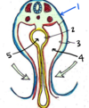
What is #1 pointing to?

A

Syncytiotrophoblast

11
Q

What is #2 pointing to?

A

Amniotic Cavity

12
Q

What is #3 pointing to?

A

Ectodermal amnion

13
Q

What is #4 pointing to?

A

Epiblast/Ectoderm

14
Q

What is #5 pointing to?

A

Hypoblast/Endoderm

15
Q

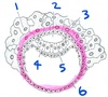
What is #6 pointing to?

A

Trophobolast

16
Q

The structure labeled by which number will eventually be in direct contact with maternal blood?

A

1 - the synytiotrophoblast

This layer will form the outermost part of the villious chorion that invades the endometrium and forms the fetal component of the placenta. It will be in direct contact with maternal blood from decidua basalis.

17
Q

Name the 4 extra-embryonic membranes and their functions

A

Chorion: Villous forms the placenta, cmooth covers the amnion

Amnion: Surroudns embryo, will eventually fold down to form the cylinder

Yolk Sac: Provides early nutrition. Will become the first source of embryonic blood cells

Alantois: Vestigal membrane in humans

*Yolk Sac + Alantois become the umbilial cord

18
Q

What is the final step in the formation of the 3 major extraembryonic membranes?

A

Extraembryonic mesoderm coats the old blastocyst cavity. Everything gets an “extra layer”

Trophoblast -> Chorion

Primary Yolk Sac -> Yolk Sac

Primary Amnion -> Amnion

19
Q

Which embryonic structure forms the placenta?

A

The chorion (which came from the trophoblast)

20
Q

Which embryonic structure forms the umbilical cord?

A

The yolk sac and the alantois

21
Q

Which embryonic structure(s) form(s) the afterbirth?

A

The villous chorion (placenta) and the cytotrophoblastic shell

22
Q

What is the major event of week 3 of embryogenesis?

A

Gastrulation (and subsequent formation of the intraembryonic mesoderm)

23
Q

Describe the formation of the intraembryonic mesoderm

A

Induced by the primitive knot/node, which forms the primitive streak

Epiblast cells migrate through the invaginating primitive streak to first migrate into the hypoblast (displacing it), then create a layer of mesoderm in between the endoderm and ectoderm

24
Q

From where does the intraembryonic mesoderm originate?

A

The primitive streak (epiblast cells migrate through it)

25
Q

Describe the organization of the mesoderm

A

Middle = Notochord (induces neural tube)

Paraxial Mesoderm/Column (lateral to notochord)

Intermediate Mesoderm/column (lateral to paraxial)

Most lateral = Lateral Plate

26
Q

What is the fate of the notochord?

A

Induces the neural plate (which will become the neural tube)

The notochord itself becomes the nucleus pulposis, the gelatinous interior of vertebral discs

27
Q

What is the fate of the paraxial mesoderm?

A

The paraxial mesoderm forms somites, which eventually become bone, muscle, and dermis

28
Q

What is the fate of the intermediate mesoderm?

A

The intermediate mesoderm becomes the urogenital system (kidneys and gonads)

29
Q

What is the fate of the lateral plate mesoderm?

A

The lateral plate mesoderm contributes to the splanchnopleure and the somatopleure

Splanchnopleure = endoderm + mesoderm from lateral plate -> Walls of gut tube, visceral pleruae, visceral peritoneum, supporting mesenteries (suspend sheets of visceral peritoneum)

Somatopleure = ectoderm + mesoderm from lateral plate-> Lateral and ventral body wall, parietal pleura, parietal peritoneum

Lateral plate + cardiogenic plate -> heart

30
Q

What is the major event in week 4 of embryogenesis?

A

Shaping of the gastrula: Turning the trilaminar disk into a cylinder

31
Q

How does the neural plate form?

A

Induced by the notochord; thickened ectoderm

32
Q

What forms the neural tube?

A

Ectoderm is induced by the notochord to form the neural plate. The neural plate folds in to form the neural tube

33
Q

What does the neural tube become?

A

Most of the central nervous system; all cell bodies in the CNS are derived from the neural tube

34
Q

What forms the neural crest?

A

Cells from the ectoderm break off from the neural crest during neural tube formation; these are neural crest cells

35
Q

What does the neural crest become?

A

The peripheral nervous system: all cell bodies in the PNS came from the neural crest

(Dorsal root ganglia, collateral ganglia, sympathetic chain ganglia, post-synaptic autonomic cell bodies)

36
Q

What 3 parts do somites differentiate into?

What do they become?

A

Myotome -> Muscle

Dermatome -> Skin/surface ectoderm

Sclerotome -> Bone

37
Q

Describe the formation of the intra-embryonic celom

A

Lateral plate mesoderm and cardiogenic mesoderm form a U-shaped tube in the trilaminar disk.

When the disk turns into a cylinder, this becomes the intra-embryonic coelom

Eventually, this will divide into the pleural cavity, pericardial cavity, and peritoneal cavity

38
Q

What is the origin of the primitive knot/primitive streak?

A

Appears in the epiblast/ectoderm to initiate gastrulation. Moves from caudal to cranial end

39
Q

What is the origin of the cardiogenic plate?

A

The cardiogenic plate is derived from splanchnopleuric mesoderm

40
Q

Which embryonic structure forms the somite?

A

Paraxial mesoderm/columns

41
Q

Which embryonic structure forms the urogenital system?

A

Intermediate mesoderm/columns

42
Q

Which adult tissues are derived from the surface ectoderm?

A

Skin, hair, nails

Lining of mouth, anal canal

Think: most things in contact with the outside world

43
Q

Where does amniotic fluid come from?

A

Early: diffusion from maternal tissues + contriubtion from amniotic cells and the embryo

Later: When the urogenital system forms, the amniotic fluid is mostly fetal urine that the fetus swallows and recycles

44
Q

What is the function of amniotic fluid?

A

Protect the fetus, allow for movement

45
Q

What conditions can result in polyhydroaminos?

A

Polyhydroaminos = too much amniotic fluid

Causes: inhibition of swallowing (anecephaly) or absorption (transesophageal fistula)

46
Q

What conditions result in too little amniotic fluid?

A

Renal agenesis (newborn is missing one or both kidneys)

Other conditions resulting in lack of kidney function/urine production

47
Q

Describe the structure of the placenta

A

All 3 layers of the chorion (which came from the trophoblast) form a finger-like projection (villous) that invades the endometrium.

The villous chorion has the outer syncytiotrophoblast, the extra-embryonic mesenchyme conenctive tissue core (blood vessels), and the cytotrophoblast in between them. The cytotrophoblast eventually disappears.

The decidua basalis of the endometrium is basically an open blood vessel that bathes the villous chorion in maternal blood.

48
Q

What is the structure labeled #1?

A

Amnion/Ectoderm

The ectoderm will form the skin/outer layer of the body. The amnion will be wrapped around the ectoderm/developing fetus until birth

49
Q

What is the structure labeled #2?

A

The splanchnopleure

50
Q

What is the structure labeled #3?

A

The somatopleure

51
Q

What is the space labled #4?

A

The intraembryonic coelom

52
Q

What is the space labeled #5?

A

The gut tube

53
Q

What is the yellow layer in this picture?

A

The endoderm

54
Q

What is the cloacal membrane?

A

A membrane present at the posterior end of the embryo after gastrulation

It will eventually form the anus

55
Q

When do the cloacal and oral membranes arise?

A

After gastrulation

56
Q

What is mesenchyme?

A

Loose connective tissue in the mesoderm

Mesenchyme gives rise to most of the body’s connective tissue

57
Q

What is the source of blood vessels in placenal villus?

A

Extraembryonic Mesoderm

58
Q

What is the source of cardiogenic mesoderm?

A

Primitive streak

59
Q

What gives rise to the chorion?

A

Trophoblast

60
Q

When is the embryonic period?

A

2-8 weeks (biological)

61
Q

What is the difference between biological age and clinical age of an embryo?

A

Biological age = Starts at fertilization of the egg

Clinical age/gestational age= Starts with the first day of the last normal menstrual period (add 2 weeks to biological time). This is used because the exact day of fertilization is usualy not known

62
Q

Which embryonic structure forms the vertebrae?

A

The paraxial mesoderm (vertebrae come from somites, which come from paraxial mesoderm)

63
Q

When does patterning occur in human development?

A

In weeks 4-8

64
Q

What is the end of week 8 a significant time point in embryonic develpment?

A

At the end of week 8, all of the organs are formed and axes are established. The embryo is now a fetus (anything later than week 8 is the fetal period)

65
Q

Which mechanism establishes the anterior/posterior axis in the embryo?

A

HOX genes

66
Q

Describe the organization of the HOX gene complex

A

HOX genes with lower numbers are at the 3’ end of the chromosome, while genes with higher numbers are at the 5’ end

67
Q

Describe temporal colinearity of HOX genes

A

Genes at the 3’ end of the complex (lower numbers) are expressed earlier in development

Genes at the 5’ end of the complex (higher numbers) are expressed later in development

68
Q

Describe spatial colinearity of HOX genes

A

Genes at the 3’ end of the HOX complex (lower numbers) have expression that extends more anteriorly

Genes at the 5’ end of the HOX complex (higher numbers) have expression that ends more posteriorly

69
Q

Describe posterior prevalence

A

More HOX genes are expressed in posterior regions (Lots of overlap posteriorly)

70
Q

Describe posterior dominance

A

If several HOX genes are expressed in a segment, the one whose expression ends furthest poteriorly determines the segment phenotype

For example: In the segment underlined in blue, HOX-B5 determines the phenotype for that segment

(HOX-B4 and HOX-B3 have expression that ends more anteriorly, so they do not determine the segement phenotype)

71
Q

What is a homeotic gene?

A

Any gene that determines the fate or identity of body segments

72
Q

Describe redundancy in HOX genes

A

There is considerable redundancy in function between paralogous HOX genes in different complexes

Ex: HOX-A4 = HOX-B4 = HOX-C4 = HOX-D4

73
Q

Where do HOX genes exhibit colinear expression?

A

Along the primary anterior/posterior axis

Along regional/organ-specific axes (limbs, GI tract, female reproductive tract)

74
Q

Which protein establishes the dorsal/ventral axis in the central nervous system and somites?

A

Sonic Hedgehog (Shh)

75
Q

Describe the Shh signaling pathway

A
  • The Shh receptor is Patched (Ptc)
  • If no Shh, Ptc inhibits the signaling pathway
  • When Shh binds to patched, the pathway is activated
  • The signaling pathway activates the GLI family of transcription factors
    • GLI1 = transcription activator
    • GLI2 = transcription activator or repressor
    • GLI3 = transcription repressor
76
Q

What is an organizing (aka patterning) center?

A

A region that secretes Shh

77
Q

Name 2 organizing centers in human embryos

A
  1. Notochord
  2. Zone of Polarizing Activity
78
Q

Which axes are established by Shh signaling?

A
  • Dorsal/Ventral axis in CNS and somites
  • Anterior/posterior axis in developing limbs
79
Q

Which axis is established by HOX gene signaling?

A

Anterior/Posterior

Hox genes determine the fate or identity of body segments along the anterior/posterior axis

80
Q

Describe Shh signaling of dorsal diffusion from the notochord

A

Establishes Dorsal/Ventral axis in CNS

  • Shh is secreted from the notochord -> Dorsal diffusion
    • High concentration -> induces neural tube
    • Low concentration -> induces sensory ganglia/neurons (from the neural crest)
81
Q

Describe Shh signaling of lateral diffusion from the notochord

A

Establishes Dorsal/Ventral axis in Somites

  • Shh is secreted from the notochord -> Lateral diffusion
    • High concentration (ventral-medial somites) -> Sclerotome -> vertebral bodies, ribs
    • Low concentration (dorsal-lateral somites) -> Dermomyotome -> Dermis, musculature
82
Q

Describe Shh signaling right above the notochord

A

Highest concentration:

Induces the ventral floor plate, motor neurons in the ventral neural tube

83
Q

Describe Shh signaling in developing limbs

A

Shh is secreted from the Zone of Polarizing Activity in the proximal-posterior aspect of the limb bud

  • High concentration -> 5th digit/posterior side
  • Low concentration -> 1st digit/anterior side
84
Q

In a limb bud, high concentrations of Shh induce the ________ side

A

5th digit/posterior

(Recall that the ZPA is in the proximal-posterior aspect of the limb bud)

85
Q

Where is the Zone of Polarizing Activity (ZPA) located in the limb bud?

A

The proximal-posterior aspect of the limb bud

86
Q

What mutation results in synpolydactyly?

A

HOX-D13

87
Q

Describe the effect of a HOX-D13 mutation

A

Synpolydactyly (distal metacarpals take on a more proximal/carpal phenotype)

Heterozygotes: “branching”

Homozygous for the mutated allele: All metacarpals have a carpal phenotype

88
Q

What mutation results in holoprosencephaly?

A

Shh LOF

The signaling pathway is never activated since Shh cannot bind to Ptc to remove pathway inhibition

89
Q

Describe the phenotypic result of an Shh LOF mutation

A

Holoprosencephaly

This is the result of abnormal septation of the hemispheres of the brain and resultant anormal CNS patterning

In this LOF mutaiton, Shh cannot inacivate Ptc to turn on the signalling pathway

(eyes are close together or there is only 1 eye)

90
Q

What mutation results in Basal Cell Nevus Syndrome?

A

Patched1 inactivation

This results in constitutive signaling pathway activation

91
Q

Describe the phenotypic result of a Patched1 mutation

A

A mutation in Ptc results in constitutive activation of the Shh signaling pathway

The result of a Patched1 mutation is Basal Cell Nevus Syndrome, characterized by large head, wide-spaced eyes, rib and abdomen abnormalities, and a predisposition to cancer

92
Q

What mutation causes cephalopolysyndactyly?

A

GLI3 Mutation

Normally, when GLI3 is activated itrepresses transcription

A GLI3 mutation results in abnormal activation, which results in extra fingers

93
Q

Describe the phenotypic result of a GLI3 mutation

A

Cephalopolysyndactyly (aka polydactyly)

Extra fingers or toes

94
Q

What is the difference between a transgenic mouse and a knockout mouse?

A

Trangenes: expressed at the wrong time or wrong place during development

Knockout: The functional gene or protein is removed

95
Q

What is the expected phenotype with inapproporate loss of expression of a HOX gene?

A

The segment in which expression is lost will have a more anterior phenotype than normal

96
Q

What is the expected phenotype wiht inappropriate ectopic anterior expression of a HOX gene?

A

The segment with ectopic anterior expression will have a more posterior phenotype than normal

97
Q

What characteristic defines a tissue with organizing properties?

A

It affects the development of neighboring regions

98
Q

What is the fate of the lateral dermomyotome?

A

Lateral dermatome -> skin of ventral wall, limb

Lateral myotome -> Muscle of ventral wall, limb

99
Q

What is the fate of the medial dermomyotome?

A

Medial Dermatome -> Skin of back

Medial Myotome -> Muscle of back

100
Q

What is the fate of teh sclerotome?

A

Vertebral bodies, ribs

101
Q

Monozygotic twins have share ________ and have their own ______

A

Share: Chorion, Placenta

Have their own: Amnion, yolk sac

(So there will be 1 chorion, 1 placenta, 2 amnions, and 2 yolk sacs in a pregnancy of monozygotic twins)

102
Q

What causes the separation of the pleural and peritoneal cavities?

A

The septum transversum: A condensation of primitive streak mesenchyme

103
Q

What is the septum transversum? What does it do?

A

The septum transversum is a condensation of primitive streak mesenchyme

It separates the pleural and peritoneal coelom

104
Q

What is a malformation?

A

Poor formation of tissue due to an intrinsically abnormal developmental process

105
Q

What is a deformation?

A

Unusual (mechanical) forces on normal tissue cause abnormal form or position of a body part

106
Q

What is a disruption?

A

Breakdown of normal tissue

107
Q

Describe the timing of a malformation

A

The gestational age at which the distubance in morphogenesis was caused is between 2-8 weeks

108
Q

Which of the three types of congenital anomolies are caused during the fetal period?

A

Deformations and disruptions

The fetus is exposed to the cause of abnormal morphogenesis sometime after 8 weeks

109
Q

Which of the three types of congenital abnormalities may be spontaneously correted?

A

Deformations

110
Q

Give some examples of malformations

A

Cleft lip

Polydactyly

Syndactyly

Ectrodactyly

Radial hypoplasia

Spina Bifida

111
Q

Give some examples of deformations

A

Club foot

Mandibular asymmetry

Potter’s sequence

Plagiocephaly

Micrognathia

112
Q

Give some examples of disruptions

A

Amputated fingers

Amniotic bands

Encephalocele

113
Q

What are some common causes of deformations?

A
  • Amniotic tear -> oligohydraminos -> compression on the fetus
  • Twins
  • Unusual implantation site
  • Uterine malformaiton
  • Malformations (ex: Spina bifida -> renal agenesis -> oligohydraminos)
  • Functional causes (muscualr disturbance -> decreased movement -> Deformation)
114
Q

What are the common causes of malformations?

A

Single gene

Chromosome abnormality

Multifactorial trait

Maternal influence

Unknown

115
Q

What is a “sequence?”

A

Condition in which there are several abnormalities arising as secondary consequences of a single underlying problem

116
Q

What is a “syndrome?”

A

Condition in which there are several abnormalities of apparently independent origin

117
Q

What is the Pierre-Robin Sequence?

A

Malformation sequence: Mandibular hypoplasia -> failure of tongue descent -> cleft palate

Deformation sequence: Mandibular constraint -> failure of tongue descent -> cleft palate

118
Q

A minor anomoly is usually of no functional significance to the individual. Why then, are clinicians concerned with identifying them at birth?

A

When there is one, there is a high probability that there is another that may be major

  • Newborns with 3+ minor anomalies have a 90% chance of having a major anomaly
  • 42% of patients with idiopathic mental retardation have 3+ anomalies (80% of which are minor)
  • Minor anomalies are present in many congenital abnormality syndromes
119
Q

What is the difference between a teratogen and a mutagen?

A

A teratogen causes developmental toxicity that can alter viability or a specific organ system

A mutagen induces a change in DNA (at any change in life)

120
Q

What are the 4 main categories of teratogens?

A

Maternal factors

Drugs (Chemical Factors)

Infectious Agents (TORCH)

Physical agents (Ionizing Radiation)

121
Q

What factors affect the dose of a teratogen that reaches the embryo?

A

Fetal or maternal genotype, other drugs, concomitant disease

122
Q

What is a teratogen?

A

A chemical, physical, or infectious agent that affects viability of the embryo or specific organ systems.

123
Q

Describe specificity, as it relates to teratogens

A

Each teratogen produces a consistent, specific malformation* at a specific time during development

Ex: Phenytoin -> nail and 5th finger hypoplasia

Valproic acid -> neural tube defect

*To cause its specific malformation, a teratogen must act at 2-8 weeks gestational age

124
Q

What is the “all or none” period? What is its significance?

A

0-2 weeks (this is prior to implantation)

At this stage, exposure to a teratogen will kill the embryo

If the embryo doesn’t die, it was probably not exposed

125
Q

What happens if a developing fetus is exposed to a teratogen during the fetal period?

A

By definition, a teratogen produces a specific malformation that affects viability of the embryo or specific organ systems.

In the fetal period, teratogens can affect fetal growth, and size and function of a specific organ. However, they will not “act” to cause their characteristic malformation

*Note: I am basing this off of the guiding questions/answers to Charrow’s learning guide. Please let me know if this information is wrong!

126
Q

What maternal factors can cause teratogenic conditions?

A

Maternal Diabetes Melitus

Maternal Phenylketonuria (PKU)

127
Q

How might maternal PKU affect gestation?

A

If PKU is well managed and phenylalanine levels remain low, there may be no effect

If PKU is not well-managed, fetal exposure to high phenylalanine levels results in malformation in >90% of infants

  • Mental retardation

Microcephaly, congenital heart defects

128
Q

What specific malformations are associated with maternal diabetes melitus?

A
  • Caudal regression syndrome
  • Spine/lower extremity malformation
  • Congenital heart malformation
  • Brain malformation

Note: Effects are more likely if blood glucose is poorly controlled

129
Q

What physical factors are teratogenic?

A

Ionizing radiation

(doses used for diagnostic purposes are usually negligible)

130
Q

What specific malformations are associated with ionizing radiation?

A

Exposure 2-5 weeks post conception ->

  • Intrauterine growth retardation
  • CNS damage
  • Microcephaly
  • Ocular defects
131
Q

What specific malformations are associated with Warfarin exposure?

A

Exposure during weeks 6-9 ->

  • Nasal Hypoplasia
  • Stippling (punctate calcification aka bone dots in cartilage)
  • Shortening of distal phalanges, small nails
  • Intrauterine growth retardation
  • Deafness
  • CNS, opthalmologic, cardiac abnormalities
132
Q

What is the mechanism of action of the teratogenic effects of Warfarin?

A

Warfarin (and other coumarin derivatives) inhibit vitamin-K dependent clotting factor activation

  • They interfere with the gamma-carboxylation step, which eventually results in deficient arylsulfatase E

This results in the characteristic phenotype (small nose, stippling)

133
Q

What are the common teratogenic infectious agents?

A

TORCH

  • T = Toxoplasmosis
  • O = Syphillis
  • R = Rubella
  • C = Cytomegalovirus
  • H = Herpes
134
Q

What is a critical period?

A

The time during gestation when the development of a specific organ can be altered