HNS 1 Flashcards

(127 cards)

1
Q

What structure is being pointed at here?

A

bregma

2
Q

What is the biggest venous sinus?

A

Superior saggital sinus

3
Q

Advantage of the anterior fontanelles?

A

Bones haven’t fused yet, gives skull a little flexibility in case the birth canal is too tight

4
Q

Which suture does the sphenoid bone articulate with the squamous part of the temporal bone?

A

Sphenosquamous suture

5
Q

Which 2 main artery systems form the Circle of Willis?

A

Internal carotid arteries

Vertebral arteries

6
Q

Which bones articulate with the squamous part of the temporal bone?

A

Greater wing of sphenoid

Parietal bone at the squamous suture

7
Q

Which bone does the temporal bone articulate posteriorly with?

A

Occipital bone

8
Q

Which foramen does the facial nerve and vestibucochlear nerve + where is it on the diagram?

A

Internal acoustic meatus

9
Q

What are the 8 branches of the external carotid artery?

A

Superior thyroid artery
Ascending pharyngeal artery
Lingual artery
Facial artery
Occipital artery
Posterior auricular artery
Maxillary artery
Superficial temporal artery

10
Q

Describe characteristics of the pia mater

A

Innermost, thin + delicate layer

11
Q

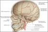
How does this artery enter the cranial fossa?

A

Foramen spinosum

12
Q

Which foramen is associated with mandibular nerve + where is it on the diagram?

A

Foramen ovale

13
Q

Which layer is closely adherent to the brain?

A

The pia mater

14
Q

Which arteries supply the anterior cranial fossa + where is on the diagram?

A

Anterior meningeal arteries

15
Q

Which structures of the brain are found within the posterior cranial fossa?

A

Cerebellum and brainstem

16
Q

Which bones form the floor of the posterior cranial fossa?

A

Occipital bone

Temporal bone

17
Q

How can you identify an extradural haematoma on a CT scan?

A

More focal

Does not extend beyond suture

Bi-convex (lens shaped)

18
Q

What is the falx cerebri?

A

Falx cerebri is a crescent shaped downward projections of meningeal dura mater from the dura lining the calva that passes between the two cerebral hemispheres towards the corpus callosum.

Helps stabilise the brain within the cranial cavity

19
Q

What structure is being pointed to?

A

Pterion

20
Q

Name the different coloured structures

A

Green = frontal bone

Blue = sphenoid bone

Dark pink = nasal bone

Light pink = maxilla

Orange = zygomatic bone

Purple = mandible

Red = vomer

21
Q

What is the highlighted bone?

A

Zygomatic process

22
Q

Which foramen does the accessory nerve pass through?

A

Jugular foramen

23
Q

Which part of the temporal bone has a flat plate appearance and forms the superior regions of the temporal bone?

A

Squamous part

24
Q

What is a subdural bleed?

A

Deep to the dura

25
Role of the 2 posterior communicating arteries?
Connects internal carotid artery with posterior cerebral artery
26
What structures pass through the foramen ovale?
Mandibular nerve Accessory meningeal artery Lesser petrosal nerve (branch of the glossopharyngeal nerve) Emissary vein connecting through cavernous sinus
27
Which bones do the body of the sphenoid bone articulate with?
Ethmoid, vomer and palatine bone
28
Label the parts of the maxilla: - Zygomatic process - Orbital surface - Infra-orbital foramen - Frontal process - Alveolar process - Anterior nasal space
29
What is the junction between the saggital and coronal suture?
Bregma
30
Upon CSF drainage which layer is collapsed onto surface of the brain?
Arachnoid layer
31
Identify structures on the diagram: - optic canal - superior orbital fissure - foramen rotundum - foramen spinosum - foramen lacerum - foramen ovale - carotid canal
32
Which 2 branches arise from the middle meningeal artery + where do they go?
Anterior and posterior branch of meningeal artery Anterior branch crosses sphenoid's great wing reaching groove of parietal bone Posterior branch curves back toward temporal bone before reaching back portions of parietal bone
33
Name of the 2 cranial foramina found on greater wing of sphenoid bone?
Foramen ovale Foramen spinosum
34
What bone forms the forehead?
Frontal bone
35
What does the frontal bone form anteriorly?
Forehead, and the superior part of the rim of each orbit Superior to the rim of the orbit on each side are the raised superciliary arches
36
Which foramen is circled in red + what nerve is associated with it? What is its position to the superior orbital fissure?
Optic canal and optic nerve Medial to the superior orbital fissure
37
Describe the 2 layers of the dura mater
Periosteal is very closely adherent to inner surface of the bone Meningeal is close to the arachnoid
38
What is the tentorium cerebelli?
Horizontal projections of meningeal dura mater that covers and separates cerebellum in posterior cranial fossa from posterior parts of cerebral hemisphere Provides structural support for brain in event head is shaken or hit
39
What meningeal layer lies beneath the dura?
The arachnoid mater
40
What structure is highlighted in the picture and what nerve passes through it?
Foramen lacerum + greater petrosal nerve (facial nerve)
41
What are the 5 layers that form the scalp + label them on the diagram
Skin Connective layer Aponeurotic layer Loose connective tissue Pericranium
42
Which nerve bundles is associated with the cribiform plate?
Olfactory nerve bundles
43
Which artery forms upon the union of the 2 vertebral arteries?
Basilar artery
44
Which carotid artery supplies the face?
External carotid artery
45
Role of anterior communicating artery
Connects the left and right anterior cerebral arteries
46
Which 4 cranial nerve pass through the superior orbital fissure + which structure is it?
Oculomotor nerve (CN3) Trochlear nerve (CN4) Abducens nerve (CN6) Opthalmic nerve (first branch of the trigeminal nerve (CN5)
47
What structure is highlighted and what bones articulate with it?
Lamboid suture Laterally, parietal bones and posteriorly, occipital bone
48
What is the glabella?
Depression within the raised supercilliary arches (bony point present on the frontal bone between two superciliary arches)
49
What structure is being pointed to and what bone is it located on?
Mental foramen and mandible bone
50
Function of the CSF
Helps to protect the brain against movement Nutrient role in terms of supplying nutrients to the brain
51
What structure is being pointed to and what bone is it located on?
Infraorbital foramen and maxillary bone
52
What projections are found in the subarachnoid space?
Spider-like projections of the arachnoid mater
53
When do venous sinuses form?
When the two layers of the dura mater open up, they form venous sinuses where venous blood circulates within the cranial cavity
54
Name the highlighted structure
Sphenoid bone
55
Which structures is formed from the fold and union of the two dura mater layers?
Falx Celebri
56
What are the 3 cranial fossa? Identify them on the diagram
Purple = anterior cranial fossa Blue = middle cranial fossa Green = posterior cranial fossa
57
What parts of the brain does the anterior cranial fossa contain?
- Frontal lobe of cerebral cortex - Olfactory bulb - Olfactory tract - Orbital gyri
58
What is the large membranous and unossified structure between the bones of the skull found in infants?
Anterior fontanelles
59
How can you identify a subdural haematoma on a CT scan?
Usually large, crescentic + indirect (not a site of impact)
60
Describe the qualities of the arachnoid mater
Elastic and has spider-like projections
61
What structure is being pointed to?
Foramen magnum
62
Arteries within the circle of willis?
Left and right anterior cerebral artery Anterior communicating artery Left and right internal carotid artery Posterior cerebral artery (left + right) Posterior communicating artery (left + right)
63
Bones forming the anterior cranial fossa?
Frontal, ethmoid and sphenoid bones
64
Which bone does the zygomatic process anteriorly articulate with?
Zygomatic bone
65
Which artery resides deep to the pterion?
Anterior branch of middle meningeal artery
66
Which dura layer is adherent to the inner surface of the bone
Periosteum
67
Which suture does the unpaired frontal bone pair up with the parietal bones?
Coronal suture
68
Which foramen is characterised by its minute hole and where is it on the diagram?
Foramen spinosum
69
What are the different bones that form the pterion (colour)?
Red - frontal bone Orange - parietal bone Purple - temporal bone Yellow - greater sphenoid wing
70
Which cranial nerves pass through the jugular foramen?
Glossopharyngeal nerve (IX) Vagus nerve (X) Accessory nerve (XI)
71
Which arteries arise from the internal carotid arteries?
Ophthalmic artery Posterior communicating artery Middle cerebral artery Anterior cerebral artery
72
Which carotid artery supplies the cranial cavity?
Internal carotid artery
73
Which foramen does the hypoglossal nerve pass through?
Hypoglossal canal
74
Which foramen are located on the superior rim of the orbit?
Supraorbital foramen
75
Which foramen does the vagus nerve pass through?
Jugular foramen
76
Four main parts of the temporal bone and identify them on unlabelled diagram
Squamous part Mastoid process Zygomatic process External acoustic meatus (timpanic part)
77
Which bones form the pterion?
Point where the greater sphenoid wing, temporal, parietal and frontal bones meet
78
What passes through the foramen spinosum?
Middle meningeal artery
79
Junction between the sagittal and lamboid suture?
Lambda
80
Which is the main foramen that can be seen on the inferior aspect of the skull?
Foramen magnum
81
What is the unlabelled structure called?
Anterior fontanelles
82
Which structure is highlighted and what does it contain?
Foramina of the cribiform plate (within the ethmoid bone) Contains the olfactory nerve bundles
83
Which formamen does the hypoglossal nerve pass through?
Hypoglossal canal
84
Which two cranial nerves pass through the internal acoustic meatus?
Facial nerve (CN7) Vestibulocochlear nerve (CN8)
85
Which types of bleed are associated with damage to the pterion?
Intracranial bleeds
86
What does a rupture of middle meningeal artery at the pterion typically lead to?
Epidural haematoma
87
Label the structures missing on the diagram - Falx cerebelli - Tentorium cerebelli
88
How far does the falx cerebri go?
Down to the corpus collosum
89
What nerve goes through optic canal and where does it go?
Optic nerve and goes through the back of the eye to supply the eye
90
Which foramen does the abducens nerve pass through?
Superior orbital fissure
91
Which artery supplies the posterior cranial fossa?
Posterior meningeal artery
92
What structure is highlighted in the diagram and what bone does it articulate with?
Saggital suture and the paired parietal bone
93
Which lobe of cerebrum resides within middle cranial fossa + what area of MCF?
Temporal lobe Lateral part
94
How do the 2 posterior cerebral arteries form?
Bifurcation of basilar artery
95
Which part of the temporal bone forms part of the surface of the external acoustic opening?
Tympanic part
96
What is the largest artery supplying the dura mater?
Middle meningeal artery (branch of maxillary)
97
Which foramen does the maxillary nerve pass through + what structure is it on the diagram?
Foramen rotundum
98
What are the labeled structures?
99
Which meningeal artery passes through jugular foramen?
Posterior meningeal artery
100
Which foramen does the internal carotid artery pass through + where is it on the diagram?
Carotid canal
101
Which bone does the pituitary gland reside within?
Sella turcica of the sphenoid bone
102
Identify: - Lesser wing - Greater wing - Foramen ovale - Foramen rotundum - Foramen spinosum - Superior orbital fissure
103
Structures associated with optic canal?
Opthalmic artery + optic nerve
104
Which arteries supply the middle cranial fossa + what foramen does it come out of?
Middle meningeal artery and it enters through the foramen spinosum
105
What is the highlighted structure?
Supraorbital foramen
106
Which foramen does the trochlear nerve pass through?
Superior orbital fissure
107
What does the subarachnoid space contain?
Spider-like projections of the arachnoid mater Cerebral spinal fluid
108
What is the structure being pointed to?
Vomer bone
109
Which artery is deep to the pterion?
Middle meningeal artery
110
Describe the dura mater
Outermost layer Thick + inelastic layer Has 2 layers (periosteal and meningeal)
111
Which part of the skull is the weakest?
pterion
112
What bones form the middle cranial fossa?
Sphenoid and temporal bone
113
What is the extra-dural space?
Generated within vertebral columns Spinal dura mater is adherent to the foramen magnum Periosteal mater lines the vertebrae This produces a separation which is the extra-dural space Epidural anaesthesia is injected within this space
114
What is the structure highlighted?
Sphenosquamous suture
115
What cranial nerve is the maxillary nerve from?
One of the three branches of the trigeminal nerve (CN5)
116
Structures passing through superior orbital fissure?
Oculomotor nerve Trochlear nerve Opthalmic nerve Abducens nerve Opthalmic vein (superior divisions)
117
Structures forming the brainstem + label them on the diagram?
medulla pons cerebellum
118
What are the 6 main anterior bones of the skull?
Frontal Nasal Lacrimal Maxilla Zygomatic Mandible
119
Which artery provides afferent information regarding vision?
Internal carotid artery
120
What are the 3 meningeal layers?
Dura mater Arachnoid Pia mater
121
Which are the six main bones on the lateral view of the skull and what colour do they correspond to?
Frontal bone - light yellow Parietal bone - red Occipital bone - orange Temporal bone - pink Zygomatic bone - blue Sphenoid bone (greater wing) - purple
122
What is the effect of the haematoma on intracranial pressure?
Intracranial pressure increases
123
What structure is being pointed to and what bone is it located on?
Supraorbital foramen and frontal bone
124
Name of highlighted bone
Palatine bone
125
Which foramen does the glossopharyngeal nerve pass through?
Jugular foramen
126
Which artery provides impressions on the cranial cavity?
Middle meningeal artery
127
Which sinus is lateral to the sella turcica?
Cavernous sinus