HNS 2 Flashcards

(209 cards)

1
Q

What are the 3 main functions of the neck?

A

Structural: supports and moves the head

Visceral: contains the trachea and oesophagus

Conduit for blood vessels and nerves

2
Q

Principle arterial supply of the head and neck?

A

Carotid arteries

3
Q

What is fascia?

A

A connective tissue mainly composed of collagen fibres

Organises the body into different compartments

4
Q

Why is the fascia clinically important?

A

Permits the spread of infection within compartments

5
Q

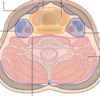
What is the most external fascial layer in the neck and what does it contain?

A

Superficial fascia

Contains the platysma muscle at the front of the neck

6
Q

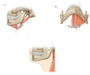
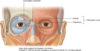
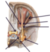
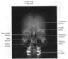
Where on this diagram is the platysma muscle?

A

Anterior to the neck

7
Q

What is the platysma innervated by?

A

Cervical branch of facial nerve

8
Q

What are the 3 main compartments of the neck?

A

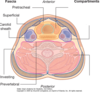
Visceral compartment

Vertebral compartment

Vascular compartment

9
Q

What is deep to the superficial fascia?

A

Deep fascia

10
Q

What layers are the deep fascia divided into?

A

Pretracheal fascia

Carotid sheath

Investing fascia

Prevertebral fascia

11
Q

What does the pretracheal fascia surround?

A

Some of the visceral components of the neck

We can find some of the components of the digestive system and respiratory system + endocringe glands

12
Q

Where is oesophagus in relation to trachea?

A

Oesophagus is posterior to the trachea

13
Q

Which structures reside within the visceral compartments?

A

Oesophagus, trachea, pharynx + thyroid gland

14
Q

What is deep fascia?

A

Dense, organised connective tissue deep to the superficial fascia

Organised into distinct layers:

  • Carotid sheath
  • Pretracheal fascia
  • Investing fascia
  • Prevertebral fascia
15
Q

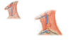
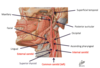
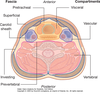
What is the carotid sheath and where is it on the diagram?

A

Fascia that surrounds the:

  • common carotid artery
  • internal jugular vein
  • internal carotid artery
  • vagus nerve
16
Q

What does the investing layer of fascia contain?

A
  • Sternocleidomastoid muscle
  • Trapezius muscle
  • Infrahyoid muscles
17
Q

What structures are contained in the prevertebral layer + function?

A
  • Spinal cord
  • Deep muscles of the back
  • Anterior, posterior + middle scalene muscles

Contains a number of muscles which help move + stabilise the ehad

18
Q

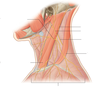
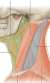
What are the 2 main triangles of the neck?

A

Anterior triangle

Posterior triangle

19
Q

What divides the 2 muscles of the neck + where does it go from and to?

A

Sternocleidomastoid muscle

From the skull down to the sternum + clavicle

20
Q

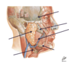
What are the boundaries of the anterior triangle?

A
  • Inferior margin of the mandible (superior)
  • Anterior border of the sternocleidomastoid muscle (posterior)
21
Q

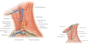
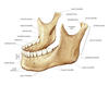
Label diagram of the neck

A
22
Q

Boundaries of the posterior triangle?

A

Posterior aspect of the sternocleidomastoid muscle

Anterior aspect = anterior aspect of the trapezius muscle

23
Q

What muscles are contained in the anterior triangle of the neck?

A

Platysma muscle

Suprahyoid muscles (mylohyoid, geniohyoid, digastric + stylohyoid muscles)

Infrahyoid muscles (omohyoid, sternohyoid, thyrohyoid + sternothyroid muscle)

24
Q

What blood vessels are contained in the anterior triangle of the neck?

A

Internal jugular vein

Common carotid artery

Internal carotid artery

25
Label the diagrams
26
Which cranial nerves pass through the anterior triangle of the neck?
Vagus nerve Glossopharyngeal Accessory Hypoglossal Facial nerve
27
What structures are mainly contained in posterior triangle of the neck?
Mainly blood vessels and nerve
28
What blood vessels and nerves are found in posterior neck?
External jugular vein Subclavian artery Subclavian vein Trunks of brachial plexus Phrenic nerve Vagus nerve Spinal accessory nerve
29
Which organ does the phrenic nerve supply?
Diaphragm
30
Where does the vagus nerve have functions?
Respiratory, cardiovascular + abdominal or GI tract
31
Label the diagram in posterior triangle
32
Label this diagram of nerves in the posterior triangle of the neck
33
Name cranial nerve XI and what muscles does it supply?
Accessory nerve Sternocleidomastoid and trapezius muscle
34
Label the diagram of principal artery supply to the neck
35
What does the common carotid artery divide into?
External carotid and internal carotid
36
What does: - external carotid artery - internal carotid artery
- External carotid artery supplies the face - Internal carotid artery goes into cranial cavity to supply brain
37
What are the principle branches of the external carotid artery?
- Superior thyroid - Lingual - Ascending pharyngeal - Occipital - Facial - Posterior auricular (emerges behind the ear and supplies the skin on the back of the ear) - Superficial temporal - Maxillary (has a branch that goes into cranial cavity to support the meninges)
38
Which nerve innervates the posterior belly of the digastric muscle?
Facial nerve
39
Which vein is superficial to the sternocleidomastoid muscle?
External jugular vein
40
What do the muscles of facial expression do?
They can act to dilate or constrict the orifices of the face e.g. eyes and mouth
41
What groups can the muscles of facial expression be divided into and what are they innervated by?
Orbital, nasal and oral muscles Innervated by cranial nerve 7 (facial nerve)
42
Label diagram of orbital muscles
43
44
What do the oral group of muscles act on?
Mouth and lips
45
What do auricular muscles move?
Move the ear
46
What nerve are the facial musles innervated by?
Cranial nerve number 7
47
Does the facial nerve innervate the parotid gland?
No
48
Why is the relationship between the facial nerve and the parotid gland clinically relevant?
surgery to remove the parotid gland, can also impact upon the function of the muscles of facial expression if damage to the cranial nerve occurs during surgery (facial nerve runs through parotid gland but doesn't innervate it) This can leave a patient with paralysis of the facial muscles on one side of the face
49
What are the branches of the facial nerve?
Zygomatic, buccal, mandibular, temporal and cervical branches
50
Label this diagram of the facial nerves
51
Joint between mandible and temporal bone?
Temporomandibular joint (TMJ)
52
Where do the ramus and inferior border of the mandible meet?
Angle of the mandible
53
What is the temporomandibular joint
Pair of joints connecting the mandible to the skull Bilateral synovial articulation between the temporal bone and mandible, assisting in mastication Muscles of mastication move the mandible
54
What part of the mandible forms the joint with the temporal bone?
Condylar process
55
What are the TMJ?
Temporomandibular joints are a pair of joints on the side of the head Allow for opening and closing of the mouth for chewing (mastication)
56
What are the muscles of mastication innervated by?
Mandibular branch of the trigeminal nerve (CN5)
57
What does the movement of the TMJ depend on?
Dependent upon the level to which the mandible is open
58
What happens when the jaw is slightly open?
Hinge action predominates (opening + closing of mouth)
59
What happens when jaw is opened widely?
Hinge + gliding action occurs
60
What do movements of the mandible include?
Protrusion - jaw moves forwards Retraction - jaw move backwards Elevation - jaw moves upwards Depression - jaw moves downwards
61
What muscle facilitates movements of the jaw (protrusion, retraction, elevation + depression)?
Facilitiated by the superficial muscles
62
What are the superficial muscles that facilitate movements of the jaws?
Temporalis muscle and masseter muscle
63
Where does temporalis muscle originate from?
Temporal fossa
64
Where does the temporalis muscle insert into?
Coronoid process of the mandible
65
What is the main function of the temporalis muscle?
Elevates + retraction of the mandible
66
Where do masseter muscles attach to?
The ramus and angle of the mandible
67
Where does masseter muscle originate from?
From the zygomatic arch
68
What does masseter muscle cause the jaw to do + when is it used for?
Causes elevation of the mandible + forced closure of mouth is required
69
What are the deeper muscles that move the mandible?
Lateral + medial pterygoid muscles
70
Label this diagram
71
Label this diagram
72
What is the lateral pterygoid muscle attached to and what process does it bring about?
Sphenoid bone, lateral pterygoid plate and neck of mandible Depresses and protracts mandible to open the mouth
73
What is the medial pterygoid muscle attached to and what process does it bring about?
Lateral pterygoid plate, maxilla, palate and attached to the angle of the mandible Bring about elevation, protraction and when used in isolation, brings about side to side movement for grinding
74
What are the orbits of the eye?
Bilateral structures which contain the eyeball, the muscles that move the eye, extraocular muscles, the optic nerve and other nerves and vessels
75
What is the roof of the orbit made of?
Orbital plate of the frontal bone
76
What is the floor of the orbit made up of?
Orbital plate of the maxilla
77
What is the medial wall of the orbit made up of?
Ethmoid and lacrimal bones
78
What is the lateral wall of the orbit made up of?
Lateral wall made up of the zygoma
79
What are the 3 main foramina of the orbit?
Optic canal Superior orbital fissure Inferior orbital fissure
80
Label this diagram containing the orbital foramina
81
What passes through the superior orbital fissure?
Opthalmic branch of trigeminal nerve (V1) Oculomotor nerve (III) Trochlear (IV) Abducens (VI) Opthalmic vessels Sympathetic fibres
82
Where is the optic canal positioned in orbit?
More medially
83
What passes through the optic canal?
The optic nerve + opthalmic artery
84
Where does the inferior orbital fissure sit in the orbit?
Inferiorally in the orbit
85
What passes through the inferior orbital fissure?
Maxillary division of the trigeminal nerve + infraorbital vessels
86
Where is the medial and lateral side of the orbit retrospective to the rest of the face?
Medial = towards the nose Lateral = towards the ear
87
What are the 6 extraocular muscles of the eye + the extra facial muscle?
Superior rectus Medial rectus Lateral rectus Inferior rectus Superior oblique Inferior oblique Levator palpebrae superioris
88
What do each of the extraocular muscles do?
Medial rectus -\> adduction of eye Lateral rectus -\> abduction of eye Superior rectus -\> elevation of eye Inferior rectus -\> Depression of eye Superior oblique -\> Incyclotorsion (inward, torsional movement of the eye) Inferior oblique -\> Excyclotorsion (outward, torsional movement of the eye) Levator palpebrae superioris -\> Elevation/retraction of upper eyelid
89
What is the common origin of the rectus muscles and how far do the rectus muscles insert into the eye?
Common tenderness + insert about 5mm into each eyeball
90
What are each of the rectus muscles innervated by?
The oculomotor nerve (CN3) (except LR) Lateral rectus muscle is innervated by abducens nerve (CN6)
91
Where do superior and inferior oblique muscles arise from respectively?
Body of sphenoid (superior) Orbital surface of maxilla (inferior)
92
Where do the superior and inferior oblique muscles insert into?
Outer posterior quadrant for the inferior surface of the inferior oblique and the superior surface for the superior oblique
93
What are the inferior and superior oblique innervated by respectively?
Oculomotor nerve (CN3) for inferior oblique Trochlear nerve for superior oblique
94
What muscle elevates the upper eyelid?
Levator palpebrae superioris
95
Explain the innervation of the levator palpebra superioris
Smooth muscle fibres innervated by sympathetic fibres Voluntary muscles fibres innvervated by oculomotor nerve (CN3)
96
Label this diagram of the eye
97
What is the action of the lateral rectus muscle in isolation?
Lateral rectus moves the eye laterally
98
What is the action of the medial rectus muscle in isolation?
Medial rectus moves the eye medially
99
What is the action of the inferior rectus muscle in isolation?
Moves the eye inferiorly and medially
100
What is the action of the superior rectus muscle in isolation?
Superior rectus moves the eye medially and superiorly
101
What is the action of the superior oblique muscle in isolation?
Moves the eye inferiorly and out laterally (down + out)
102
What is the action of the inferior oblique muscle in isolation?
Moves eye superiorly and laterally (up + out)
103
What is important to note when testing the muscles clinically?
The isolated action of the muscles is slightly different to the way the muscles are tested clinically
104
How would you test the lateral rectus muscle?
Ask someone to look laterally e.g. left eye look left + right eye look right
105
How would you test the medial rectus?
Look towards the nose (test each eye separately)
106
How would you test the superior and inferior rectus?
Ask patient to respectively look upwards and look downwards You must position the eye to give these muscles the advantage as more than one muscle contributes to movement of the the eye upwards and downwards For superior rectus muscle - ask them to look outwards and then upwards and then downwards to test the inferior rectus muscle
107
In a clinical test how would you distinguish between the rectus and oblique muscles in the eye?
Ask the patient to look towards their nose (medially -\> adducted position) then test superior oblique by asking them to look downwards for the inferior oblique ask them to look superiorly with the eye already placed in a medial position (adducted position)
108
Label this diagram of the nerves of the orbit + what view is this diagram in?
Superior
109
What is in the optic chiasm?
A proportion of fibers coming from the eye will cross to the other side of the body
110
What does the trochlear nerve innervate in the eye?
Carrying fibres that will supply the superior oblique muscle
111
What does the oculomotor nerve innervate?
Skeletal muscle of the levator palpebrae superioris, superior rectus, medial rectus, inferior rectus and inferior oblique muscles Also carries autonomic fibres that alter the size of the pupil and thickness of the lens
112
What are the 3 divisions of the trigeminal nerve?
The opthalmic division (V1) Maxillary division (V2) Mandibular division (V3)
113
What divisions of the opthalmic division carry sensation?
The frontal and lacrimal divisions (lacrimal receives fibers that supply the lacrimal gland)
114
Which ganglion contains the cell bodies of the oculomotor nerve?
115
What is the ciliary ganglion?
Ganglion of the third or oculomotor nerve through which autonomic parasympathetic fibres get to the eye and serve functions such as changing diameter of the pupil and lens thickness
116
Which fibres cause the pupil to dilate and constrict respectively?
Sympathetic (dilate) and parasympathetic (constrict)
117
How do parasympathetic fibres change the thickness of the lens?
Innervate some smooth muscles (thickening of lens is called accommodation)
118
Which paired dural venous sinus is located within the cranial cavity?
Cavernous sinus
119
Which cranial fossa is the cavernous sinus located within?
Middle cranial fossa on either side of the sella turcica of the sphenoid bone
120
Which veins supply the cavernous sinus?
Cerebral and opthalmic veins
121
Why is the cavernous sinus clinically relevant?
Spread of infection through this to the cranial cavity from facial injuries
122
What is the lacrimal system?
System involved in production, movement and drainage of fluid from surface of the eye
123
Where is the lacrimal gland located?
In the supralateral quadrant of the orbit
124
What are the 4 structures of the lacrimal system?
Lacrimal gland Canaliculi Sac Nasolacrimal duct
125
Through what nerve does the lacrimal gland receive parasympathetic secretor motor fibres?
CN 7 facial nerve
126
Where does excess fluid drain into (eye)?
Into the nasal cavity via nasolacrimal duct
127
Label this diagram of the lacrimal system
128
Where does the lacrimal canaliculi open into?
Open into the medial corner of the eye
129
Where does fluid from the nasolacrimal duct eventually drain into?
The nasal meatus
130
What are the nasal cavities?
Two wedge shaped cavities that are the uppermost part of the respiratory tract
131
What are the 2 nasal cavities separated by?
They are separated by a midline septum
132
What separates the nasal cavities from the oral cavity below?
Separated from oral cavity by hard palate
133
What separates the nasal cavity from the cranial cavity?
Parts of a number of bones including the frontal, ethmoidal + sphenoid bones
134
What are the bones that stick out into the nasal cavity?
Conchae
135
What are the different types of conchae?
Superior concha, middle concha + inferior concha
136
What are the main functions of the nasal cavity?
Warms + humidifies the inspired air Removes and traps parthogens and particular matter from inspired air Responsible for sense of smell Drains and clears the paranasal sinuses and lacrimal ducts
137
What are the nasal conchae?
Projecting out of the lateral walls of the nasal cavity. Inferior, middle and superior conchae Project into the nasal cavity which results in the formation of 4 pathways for airflow The passages are called meatuses
138
What are the 3 meatuses?
Inferior meatus Middle meatus Superior meatus
139
Where is the superior meatus?
Between the middle and superior concha
140
Where can the middle meatus be found?
Between the middle and inferior conchae
141
Where is the inferior meatus?
Inferior to inferior conchae
142
What do the conchae do?
Increase the surface area of the cavity and thought to warm and humidify air + trap pathogens
143
Label this diagram of the nasal cavity
144
What is the nasal cavity innervated by + what do each of the nerves innervate?
The olfactory nerve -\> olfaction (sense of smell) The trigeminal nerve -\> general sensation of the nasal cavity via the opthalmic division in anterior region (v1) + maxillary division of the trigeminal nerve in posterior region(v2) The facial nerve -\> supplies parasympathetic fibers to the glands of the nasal cavities Sympathetic nerves (from T1) -\> supply vascular smooth muscle of the nasal cavities
145
Why do the nasal cavities have a rich blood supply?
They aid in humidifying and changing the temperature of the respired air
146
Which arteries supply the nasal cavities?
Internal carotid artery and branches of the external carotid artery
147
Why is the nasal cavity susceptible to nose bleeds?
Great degree of anastomoses between the arteries supplying the nasal cavities
148
Why is there a risk of infection with the nasal cavity?
Risk of infection being taken from the nasal cavity into the cranial cavity Because some of the blood vessels which drain the nasal cavity pass through the cavernous sinus into the cranial cavity
149
Label this diagram of the arterial supply to the nose
150
What are the 4 main paranasal sinuses in the skull ?
Frontal sinuses Ethmoid air cells Sphenoid sinuses Maxillary sinuses
151
What do the facial sinuses do?
Helps to reduce the weight of the skull Acts as a crumple zone in the case of facial trauma Increase the resonance of the voice and humidifying + heating inhaled air
152
What are paranasal sinuses?
Air filled extensions of the respiratory regions of the nasal cavity
153
What do paranasal sinuses secrete and why are they clinically relevant?
Mucous Clinically relevant as they can also become infected
154
Which nerve innervates the paranasal sinuses?
Branches of the trigeminal nerve
155
Which cranial nerve innervates the frontal sinus?
The opthalmic division (v1)
156
What cranial nerve innervates the ethmoid air cells?
Opthalmic and maxillary divisions
157
What cranial nerve innervates the sphenoid sinuses?
Opthalmic and maxillary
158
What cranial nerve innervates the maxillary sinuses?
Maxillary branch of the trigeminal nerve
159
Label this diagram
160
Label this diagram of the paranasal sinuses
161
Label this diagram of the x ray
162
Where does the sphenoid sinus drain into?
Drains into the sphenoidal recess superior to the superior concha
163
What is inferior to the sphenoid sinus and to the superior concha?
Opening of the ethmoidal cells into the lateral wall of the superior meatus
164
What is anterior to the ethmoidal cells?
The opening or drainage point of the frontal sinus
165
What is inferior to the ethmoidal cells?
The opening of the maxillary sinus
166
What is under the inferior concha?
The opening of the nasolacrimal duct
167
Where is the opening of the nasolacrimal duct + function?
Inferior to the inferior concha + where excess fluid from the lacrimal system is drained
168
What is posterior to the nasolacrimal duct?
The opening of the pharyngotympanic tube
169
Label this diagram
170
What three regions is the ear divided into?
External, middle + internal ear
171
What is the purpose of the outer ear?
To transmit sound towards tympanic membrane or the eardrum
172
What does the tympanic membrane have + function?
Small ossicles or bones attached which transmits sound into the inner ear
173
What is located in the inner ear?
Sense organ for both hearing, cochlear, balanced and the semicircular canals are located
174
Which part of the CN VIII is for hearing and which part is for balance?
Hearing -\> cochlear part (brings information about sound) Balance and orientation -\> vestibular part (signals relating to balance and orientation from the vestibular system)
175
What nerve runs through the middle ear?
Facial nerve
176
Which canal forms the superficial surface of the external ear?
External acoustic meatus
177
What is the lateral projection from the head of the external ear?
Auricle
178
What is the auricle made of?
Cartilage
179
What is the outer surface of the ear?
External auricular rim = helix
180
What is the hollow centre of the auricle?
Concha of the auricle
181
What structure is found anterior to the opening of the external acoustic meatus?
Tragus
182
What structure resides opposite to the tragus?
Anti-tragus
183
What does the pharyngotympanic tube connect?
Connects the middle ear with the nasopharynx
184
What does the pharyngotympanic tube connect?
Connects the middle ear with the nasopharynx
185
What is the role performed by the pharyngotympanic tube?
Equalises pressure on both sides of tympanic membrane Equalises pressure between the middle ear and atmosphere
186
What is the middle ear continuous with?
Opening into the nasal cavity known as pharyngotympanic tube
187
Where does the pharynogotympanic tuber enter?
Enter the nasopharynx, posterior to the inferior meatus of the nasal cavity
188
What does the facial nerve innervate in the ear?
Stapedius
189
What motor functions does the facial nerve have?
Muscles of facial expression, stapedius, digastric (posterior belly) + stylohyoid
190
What does the sensory component of the facial nerve innervate?
Gives rise to the sensation of taste from the anterior 2/3s of the tongue, contains parasympathetic fibers which go to the lacrimal glands, the muscous glands of the nasal cavity, hard and soft palates and sublingual + submandibular glands (saliva) General sensation from external acoustic meatus and deeper parts of the auricle
191
Label this diagram
192
What can happen if a patient has a serious middle ear infection?
Can be transmission of infection from middle ear into cranial fossa, which can result in serious complications such as meningitis or inflammation of the meninges surrounding the membrane.
193
What is the vasculature of the middle ear?
Branches arise from the ascending pharyngeal artery (Branch of the external artery), and from two branches o fi to the maxillary artery (Middle meningeal artery, and the artery of the pterygoid canal)
194
What is the venous drainage of the middle ear?
Pterygoid plexus of veins in the infra temporal fossa
195
Which plexus innervates the mucous membrane lining the pharyngotympanic tube?
Tympanic plexus
196
Which nerve innervates tympanic plexus?
Glossopharyngeal nerve [IX]
197
What can happen if a patient has a serious middle ear infection?
Can be transmission of infection from middle ear into cranial fossa, which can result in serious complications such as meningitis or inflammation of the meninges surrounding the membrane.
198
Label diagram + what type of muscles are they?
Strap muscles
199
Label this diagram
200
What is the chorda tympani?
The chorda tympani is a branch of the facial nerve that originates from the taste buds in the front of the tongue, runs through the middle ear, and carries taste messages to the brain.
201
What is the geniculate ganglion?
The geniculate ganglion is a collection of pseudounipolar sensory neurons of the facial nerve located in the facial canal of the head.
202
What is the stylomastoid foramen?
The stylomastoid foramen is a rounded opening at the inferior end of the facial canal. between the base of the styloid process and the mastoid process of the temporal bone
203
How does facial nerve work in the ear?
The facial nerve [VII] is closely associated with the vestibulocochlear nerve [VIII] as it enters the internal acoustic meatus of the temporal bone. Traveling through the temporal bone, its path and several of its branches are directly related to the internal and middle ears. The facial nerve [VII] enters the internal acoustic meatus in the petrous part of the temporal bone. The vestibulocochlear nerve and the labyrinthine artery accompany it. At the distal end of the internal acoustic meatus, the facial nerve [VII] enters the facial canal and continues laterally between the internal and middle ears. At this point the facial nerve [VII] enlarges and bends posteriorly and laterally. The enlargement is the sensory geniculate ganglion . As the facial canal continues, the facial nerve [VII] turns sharply downward, and running in an almost vertical direction, it exits the skull through the stylomastoid foramen.
204
What connects the middle ear with the nasopharynx and where is the nasopharynx in relation to the inferior meatus?
Pharyngotympanic tube Posterior
205
Label this diagram
206
207
Where does the masseter originate and insert from? What does it do?
Origin: zygomatic arch and maxillary process of zygomatic bone Insertion: lateral surface of ramus of mandible Elevation of mandible
208
Where does temporalis originate and insert from? Function of temporalis?
Origin: bone of temporal fossa + temporal fascia Insertion: coronoid process of mandible and anteriori margin of ramus function: elevation + retraction of mandible
209