Lab 4 Flashcards

(91 cards)

1
Q

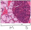
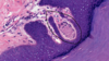
Skin layers - epidermis, dermis (Papillary Layer: Loose CT, Reticular Layer: Dense irregular CT), hypodermis

A
2
Q

Epidermis (E). Papillary layer of dermis (P), reticular layer of dermis (R). Hypodermis (H)

A
3
Q

Dermal papillae, epidermal ridge or interpapillary peg

A
4
Q

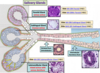
Eccrine sweat glands in reticular layer of dermis

A
5
Q

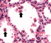
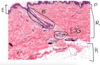
Pacinian corpuscle at border between hypodermis and dermis – detects vibration and pressure lower down in the skin. There are layers of Schwann cells with fluid filled spaces in between, and centre has an axon

A
6
Q

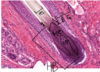
Meissner’s corpuscle in papillary layer of dermis - have barrel shape that have unmyelinated axon and groups of schwann cells that form layers. The nuclei on the outer edge of meissner is schwann cell nuclei, there are some fibroblasts too.

A
7
Q

Basement membrane under epidermis (wrong), points to granulosum layer
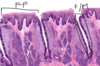
A
8
Q

Layers of epidermis

A

Stratum corneum has keratin (no nuclei, has some holes). Stratum basale (forms stem cells that divide, mitosis occurs here, has merkel cells). Stratum spinosum (cells with nice nucleolus and lots of euchromatin, prickle areas between adjacent cells where desmosomes bind the intermediate filaments from one cell to the next cell). Stratum granulosum (keratohyalin granules, 2-3 cell layers thick, cells make proteins that cross link the keratin). Stratum lucidum (cells from stratum granulosum that died, has keratin flakes).

9
Q

Thin skin characteristics

A

Epidermis with thin keratin layer (stratified squamous keratinized epithelium), papillary layer of dermis, reticular layer of dermis. Sweat glands, hair, sebaceous glands. Arrector pili muscle (smooth muscle that attaches to hair follicle, causes goose bumps, if the muscle contracts then it squishes the sebaceous gland to release sebum for hair lubrication)

10
Q

Thin skin - thin epidermis (due to thin stratum corneum and thin stratum spinosum), adipose tissue at hypodermis. Reticular layer of dermis. Papillary layer of dermis (Lighter collagenous layer). Eccrine sweat glands. Hair follicle.

A
11
Q

Thin skin – thin stratum corneum, stratum basale (cells with brown pigments, melanocytes with white halo due to golgi apparatus making melanosomes)

A
12
Q

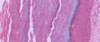
Thin skin – bundles of elastin within reticular layer of dermis (elastin changes with age, decreases with age)

A
13
Q

Scalp is thin skin – lots of hair follicles in cross section. Sebaceous glands, sweat glands.

A
14
Q

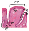
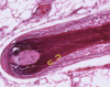
Hair follicle – hair bulb, dermal papilla. Hair matrix cells. Hair shaft (inside to out: medulla, cortex, hair cuticle), internal root sheath (inside to out: internal root sheath cuticle, Huxley layer, Henle layer), external root sheath (continuous with epithelium)

A
15
Q

Hair follicle cross section

A
16
Q

Scalp – hair follicles in hypodermis; hair shaft, internal root sheath, external root sheath (multicellular layer), glassy membrane (is basement membrane continuous with epidermis). Arrector pili muscle

A
17
Q

Hair shaft longitudinal – hair bulb, dermal papillae, matrix cells, hair shaft, cuticle, internal root sheath, external root sheath, glassy membrane
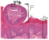
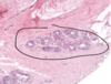
A
18
Q

Sebaceous gland

A

big grouping is one sebaceous gland

19
Q

Sweat gland, Simple tubular eccrine sweat gland

A

Sweat glands in clumps, Simple tubular eccrine sweat gland is lighter colored and encircled with black outline

20
Q

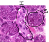
Eccrine sweat gland of Thick skin with resorptive duct and secretory coil – 2 profiles: darker stained is duct portion (modifies secretions), lighter stained is secretory portion (secretes components of sweat: proteins, electrolytes, ammonia, urea). Darker stained duct portion has STRATIFIED CUBOIDAL epithelium with lumen (2 layers of cells). Lighter stained secretory portion has PSEUDOSTRATIFIED EPITHELIUM around the gland and also a ring of darker eosinophilic cells called MYOEPITHELIAL cells on the outside.

A
21
Q

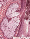
Scalp – sebaceous gland (branched acini) around hair follicle.

A

Holocrine secretion occurs in sebaceous gland: cells at basal part are on basement membrane and are a bit basophilic due to protein and lipid production. Basal part cells divide and daughter cells are pushed to centre of sebaceous gland, filling with lipid droplets. After cells are filled up, apoptosis occurs, releasing oil to hair follicle.

Merocrine secretion is exocytosis of secretory vesicles in eccrine sweat glands and apocrine sweat glands. Apocrine secretion is in mammary glands and some male reproductive glands.

22
Q

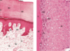
Left: Human thick skin, x132. Right: Monkey thick skin, x540.
Abbreviations: BV: Blood vessels; D: dermis; DR: dermal ridges; E: epidermis;
ER: epidermal ridges; KG: Keratohyalin granules; PL: papillary layer; RL: reticulum layer; S:squames; SC: stratum corneum; SG: stratum granulosum; SS: stratum spinosum; Tc: transitional cells; d: duct of sweat gland.

A
23
Q

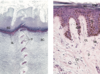
Human thick (left, x132) and thin (right, x270) skin sections. Abbreviations: CF: collagen fibre; CL: capillary loops; D: dermis; SB: stratum basale; SC: stratum corneum; SG: stratum granulosum; SL: stratum lucidum; SS: stratum spinosum; sDR: secondary dermal ridges; d:duct of a sweat gland.

A
24
Q

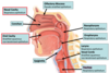
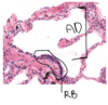
Epithelium of Nasal Cavity, conchae, Oral Cavity, epiglottis, Olfactory Mucosa, Nasopharynx, Oropharynx, Larynx, Vocal Cords, Trachea

A

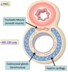
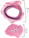
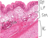
Respiratory epithelium is pseudostratified columnar epithelium with goblet cells

25
Nasal Conchae and Palate – nasal cavity (pseudostratified columnar with cilia epithelium with hard-to-see goblet cells) on top, oral cavity (stratified squamous epithelium non keratinized) on bottom
26
Hyaline cartilage (around lumen to keep it open), Submucosa (has submucosal glands to make mucus and serous secretions), Trachealis muscle (smooth muscle)
27
Trachea, esophagus
28
Trachea – Hyaline cartilage, Trachealis muscle (thick bundles of smooth muscle with worm-like nuclei)
29
Trachea – hyaline cartilage (isogenous groups and chondrocytes in lacunae), submucosa, sero-mucous glands (patchy appearance ones secrete mucus, eosinophilic ones have serous secretions), muscularis mucosa (smooth muscle), lamina propria, respiratory epithelium with cilia (cilia pushes debris up respiratory tract to leave tract, goblet cells make mucus to trap debris)
30
Isogenous groups, chondrocytes in lacunae
31
Trachea longitudinal – many cartilage rings
32
Trachea longitudinal - hyaline cartilage, submucosa with submucosal glands (mucus glands and serous glands), muscularis mucosa (smooth muscle), lamina propria, basement membrane, respiratory epithelium with pseudostratified columnar and cilia and goblet cells.
33
Hyaline cartilage, submucosa, submucosal glands, lamina propria, respiratory epithelium
34
Lung – Alveoli made of simple squamous epithelium
35
Intrapulmonary bronchi in lung – hyaline cartilage
36
Intrapulmonary bronchi in lung – hyaline cartilage, submucosal glands in submucosa, muscularis mucosa (smooth muscle), lamina propria, basement membrane, respiratory epithelium
37
Bronchiole (less glands, less cartilage, no respiratory epithelium, no goblet cells) – simple cuboidal epithelium with cilia, club cells (secretory cells that do not secrete mucus but secrete surfactant-like material that lubricates the lumen of the bronchiole and prevents the lumen from sticking together and it helps keep the lumen open b/c no more cartilage)
38
RESPIRATORY BRONCHIOLE with simple cuboidal epithelium on one end and ALVEOLAR DUCT (which is continuous with alveolar tissue) with simple squamous epithelium on the other end
39
RESPIRATORY BRONCHIOLE with simple cuboidal epithelium on one end and ALVEOLAR DUCT (which is continuous with alveolar tissue) with simple squamous epithelium on the other end
40
Alveolar duct (collection of alveoli that form elongated shape), alveolar sac (collection of alveoli form a circular shape), atrium (spaces where one side is termination of alveolar duct and , pore of Kohn
41
Lung – alveolar septum (wall that separates air space of diff Alveoli), squamous simple epithelium made of type I pneumocytes on either side of alveolar septum, capillaries (RBC) in between type I pneumocytes, endothelial cells are nuclei surrounding RBC. Blood-air barrier is made up of type I pneumocyte, ‘basement membrane shared between type I pneumocyte & endothelial cell’ and endothelial cell. Type II pneumocyte is bigger and more cuboidal and found at junction or corners between alveoli. Alveolar macrophages are large and found in lumen of alveoli, they phagocytose foreign material (dust, bacteria) to push out respiratory tract using mucus and cilia
42
Lung - Alveolar duct, alveolar sac
43
Olfactory Mucosa - olfactory epithelium, duct of olfactory glands, olfactory glands, olfactory nerves, ethmoid bone
Photomicrograph of olfactory epithelium. The olfactory epithelium is a pseudostratified epithelium, which contains olfactory cells, supporting cells, and basal cells. The olfactory cells contain specialized cilia, which are non-motile. The underlying connective tissue connects to the ethmoid bone and is largely occupied by olfactory (Bowman’s) glands, olfactory nerves, and blood vessels. The ducts of the olfactory glands extend from the secretory portion of the gland to the epithelial surface.
44
Pharynx and larynx diagram
45
Terminal bronchioles
Smallest conducting bronchioles are terminal bronchioles (TB). Have very small diameters, and lumen lined with simple cuboidal epithelium (E) with Clara cells (CC) or club cells. Terminal bronchioles give rise to respiratory bronchioles (RB) whose walls resemble those of the terminal bronchioles, except there is also the presence of alveoli to allow gas exchange.
46
Mouse tongue – skeletal muscle in all orientations, stratified squamous epithelium with keratin at bottom of tongue (no lingual papillae but yes dermal papillae at bottom of tongue) (human tongue is stratified squamous non keratinized), filiform papillae (no taste buds) on top of tongue to make it grippy
47
Mouse tongue
48
Human tongue
49
Human tongue cross section – stratified squamous epithelium with no keratin, filiform papillae without taste buds but with some keratin
50
Human tongue cross section – glandular tissue (aka lingual glands) (aka salivary glands): serous glands (serous acini) and mucus glands (mucus acini)
51
Tongue – fungiform (can see dorsal taste bud), stratified squamous mostly non keratinized epithelium
52
Sides and back of tongue - Foliate papillae, furrows aka moats
53
Cell types in lateral taste bud of foliate papillae – taste bud, taste pore, basal stem cells (differentiate into other cell types), sensory cells (have receptors that bind to particles) (have microvilli that extend into the taste pore), supporting cells (provide metabolic support to sensory cells) (have microvilli that extend into the taste pore)
54
Circumvallate papillae, dermal papillae, lateral taste buds, stratified squamous non keratinized epithelium, von Ebner gland (mucus or serous) (secretes into moats between circumvallate papillae to solubilize the particles you taste to help it interact with taste buds receptors), moat
55
Filiform papillae, circumvallate papillae, fungiform papillae, foliate papillae
56
Parotid Gland - serous acinus
57
Sublingual Gland - mucous acinus
58
Striated Duct (Intralobular)
59
Interlobular Excretory Duct
60
Submandibular Gland - mixed acinus with serous demilune
61
Salivary glands diagram
62
Parotid gland (close to ear) (mostly serous) – serous acini, striated duct (columnar epithelium)
63
Parotid gland (close to ear) (mostly serous) – serous acini, intercalated duct (cuboidal epithelium)
64
Parotid gland (close to ear) (mostly serous) – lobules separated by connective tissue
65
Parotid gland (close to ear) (mostly serous) – interlobular excretory duct
66
Parotid gland (close to ear) (mostly serous) – intercalated duct, serous acini
67
Sublingual gland (beneath tongue) (mostly mucus) – right side is mucus acini, left side if mix of mucus and serous acini
68
Sublingual gland (beneath tongue) (mostly mucus) – mucus acini, lobules separated by connective tissue, interlobular excretory duct (low columnar or tall cuboidal cells)
69
Submandibular gland (beneath jaw bone) (mix of serous acini and mucus acini) – serous demilune (preparation artifact: when this section is prepared, the serous cells that are part of the acini get push to the outside and wrap around the mucus cells), striated ducts, serous acini, mucus acini
70
Submandibular gland (beneath jaw bone) (mix of serous acini and mucus acini) – interlobular duct
71
Submandibular gland (beneath jaw bone) (mix of serous acini and mucus acini) – intercalated duct
72
Type I pneumocyte
73
interpapillary peg or epidermal ridge
74
Circumvallate papilla
75
eccrine sweat gland
76
submandibular gland
77
Alveolar macrophage
78
dermal papilla of hair follicle
79
olfactory mucosa
80
Taste pore
81
stratum granulosum of thick skin
82
Filiform papilla
83
Respiratory epithelium lines this structure
84
Thick skin
85
Alveolus or alveolar sac
86
eccrine sweat gland does merocrine secretion
87
Parotid gland
88
Internal root sheath of hair follicle
89
Clara cells secrete surfactant-like material
90
Stratum lucidum
91
Von ebner's gland