Lab 6 Flashcards

(149 cards)

1
Q

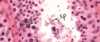
Endocrine pancreas cells

A

Pancreatic acini (with exocrine cells) - dark purple

Pancreatic islet (with endocfine cells) - light purple

2
Q

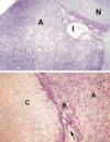
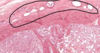
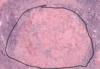
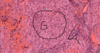
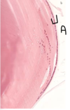
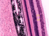
Pineal gland - identifying feature

A

Brain sand

3
Q

Pineal gland - general

A
4
Q

Neurohypophysis vs pineal gland

A

Neurohypophysis:

• Hormone release (oxytocin, ADH)

  • Produced in Hypothalamus
  • Pituicytes
  • Herring Bodies

Pineal Gland

• Hormone production and release (Melatonin)

  • Pinealocytes
  • Brain Sand (corpora arenacea)
5
Q

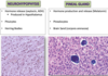
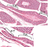
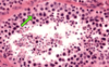
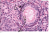
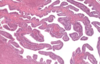
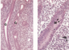
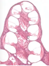
Islet of Langerhans (endocrine pancreas), surrounded by serous acini (exocrine pancreas)

A

Assume that most cells in middle of islet are beta and cells at periphery of islet is alpha cells

6
Q

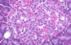
Alpha and beta cells in islet of Langerhans

A

Allows differentiation bw alpha and beta cells in islet of Langerhans. Beta cells stain more purple, alpha cells stain more pink. Most cells are beta cells, especially the ones in the middle

7
Q

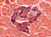
Beta cells in pancreas

A

Stains the insulin that is secreted by beta cells, beta cells stain darker

8
Q

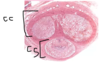
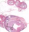
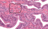
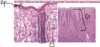
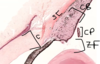
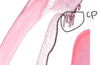
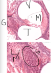
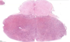
Pituitary - general parts

A

Lighter top region is pars nervosa, darker bottom region is pars distalis, middle is pars intermedia

9
Q

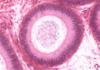
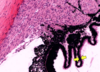
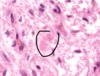
Pituitary - remnants of Rathke’s pouch

A

At pars intermedia, the circles are remnants of Rathke’s pouch

10
Q

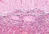
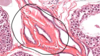
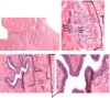
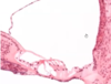
Pars nervosa of pituitary consists of neuropil, pituicytes, endothelial cells, fenestrated capillaries, Herring bodies

A

Pars nervosa: neuropil is wispy material, most nuclei belong to pituicytes, some endothelial cells near RBC, fenestrated capillaries, identifying feature: pink blob called Herring bodies

11
Q

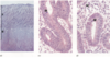
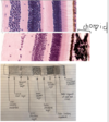
Pars distalis of pituitary: Chromophobes, Basophils, Acidophils

A

Pars distalis: Chromophobes (don’t stain very intensely, not much color, already released all their hormones). Basophils (stain very dark, basophilic), Acidophils (stains very eosinophilic). Diff regions of pars distalis will have diff proportions of these 3 cell types

12
Q

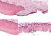
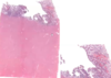
Pituitary - pars distalis and pars nervosa

A

Pars distalis is colorful. Lighter staining pars nervosa.

13
Q

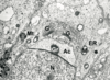
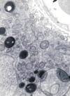
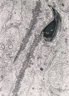
Pituitary - Pars nervosa: fenestrated capillary, neuropil, Herring body

A
14
Q

Pituitary - Pars intermedia

A

Pars intermedia: remnants of Rathke’s pouch

15
Q

Pituitary - Pars distalis: Acidophils, chromophobes

A
16
Q

Pituitary - pars distalis: basophils

A
17
Q

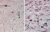
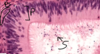
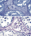
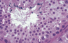
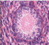
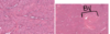
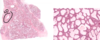
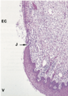
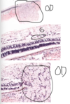
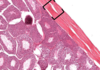
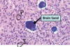
Pineal gland - pinealocytes, interstitial cells, brain sand

A

Pineal gland: made of nervous tissue, nuclei belong to one of 2 cell types. Larger nuclei that stain lighter belong to pinealocytes (make melatonin). Darker, elongated nuclei are interstitial cells. Brain sand (aggregates of calcified secretions and calcified ECM)
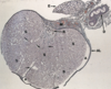
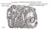
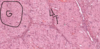
18
Q

Pineal gland - B&W

A
19
Q

Pars nervosa: pituicytes, herring bodies

A
20
Q

Pineal gland: pinealocytes, brain sand ( corpora arenacea )

A
21
Q

Pituicyte of pars nervosa

A
22
Q

Pars nervosa of pituitary gland

A
23
Q

Acidophils

A

Acidophils, secretes growth hormone and prolactin

24
Q

Pinealocytes of pineal gland

A
25
Parafollicular cells - colloid, follicles, follicular cells, parafollicular cells, blood vessels
Abbreviations: BV: Blood vessels; Cl: colloid; CT: connective tissue; F: follicles; FC: follicular cells; N: Nuclei; PF: parafollicular cells (which may be distinguished by its pale cytoplasm and larger nucleus)
26
Parafollicular cells
The lightly stained cells among the principal cells of the follicular epithelium and the rounded cells (arrowheads) in the interstitial tissue are parafollicular cells. The surrounding cytoplasm is clear. The follicles are distinguished by the presence of colloid (c).
27
Oxyphil cells vs chief cells
The parathyroid contains two major types of cell: the chief cell & the oxyphil cell. Oxyphil cells are smaller in population but larger in size. Their location are indicated by arrow on the top. They occur singly (arrow in the middle) or cluster together (arrow on the bottom).
28
Chief cells (aka principal cells) and oxyphil cells
The parenchyma of the parathyroid gland consists of two types of cells, chief cells (CC), also known as principal cells, and oxyphil cells (OC). Chief cells are more numerous and possess darker staining cytoplasm. They form small cords surrounded by slender connective tissue (CT) elements and blood vessels (BV). The nuclei (N) of connective tissue cell has an elongated appearance. Oxyphil cells stain lighter and are usually larger in size and their cell membranes are evident.
29
Pituitary gland, hypophysis, B&W
30
Pituitary gland, hypothalamus, pars tuberalis, pars anterior, pars nervosa, pars intermedia
This photomicrograph of the pituitary gland demonstrated the relationship of the gland to the hypothalamus (H) from which it is suspended by the infundibulum. The infundibulum is composed of a neural portion, the infundibular stem (IS) and the sourounding pars tuberalis (PT). The largest portion of the pituitary is the pars anterior (PA), which is glandular and secretes numerous hormones. The neural component of the pituitary gland is the pars nervosa (PN) that does not manu- facture its hormones, but stores and release them. Between the pars anterior and pars nervosa is pars intermedia (PI).
31
Pituitary gland, anterior adenohypophysis, posterior neurohypophysis, pars distalis, pars intermedia, pars nervosa
The pituitary contains two general regions: the anterior adenohypophysis (A) and the posterior neurohypophysis (N). The adenohypophysis consists of secretory cells and their blood supply. the neurohypophysis contains axons from the paraventricular and supraoptic nuclei of the hypothalamus, their supporting cells and blood vessels. the adenophypophysis is sub- divided into pars distalis (at A) and the pars intermedia (I). In bottom image, the neural characteristics of the pars nervosa ( C ) of the neurohypophysis become more evident. The pars distalis (A) is glandular in structure and the pars intermedia (B) contains collidal cysts.
32
Herring bodies
The posterior pituitary stores and secretes ADH (Vasopressin) and Oxytocin. They are synthesized by the nerve cell bodies in the hypothalamic nuclei and are transported down the axons of the hypothalamo-hypophyseal tract. The terminal regions of the axons are dilated and contain secretory granules filled with hormone. These expanded portions of the axons are known as Herring bodies (arrowhead).
33
pars intermedia, basophils, colloid-filled follicles
Situated between the pars anterior (PA) and the pars nervosa (PN) the pars intermedia of the pituitary gland is characterized by basophils (B) that are smaller than those of the pars anterior. It also contains collid (Cl)-filled follicles, lined by pale, small, low cuboidal-shaped cells (CC). Numerous blood vessels (BV) and pituicytes (P) are evident in this area of the pars nervosa.
34
Pituitary gland - pars nervosa
The pars nervosa of the pituitary gland is composed of elongated cells with long processes known as pituicytes (P) that are thought to be neuroglial in nature. These cells appear to support numerous unmyelinated nerve fibres traveling from the hypothalamus via the hypothalamo-hypothyseal tract. Neurosecretory materials pass along these nerve fibres and are stored in expanded regions at the termination of the fibres, which are known as Herring bodies (HB).
35
Testis are covered by tunica albuginea
36
Cross sections of seminiferous tubules in testis
37
seminiferous tubule in testis - myoid cells and leydig cells
A seminiferous tubule in testis. Surrounding each tubule, is MYOID cells (CT cells, with contractile function). In bw tubules in CT, are LEYDIG cells (function makes testosterone, pale cytoplasm, cholesterol is testosterone precursor).
38
seminiferous tubule in testis - sertoli cells and spermatozoa
A seminiferous tubule in testis. Lumen has small dense nuclei are SPERMATOZOA. SERTOLI cell is round nuclei with prominent nucleoli, makes blood testis barrier, supports spermatogenic cells, is epithelial cell
39
SPERMATOGONIA
SPERMATOGONIA are stem cell, on basement membrane, very dark.
40
PRIMARY SPERMATOCYTE, EARLY SPERMATIDS, LATE SPERMATIDS
PRIMARY SPERMATOCYTE has coarse textured nucleus. EARLY SPERMATIDS have smaller nucleus, round. LATE SPERMATIDS are elongated
41
Testis and Epididymis
42
Seminiferous tubule, tubulus rectus, rete testis
Testis. Seminiferous tubule (ST) on the right. Moving right to left, there is transition. Short region of simple columnar ep made up of Sertoli cells (TUBULUS RECTUS). Moving to the left again, ep becomes simple cuboidal ep (RETE TESTIS)
43
MESHWORK OF RETE TESTIS
Following rete testis further to left, there is MESHWORK OF RETE TESTIS, with lots of simple cuboidal ep lined tubules.
44
EFFERENT DUCTULE tubule and epididimys
45
EPIDIDIMYS, basal cells, principal cells, spermatozoa
EPIDIDIMYS (pseudostratified columnar ep). BASAL cells on basement membrane. PRINCIPAL cells reach from basement membrane to apical surface, which have stereocilia. Lumen has spermatozoa
46
Ductus deferens ( VAS DEFERENS )
Ductus deferens (VAS DEFERENS). Large layer of smooth muscle on outside.
47
Vas defererns, basal cells, principal cells
Epithelially-lined tubule within Vas defererns. Pseudostratified columnar ep with basal cells that don’t reach apical surface and principal cells with stereocilia reach surface
48
Vas deferens - 3 diff layers of smooth muscle. Inner longitudinal layer, Middle circumferential layer, Outer longitudinal layer
Vas deferens. Moving to smooth muscle, there are 3 diff layers of smooth muscle. Inner longitudinal layer, Middle circumferential layer, Outer longitudinal layer.
49
Vas deferens. Large blood vessel.
50
Vas deferens is in continuation with SEMINAL VESICLE
51
SEMINAL VESICLE
52
Seminal vesicle with glandular tissue and folded mucosa. Pseudostratified ep. Some basal stem cells.
53
PROSTATE GLAND - PROSTATIC CONCRETIONS
PROSTATE GLAND. Lots of glandular tissue with folded mucosa. Ep here varies from region to region. Pseudostratified columnar, simple columnar or simple cuboidal. PROSTATIC CONCRETIONS is distinguishing feature.
54
In bw the ep of the prostate gland, there is CT that has lots of smooth muscle
55
CORPUS SPONGIOSUM and CORPUS CAVERNOSUM
CORPUS SPONGIOSUM on bottom (urethra). Top is 2 of the same structure: CORPUS CAVERNOSUM. Collectively called corpora cavernosa. In centre of corpora cavernosa are central arteries.
56
URETHRA of corpus spongiosum
Corpus spongiosum. Epithelially lined URETHRA (pseudostratified columnar ep). Surrounding it is erectile tissue with blood sinusoids and some CT and some smooth muscle.
57
CENTRAL ARTERY of corpus cavernosum
Corpus cavernosum. CENTRAL ARTERY, can see layers of smooth muscle.
58
DORSAL BLOOD VESSELS above corpus cavernosum
Above corpus cavernosum. DORSAL BLOOD VESSELS. Dorsal arteries or dorsal veins
59
TUNICA ALBUGINEA surrounding corpora
Surrounding the corpora is TUNICA ALBUGINEA
60
Epididymis
61
Sertoli Cell
62
testis - seminiferous tubules, epididymis, ductus deferens, tunica albuginea, mediastinum testes
Photomicrograph of the testis, epididymis (E), and part of the ductus deferens (D). The tunica albuginea (AL), which on the posterior surface of the testis becomes thickened and forms the mediastinum testes (M). The connective tissue septae (arrows), which divide the testis into lobules containing the seminiferous tubules (S). R represents rete testis.
63
Spermatids - acrosomal vesicle, membrane bound granules
Briefly, the major steps of spermiogenesis include the formation of an acrosomal vesicle (Ac) through the elaboration of membrane bound granules (Gr) from the Golgi complex (G) of the spermatids, the development of a flagellum, changes in the size and shape of the nucleus (N) and the further conden- sation of chromatin, and the loss of residual cytoplasm around the head of the spermatozoa. In the spermatid cytoplasm shown here are microtubules (Mt), mitochondria (M) and area of rough endo- plasmic reticulum (ER).
64
Interstitial cells of Leydig
Steroid secreting cells (arrow) can be found within the interstitial spaces between the seminiferous tubules. These are testosterone secreting cells, the interstitial cells of Leydig. Leydig cells have a large, vacuolated cytoplasm in routine histological sections. Their ultrastructural components are typical of steroid secreting cells: an abundance of smooth endoplasmic reticulum, mitochondria and lipid droplets.
65
Interstitial cell of Leydig - EM
66
Seminiferous Tubules - spermatogonia, primary spermatocytes, sertoli cells, spermatids and spermatozoa
Spermatogonia (A) form the basal layer of cells. In the second layer toward the lumen are primary spermatocytes (B) and nuclei of supporting cells (Sertoli cells, arrowheads). The third alayer consists of secondary spermatocytes which rapidly divide and, as a result, cannot be identified with certainty in most histological sections. The innermost layer of cells is formed by spermatids and spermatozoa (C).
67
Seminiferous Tubules - nucleoli of Sertoli cell, primary spermatocytes, elongated heads of spermatozoa, spermatids
Elongated heads of spermatozoa (arrowhead) with their condensed complement of nuclear material are enclosed by the cytoplasm of Sertoli cells. The tails of the spermatozoa are directed toward the lumen of the seminiferous tubule. The small cells with rounded nuclei adjacent to the layer of spermatozoa are spermatids (arrows). A: nucleoli of Sertoli cell. B: primary spermatocytes.
68
Seminiferous tubules
At the lower right, the spherical nucleus (N) belongs to a fairly early spermatid. It is covered by a flattened vesicle, the developing acrosome (Ac), which contains a dense granule (Gr), the proacrosomal granule. The cap and the granule arise from the Golgi complex (G), and together they form the anterior tip of the mature sperm. Mitochondria (M) and vesicles of the endoplasmic reticulum (ER) are shown in early spermiogenesis. One spermatid (\*) appears in this micrograph. The appearance of a nearly mature sperm is illustrated at the center of the micrograph. Abbreviations: Ac’: acrosome cap; C: long bundle of filaments; Ce: centrioles; FS: fibrous sheath; M’: mitochondria; M” & ER’: mitochondria and dilated vesicles of the endoplasmic reticulum of a Sertoli cell; N’: nucleus.
69
Human prostate
70
Monkey Prostate gland
The fibroelastic connective tissue stroma (St) presents numerous blood vessels (BV) and smooth muscle cells (SM). The parenchyma of the gland is composed of columnar cells (CC) as well as short basal cells (BC). The dome-shaped apices (arrowheads) of some of the columnar cells appear to protrude into the lumen, which contain a prostatic concretion (Pc). The number of these concretions, which may calcify, increases with age
71
Uterus - oviduct, vagina, cervix, myometrium, endometrium
72
Ovary - medulla, cortex
MEDULLA is lighter inside, CORTEX is darker outside
73
Ovary - germinal epithelium, tunica albuginea
Single layer of squamous or low cuboidal. GERMINAL EP. CT layer under is TUNICA ALBUGINEA.
74
Ovary - primordial follicle, squamous follicular cells ( granulosa cells )
PRIMORDIAL FOLLICLE surrounded by single layer of SQUAMOUS FOLLICULAR CELLS aka GRANULOSA CELLS
75
Ovary - unilaminar primary follicle, granulosa cells, zona pellucida
UNILAMINAR PRIMARY FOLLICLE. GRANULOSA CELLS look cuboidal. ZONA PELLUCIDA bw oocyte and granulosa cells. Zona pellucida is glycoproteins
76
Ovary - multilaminar primary follicle, granulosa cells
MULTILAMINAR PRIMARY FOLLICLE. GRANULOSA CELLS has more than 1 layer.
77
Ovary - secondary (antral) follicle, antrum, oocyte, zona pellucida, corona radiata, cumulus oophorus, granulosa cell
SECONDARY (ANTRAL) FOLLICLE. ANTRUM in pink. Liquor folliculi is in the antrum. Large OOCYTE surrounded by ZONA PELLUCDA. CORONA RADIATA. CUMULUS OOPHORUS. Thick GRANULOSA CELL layer.
78
Ovary - outside of secondary follicle - basement membrane, theca interna, theca externa
Outside of secondary follicle. Granulosa cells sit on BASEMENT MEMBRANE. THECA INTERNA. THECA EXTERNA (circumferential layer of CT)
79
Ovary - Graafian follicle aka mature follicle
GRAAFIAN FOLLICLE. Aka mature follicle. Granulosa cells. Zona pellucida, corona radiata. Theca interna, theca externa. Small SCARS next to it.
80
Ovary - scars next to graafian follicle
81
Graafian follicle with large antrum and thin granulosa cell layer
Graafian follicle with large antrum filled with liquor folliculi. Thin granulosa cell layer.
82
Corpus albicans
Large scar
83
Corpus hemorrhagicum
84
Corpus hemorrhagicum - granulosa lutein cells, theca lutein cells
Corpus hemorrhagicum. Large pale cells are GRANULOSA LUTEIN CELLS. Darker cells are THECA LUTEIN CELLS.
85
CORPUS LUTEUM
86
Corpus luteum - granulosa lutein cells, theca lutein cells
Corpus luteum. Lighter, bigger GRANULOSA LUTEIN CELLS. Branches of darker THECA LUTEIN CELLS.
87
Corpus albicans
Scars next to corpus luteum, called CORPUS ALBICANS
88
Ovary, oviduct, corpus luteum
Ovary on bottom. Oviduct on top. Large CORPUS LUTEUM of the ovary, purpose is to support pregnancy.
89
Oviduct MUCOSAL FOLDS
90
Oviduct - ciliated cells, peg cells
Oviduct. Epithelium. Pseudostratified columnar with basal cells. CILIATED CELLS. PEG CELLS with protrusions into lumen and secretory function.
91
BASAL STEM CELL in oviduct.
92
Oviduct. LAMINA PROPRIA
Oviduct. LAMINA PROPRIA in pink
93
Surrounding oviduct is layer of CT and some smooth muscle
94
CERVICAL CANAL vs ECTOCERVIX
Right is CERVICAL CANAL. Left is ECTOCERVIX. Ectocervix transitions to vagina. Ectocervix ep is stratified squamous non keratinized ep (same ep as vagina). Cervical canal has simple columnar ep.
95
CERVICAL GLANDS
Going further into cervical canal, can see CERVICAL GLANDS with simple columnar ep. They produce mucus
96
Uterus - ENDOMETRIUM, PROLIFERATIVE PHASE: BASAL LAYER and FUNCTIONAL LAYER. MYOMETRIUM.
ENDOMETRIUM, PROLIFERATIVE PHASE. Eosinophilic region at bottom is MYOMETRIUM. ENDOMETRIUM on top; BASAL LAYER (stratum basalis) and FUNCTIONAL LAYER. (stratum functionalis)
97
Uterus - myometrium, large blood vessels
Myometrium. Bundles of smooth muscle. Large blood vessels.
98
Uterus - Endometrium, proliferative phase. ENDOMETRIAL GLANDS
Endometrium, proliferative phase. Simple columnar ep is continuous with ENDOMETRIAL GLANDS. Glands are thin and straight, characteristic of proliferative phase of endometrium.
99
Uterus - basal layer of endomterium, spiral arteries
Cross sections of arterioles. These are SPIRAL ARTERIES, which ruptures capillaries and causes functional layer to fall off during periods. Basal layer of endometrium.
100
Uterus - SECRETORY PHASE of ENDOMETRIUM
SECRETORY PHASE of ENDOMETRIUM. Myometrium is lighter stained at bottom Thinner basal layer of endometrium. Large functional layer.
101
Uterus - SECRETORY PHASE of ENDOMETRIUM - glands, spiral arteries
Glands are sacculated and wider; characteristic of secretory phase of endometrium. Some cross sections of spiral arteries
102
Uterus - MENSTRUAL PHASE of ENDOMETRIUM
MENSTRUAL PHASE of ENDOMETRIUM. Bleeding in lamina propria, due to spiral arteries contracting, causing a period.
103
Mammary gland - RESTING stage, Stage during PREGNANCY, LACTATING stage
MAMMARY GLAND. RESTING stage (has duct systems, mostly CT), pre pregnancy (1). Stage during PREGNANCY (Glandular duct systems proliferate to form alveoli. More equal ratio bw glandular tissue and CT) (2). LACTATING stage, after pregnancy (alveoli swell up to fill with secretions, not much CT) (3).
104
Mammary Gland - RESTING stage - TERMINAL DUCT LOBULE UNIT, LACTIFEROUS DUCT
RESTING stage. Lots of CT. Some Lobules. TERMINAL DUCT LOBULE UNIT. This unit has intralobular CT (in bw ducts) and interlobular CT surrounding the lobule unit. Also has some larger ducts with stratified cuboidal ep, this is LACTIFEROUS DUCT that collects secretions from terminal duct lobule unit.
105
Mammary gland - Border bw lactating and pregnancy stage - Colostrum, lactiferous duct
Border bw lactating and pregnancy stage. More glandular tissue than CT. Alveoli has COLOSTRUM (not milk yet). Organization in lobules that have intralobular CT and interlobular CT. LACTIFEROUS DUCT.
106
Mammary gland - lactating stage - lactiferous duct
LACTATING stage. Alveoli are swelled up. Not much intralobular CT, but there is interlobular CT. LACTIFEROUS DUCT with stratified cuboidal ep.
107
LACTATING mammary gland
Some cells have lipid droplets, secrete by apocrine. Some cells have proteins, secrete via merocrine secretion to release zymogen granules.
108
Proliferating endometrium - B & W
glands are straight, stroma is highly cellular
109
Proliferative endometrium - myometrium M, tratum basalis B, stratum spongiosum S and stratum compactum C.
Micrograph (a) illustrates early proliferative endometrium at low magnification. At the bottom of the field is the muscular wall, the myometrium M. The relatively thin endometrium consists of the stratum basalis B, stratum spongiosum S and stratum compactum C. The glands at this stage are fairly sparse and straight. As the gland, stroma and vessels proliferate, the endometrium gradually becomes thicker. By day 5-6 of the cycle the surface epithelium has regenerated. During the proliferative phase the epithelial cells acquire microvilli and cilia as well as the cytoplasmic organelles required for the secretory phase.
110
Proliferative endometrium - endometrial glands G, Mitotic figures M
At higher magnification in micrograph (b), the straight tubular form of the endometrial glands G can be seen. At very high magnification in micrograph (c), the proliferating glandular epithelium is seen to consist of columnar cells with basally located nuclei exhibiting prominent nucleoli. Mitotic figures M can be seen both in the epithelium and in the stroma. Note the highly cellular stroma, almost devoid of collagen fibre.
111
Proliferative endometrium - stratum functionalis F, stratum basalis B, mitotic figures M
By the late proliferative stage, shown at low magnification in micrograph (d), the endometrium has doubled in thickness. Note that in contrast to the stratum functionalis F, the appearance of the stratum basalis B is little changed when compared with the early proliferative phase. With further magnification, micrograph (e) shows that the tubular glands are now becoming coiled and more closely packed. At very high magnification in micrograph (f), mitotic figures M are more prevalent in both the glandular epithelium and the supporting stroma. The stroma is also somewhat oedematous at this stage. During the proliferative phase there is a continuum of change which makes the precise dating of the cycle inaccurate in histological specimens. Lymphocytes and occasional lymphoid aggregates are a normal feature of late proliferative phase endometrium.
112
Uterine cervix - endocervical canal (EC), vagina (V), junction (J)
The uterine cervix protrudes into the upper vagina and contains the endocervical canal linking the uterine cavity with the vagina. The function of the cervix is to admit spermatozoa to the genital tract at the time when fertilization is possible, i.e. around the time of ovulation, but at other times, including pregnancy, its function is to protect the uterus and upper tract from bacterial invasion. In addition, the cervix must be capable of great dilatation to permit the passage of the fetus during parturition. As seen in this micrograph, the endocervical canal (EC) is lined by a single layer of tall columnar mucus-secreting cells. Where the cervix is exposed to the more hostile environment of the vagina (V), the ectocervix, it is lined by thick stratified squamous epithelium as in the rest of the vagina. The cells of the ectocervix often have clear cytoplasm due to their high glycogen content (not apparent in this specimen). The junction (J) between the vaginal and endocervical epithelium is quite abrupt and is normally located at the external os, the point as which the endocervical canal opens into the vagina. The main bulk of the cervix is composed of tough, collagenous tissue containing relatively little smooth muscle. Beneath the squamocolumnar junction, the cervical stroma is often infiltrated with leucocytes forming part of the defense against ingress of microorganisms.
113
VITREOUS CHAMBER, POSTERIOR CHAMBER, ANTERIOR CHAMBER, LENS, IRIS, CORNEA
114
SCLERA, CORNEA, CILIARY BODY has muscle to pull lens, ZONULA FIBERS, CILIARY PROCESSES
115
CHOROID
116
CHOROID, outer fibrous tunic, inner fibrous tunic, retina
CHOROID at back of eye, in contact with outer fibrous tunic to the right of it and the inner fibrous tunic to the left of it called retina
117
MYOFIBROBLAST
MYOFIBROBLAST on lens have contractile function
118
CORNEAL EPITHELIUM, BOWMAN’S MEMBRANE, CORNEAL STROMA, DESCEMET’S MEMBRANE
Cornea has stratified squamous non keratinized ep, called CORNEAL EPITHELIUM, which sits on a basement membrane called BOWMAN’S MEMBRANE. Largest layer is CORNEAL STROMA made of DICT. Stroma also has lots of water. Under corneal stroma is DESCEMET’S MEMBRANE
119
CORNEAL ENDOTHELIUM on DESCEMET’S MEMBRANE. CORNEAL STROMA, BOWMAN’S MEMBRANE, CORNEAL EPITHELIUM.
120
CILIARY PROCESSES
Cornea receives nutrients from AQUEOUS HUMOUR liquid that is made by CILIARY PROCESSES. Fluid travels through pupil into anterior chamber.
121
CANAL of SCHLEMM
CANAL of SCHLEMM allows aqueous humour to drain out back into blood system
122
Ciliary processes - Outer pigmented epithelium, inner non pigmented epithelium
Ciliary processes have double layer of ep. Outer pigmented ep sits on the same basement membrane as inner non pigmented ep. These ep together make aqueous humour
123
ANTERIOR LENS EPITHELIUM
ANTERIOR LENS EP is simple cuboidal. As ep travels done, it reaches zone of proliferation. Then differentiate to zonula fibers that are long cell bodies and lose their nuclei. Crystallin is the protein made. Capsule surrounds lens, which is made of basement membrane.
124
Eye - Lens - Basement membrane, anterior lens epithelium, lose nuclei
Lens. BASEMENT MEMBRANE. ANTERIOR LENS EP. Zonula fibers also LOSE NUCLEI
125
Eye - LENS FIBER filled with CRYSTALLIN PROTEIN
126
Retina - inner limiting membrane, nerve fiber layer, gangion cell layer, inner plexiform layer, inner nuclear layer, outer plexiform layer, outer nuclear layer, outer limiting membrane, outer segment of rods and cones, retinal pigmented epithelium
127
Eye - OPTIC DISK, GANGLION CELLS
128
Inner ear - OSSEOUS LABYRINTH, MEMBRANOUS LABYRINTH, AMPULLA (Crista ampularis), UTRICLE
129
Ampulla - sensory hair cells, supporting cells
Ampulla, SENSORY HAIR CELLS at surface with stereocilia, SUPPORTING CELLS beneath
130
Inner ear - MACULA OF THE UTRICLE
MACULA OF THE UTRICLE. Hair cells with stereocilia cells and supporting cells beneath it.
131
COCHLEA
132
Inner ear - SCALA VESTIBULI (perilymph), SCALA TYMPANI (perilymph), SCALA MEDIA (endolymph), SPIRAL GANGLION, MODIOLUS
133
HELICOTREMA
134
ORGAN OF CORTI, REISSNER’S MEMBRANE
135
Tectorial membrane, cochlear nerve, outer hair cells, inner hair cells, basilar membrane, inner phalangeal cells, outer phalangeal cells
136
BASILAR MEMBRANE, 3 OUTER HAIR CELLS, 1 INNER HAIR CELL, SUPPORTING PHALANGEAL CELLS, TECTORIAL MEMBRANE
137
STRIA VASCULARIS
138
PROSTATE GLAND
139
Eye overview labelled diagram
140
Eye - ciliary body (CB), lens (L) , iris (I) , cornea (C), optic nerve (O)
141
Pars optica of the Retina
The pars optica of the retina is composed of 10 distinct layers. The pigment epithelium (1), the outermost layer, is closely apposed to the vascular and pigmented choroid (Ch). Various regions of the rods (R) and cones (C) form the next four layers. These are the lamina rods and cones (2), external limiting membrane (3), outer nucleus layer (4), and outer plexiform layer (5). The inner nuclear layer (6) houses the cell bodies of various associative glial (Muller) and functional cells. the inner plexiform layer (7) is a region of synapse formation, while the ganglion cell layer (8) houses the cell bodies of multipolar neurons and neuroglia. Fibres of these ganglion cells form the optic nerve fibre layer (9), while the inner limiting membrane (10) is composed of the expanded processes of Muller cells.
142
Epididymis
143
tectorial membrane
144
Descemet's membrane
145
brain sand
146
outer pigmented epithelium - ciliary processes
147
Osseous spiral lamina
148
tunica albuginea
149
inner plexiform layer