Oral tumours, oral surgery & stick injuries Flashcards
(50 cards)
Give an overview of oral tumours?
- Tumours can arise from bone, teeth or soft tissue structures of the lower (mandible) or upper (maxilla) jaw, or the tongue or pharynx
- Cat most common SCC
- Most tumours of the oral cavity are malignant
- Malignant melanoma and squamous cell carcinoma most common in dogs
- Squamous cell carcinoma most common in cats
- Other malignant tumours include:
- Fibrosarcoma
- Osteosarcoma
- Multilobular osteochondrosarcoma
Discuss tumour diagnosis?
You can’t just look at a tumour and decide what it is need to send off for histopath
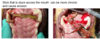
Look at some of these examples of tumours?

Look at some of these examples of tumours?

Look at some of these examples of tumours?

Look at some of these examples of tumours?

Benign tumours are also common and include (naming of benign tumours varies):
- Acanthomatous ameloblastoma (aka basal cell tumour by old vets)
- Peripheral odontogenic fibroma (aka epulis, fibrosing epulis)
Surgery is the mainstay treatment for the majority of malignant and benign tumours
Other treatments options (instead or in addition to surgery) include:
- Radiation therapy
- Chemotherapy
- Immunotherapy (there is a melanoma vaccine available but need to be qualified oncologist to order from USA and not all melanomas respond to the vaccine)
Oral tumours overview?
- Oral tumours are relative common in cats and dogs
- Benign and malignant tumours of the oral cavity account for 3-12% of all tumours in cats and 6% of all tumours in dogs
Oral tumour clinical signs?
- Presence of a mass in the oral cavity
- Increased salivation, blood in the saliva, odorous breath
- If involving alveolar bone teeth may be loose
- Swelling on the face or bulging of the eye (exophthalmos)
- Bloody nasal discharge
- Difficulty eating or pain on opening the mouth, weight loss and enlarged lymph nodes in the neck region
- Loose teeth, especially in animals with general good teeth, may be indicative of cancer-induced bone loss, especially in cats
How to diagnose oral tumours?
Physical examination
- Concomitant problems
- Size and site of oral mass
- Evaluation of regional lymph nodes
Blood tests
FNA
- Often non-diagnostic as requires the lesion to exfoliate (apart from SCC which may exfoliate)
Core biopsy
- Histopathology (bony lesions might prove difficult to obtain representative sample)
Imaging of the skull
- Conventional radiography
- Ideally, CT scan
- To assess bone involvement and degree of margins and aggressiveness
Staging
- Fibrosarcoma(local), osteosarcoma and SCC (will spread to peripheral sites e.glung)
- Conventional radiography
- Ideally, CT scan Oral tumours-diagnostics
Oral tumour treatment options?
Treatment options depend on the location of the tumour and on the type (biology) of the tumour
- benign tumours excised with 1 cm margins
- Malignant tumours excised with 2-3 cm margins
Mandibulectomy
- Unilateral rostral
- Bilateral rostral
- Segmental
- Caudal
- hemimandibuletomy
Maxillectomy
Immunotherapy for melanoma in dogs
Outline mandibulectomy surgery?

Discuss Epulis-peripheral odontogenic fibromas?
- Derived from cells of the periodontal ligament
- There is bone involvement. If you radiographed likely to see lysis around teeth root
- Benign tumour type
- Aim in removal get margin that includes local bone and teeth it is associated with
- Typically, dogs over the age of six (but can be seen at any age); rare in cats
- Common tumour type that is often misnamed as epulis when it should be called peripheral odontogenic fibromas
- Has a relatively good outcome post surgery
- Curative surgery requires taking away bone and teeth local to tumour
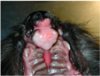
What can be seen here?


What can be seen here?


What can be seen here?


What can be seen here?


What can be seen here?


What can be seen here?


Discuss surgical aftercare for oral surgery?
- Most animals discharged 2-5 days after surgery, depending on level of surgery, comfort and ability to eat soft food
- Return for re-check 7-10 days postop
- Restrictions
- Analgesia
- Antibiotics
- Restrictive (Elizabethan) collar to prevent self-traumatisation
- Limited exercise
- Soft canned food or soaked kibble for 2-3 weeks postop
- No chews, raw hide or chewing toys for at least 3-4 weeks postop Surgical aftercare
Discuss postoperative complications?
- Incision breakdown requiring further surgery to repair
- Bleeding from the nose following maxillectomy
- Increased salivation –may persist for some weeks
- Mandibular drift following mandibulectomy
- Difficulties eating –usually not a problem in dogs but a common problem in cats (routine to put oesophagostomy tube placed at time of surgery)
- Recurrence of tumour
Discuss what to do with cat post oral surgery?
oesophagostomy tube placement

What has occurred here?