Thoracic Imaging 2 Flashcards

(63 cards)

1
Q

List imaging modalities and when they are useful?

A

•Radiography

–Important and frequently used tool

–Still in this day and age the most used tool for imaging the thorax

•Ultrasonography

–Important for heart and elsewhere

–Emergency ultrasound

–“The Chest Rad’s Companion”

–Important for the heart, but also useful elsewhere – particularly in emergency situations

–Complements radiography

•CT

–Use where radiography and ultrasonography fail to identify cause and extent of disease

–Now modality of choice for lung (if available)

–Where radiography and US doesn’t give us an answer

•Endoscopy

–Useful for larger airways and oesophagus

•MRI/scintigraphy

–Fewer indications

–At the moment it isn’t used very much, but will change as it develops

2
Q

To image or not to image? If none of these things are likely to change, no point doing the imaging as we wont see anything!

What are these things?

A

Radiographic (Roentgen) Signs

–Number

–Location

–Size

–Shape

–Margination

–Radiopacity

–(Internal architecture)

–(Function)

Ultrasonographic signs

–Number

–Location

–Size

–Shape

–Margination

–Echogenicity

–Internal architecture

–Function

N.B. Need adequate access

3
Q

Discuss normal variations?

A
  • There are loads of normal variations!
  • Positional variations
  • Dogs vs cats
  • Breed variations
  • Ageing changes
  • Take it all into consideration with radiography when interpreting it
4
Q

Structures of the same opacity which are in contact will appear asone shadow. This is known as?

A

Border obliteration

aka silhouette sign/border effacement

5
Q

Define ‘Mass Effect’?

A
  • Displacement of structures due to adjacent space-occupying lesion, e.g. mass or fluid
  • Mass displaces normal structures away from itself
  • Pic – cranial mediastinal mass, trachea dorsally displaced
6
Q

Define ‘Mediastinal shift’?

A

VD/DV view

Displacement of the midline structures due to increased or decreased volume of adjacent structures, e.g. mass, fluid or lung collapse.

Reduction in volume – normal structures move towards the area of a problem

Pic – left lung collapsed, so mediastinum and cardiac silhouette has moved towards it

7
Q

Why should a VD/DV be done before a lateral?

A
  • The dependent lung lobe will collapse rapidly when an animal is placed in lateral recumbency, particularly under GA or sedation
  • As a result, the best detail is seen in the uppermost lung as it contains more air
  • Practically - take the DV or VD radiograph first, so you can tell if any lung collapse is pathological rather than related to previous positioning
8
Q

Discuss this radiograph?

A

Rib Tumour

May be lesions explaining clinical signs of the animals

Came in with a mass, owner noticed lump. Can see the mass.

9
Q

Discuss this radiograph?

A

Subcutaneous emphysema

Can get useful info about what has happened about changes going on within the thorax, VD of cat

Can see increase opacity and cannot see cardiac silhouette – but can see air and lucencies in subcut tissues

SUBCUTAENOUS EMPHYSEMA – suggests some kind of trauma and some leaking from airways at some point

10
Q

Discuss Costochondral junctions and costal cartilage mineralisation?

A

If marked increase in opacity at one costochondral junction it might be a lesion, but if its on it all of them its normal

11
Q

What is this?

A

Nipple shadow

Can see nipples! Surrounded by air. Nodules in lung due to mets tend to be circular, nipples are more elongated in appearance as seen here

12
Q

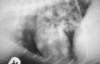
What pitfall can be seen here?
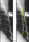
A

Skin fold

This line extends outside of thoracic cavity – SKIN FOLD!!

13
Q

What is the pleural cavity?

A

PC – virtual space

2 membranes – visceral and parietal pleura with a small amount of fluid in between

Do not see this on a normal radiograph

14
Q

What changes are seen when pleural fluid occurs?

A

you see a number of changes – fluid in pleural cavity, separating lungs from thoracic wall, soft tissue fluid opacity – see it ventrally and dorsally. If lying on side, fluid drops to bottom and then spread up the sides slightly, separates lungs from spine and sternum – get rounding up, scalloping of lung edges ventrally, see bits of fluid going in between lung lobes. Also due to this fluid, get border obliteration of Cardiac Silhouette

15
Q

What is the common pleural fluid pattern of unilateral, encapsulated, large effusions?

A

= pyothorax, (neoplastic effusion)

Normally, fluid flows freely from LHS to RHS of thorax. If thick fluid with lots of cells, this will block holes and might get unilateral effusion – puss or neoplastic effusion.

16
Q

What is common pleural fluid patterns of concurrent abdominal fluid with small effusion?

A

hypoproteinaemia, FIP, heart failure

17
Q

What are common pleural fluid patterns of the trachea (+/- oesophagus) grossly elevated and compressed, heart caudally displaced with large effusion?

A

lymphoma, thymoma

18
Q

What are common pleural fluid patterns of Large effusions?

A

•chylothorax

19
Q

Trauma signs of pleura fluid patterns?

A

•blood, lymph

20
Q

What might this suggest: Trauma signs, loss of diaphragmatic shadow, loculated gas, heart displaced and reduced abdominal contents

A

Ruptured diaphragm

21
Q

Discuss lung fields on radiographs?

A

Normal appearance – there are 6 lung lobes in dogs and cats, we don’t see individual lobes on radiographs, just see lung field

Divide into concentric circles

22
Q

Describe these concentric circles in regards to lung fields?

A

Central, middle and peripheral thirds

Central – will see airway and BV markings

Middle – BV markings

Peripheral – not much in way of lung markings at all

23
Q

Lung pattern or
normal lung markings??

A

Bronchial (parallel walls with lucent centre)

Vascular BV in transverse as circles or as branching linear structures with no air in the middle

24
Q

Describe bronchial thickening?

A

Bronchi can become thickened and will be seen further out towards the periphery, more so than usual – parallel lines called TRAM LINES and doughnuts.

25
What lung pattern is this?
Bronchial thickening
26
Describe bronchial calcification?
No thickening, walls aren’t thicker, but they are more mineral than soft tissue – more radiopaque – can see smaller airways than normal, see them extending further to the periphery
27
Mild bronchial pattern in old animals is due to?
Aging change
28
Thick soft tissue cuffs around the airways, implies acute process such as?
oedema pneumonia eosinophilic bronchopneumopathy
29
Thin soft tissue cuffs around airways, suggests chronic disease =
Chronic bronchitis
30
Very thin, mineral opacity cuffs are suggestive of?
HAC, CRF
31
Wavy tramlines, often thickened and dilated are indicative of?
bronchiectasis (enlargement of the airways)
32
Discuss alveolar pattern?
Caused by primarily the fact there is no air within the alveoli, might be fluid Increased opacity, soft tissue fluid within alveoli, so can get marked increase in opacity. Filling of alveoli can be patchy across lung field also.
33
What is an air alveologram?
On radiographs, the irregular, mottled appearance of lung tissue, caused by the contrast in density between some alveoli being filled with fluid or cellular material and others filled with air.
34
What is a bronchogram?
radiographic appearance of an air-filled bronchus surrounded by fluid-filled airspaces
35
What can be seen here?
“Fluffy” increased opacity – quite marked Border Obliteration – don’t see some of diaphragm, don’t see some of silhouette
36
Common alveolar patterns (dogs) Ventral?
pneumonia (bronchopneumonia, aspiration)
37
Common alveolar patterns (dogs) Symmetrical perihilar?
Cardiogenic oedema
38
Common alveolar patterns (dogs) Caudodorsal?
**Embolism**, systemic infections, infarcts, CNS types, FB pneumonia
39
Common alveolar patterns (dogs) Peripheral pattern ?
**Angiostrongylus (dogs mainly)**, thromboembolism
40
Common alveolar patterns (dogs) Unilateral, middle/cranial lobe?
Collapse. Lung lobe collapse will remove air from alveoli and give alveoli pattern, loss of volume
41
Look at this normal vasculature?
42
Describe normal vasculature?
Normal pulmonary vessels are clearly visible in central and middle zones Vessels taper towards the periphery In lateral view * Artery is is dorsal to bronchus and vein * Veins are ventral to bronchus and vein In DV view * Veins a medial to artery * So veins are ventral and central
43
What is normal vasculature size?
**In lateral view** Right cranial lobar artery and vein 0.5-1.0 x diameter as proximal 1/3 of 4th rib. **In DV view** Arteries and veins same diameter as 9th rib where they cross this rib (see pic)
44
Veins are VENTRAL AND CENTRAL Must not look really big and be approximately the same size BV run either side of airways
45
What does it mean when veins bigger than arteries?
Vein bigger than artery – abnormal LHS congested heart failure can be a top differential
46
What can be seen here?
47
What can cause an increase in size of both vessels?
Overperfusion of lungs can cause this
48
What can cause decreased vasculature?
Can be smaller than normal, classed as avascular pattern Thinking about hypovolaemia, something that is reducing perfusion of the lungs – some problem with outflow from the heart for example
49
What is an insterstitial pattern?
* Thickening / infiltration of supporting tissues * Two types: **–Diffuse** (can be called unstructured/fine structured) **–Nodular**
50
What is a diffuse pattern?
Some fibrosis, cellular infiltration, maybe some oedema – in interstitium On here – interstitium small part of lung so any increase in opacity will be modest compared to other increases of opacity elsewhere in the lungs
51
Why is this a diffuse pattern?
Overall hazy increase in opacity, typical for diffuse or unstructured pattern – not enough to obscure structures
52
Describe a nodular interstitial pattern?
Nodules can grow Often from blood – haematogenous spread On radiograph will only see soft tissue sphere if its diameter is about half a centimetre, wont see anything smaller than this – this means we can only see metastasis which are of a certain size
53
What can be seen here?
Multiple lung interstitial nodules – can be superimposed and see a big shadow, just lots of overlapping shadows of nodules. Can show weird shapes, but often superimposition
54
Discuss miliary lung nodulation as seen in this picture?
If you have many small individual interstitial nodules Some superimpositions can be smaller than half a cm, but as going through multiple small ones, you will see it
55
Discuss the size of insterstitial nodular patterns?
•Solitary nodule (\>5mm) – not necessarily cancer!! Margination may be helpful: –Indistinct margin, (tend to spread outside the focus of the disease) **•Focal pneumonia** **•Granuloma/abscess** **•Haemorrhage/contusion** **•Infarct/thromboembolism** **•Fluid filled bulla/cyst**
56
What do well circumscribed intersitial nodules with sharp distinct margins suggest?
**•Neoplasia** (primary or secondary) * cyst * (abscess/granuloma)
57
What do evenly rounded, discrete multiple nodules suggest?
–**Metastatic neoplasia** –Primary lung tumours –If imported from the states, fungal pneumonia can show this, but not seen in the UK really
58
What do miliary nodulations (2-5mm) suggest?
–**Metastatic neoplasia** –Granulomatous eosinophilic bronchopneumopathy –Aelurostrongylus (cat) –TB
59
What do unstructured interstitial patterns suggest?
**–Ageing changes** –Fibrous healing or chronic disease –Early/late oedema, haemorrhage, pneumonia –(Pneumonitis, diffuse metastasis e.g. lymphoma)
60
Discuss a cat specific radiography?
Cats: have big muscles under the spine, will separate out the most caudal part of lung from the spine – lung field away from spine from about caudal T11
61
What changes occur in older animals?
* Interstitial and bronchial markings increase with age * So, what may be significant in a young animal may be within normal limits for an aged one
62
What can be seen here?
**Pleural plaques/heterotopic bone** Lots of small rounded opacities in lung node smaller than 1m these are isolated these are bits of bone within pleural degenerative change, can happen in older dogs. If nodule, individual and less than 1 cm CANNOT BE METS, will be bone
63
How to choose which lung pattern?