Plantar Surface of the Foot Flashcards
(32 cards)
Dorsal View of Foot


Ventral View of Foot


Deep Fascia of Foot
Thick central portion but weaker medially and laterally
Plantar aponeurosis
Superficial ligament formed by central portion of the deep fascia
Proximal attachment to Calcaneus. Divides into 5 bands distally - continuous with fibrous digital sheaths.
Vertical intermuscular septae: Medial, Central and Lateral compartments
Compartments of the Foot
Muscles of the Dorsum of the Foot (Purple)

Extensor Digitorum Brevis
Extensor Hallucis Brevis

Compartments of the Foot
Lateral Compartment (Orange)

Muscles of the little toe:
Abductor Digiti Minimi
Flexor Digiti Minimi Brevis

Compartments of the Foot
Central Compartment (Green)

Flexor Digitorum Brevis
Muscles associated with tendon of FDL (lumbricals and quadratus plantae)
Adductor Hallucis

Compartments of the Foot
Medial Compartment (Blue)

Muscles of the great toe:
Abductor Hallucis
Flexor Hallucis Brevis

Compartments of the Foot
Interossei (Pink)

Plantar and Dorsal Interossei

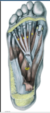
Foot - Layer 1
Abductor Hallucis (1)

Medial Tubercle of Calcaneus and flexor retinaculum to the medial base of the proximal phalynx

Foot - Layer 1
Flexor Digitorum Brevis (2)

Medial tubercle of calcaneus to the middle phalanges of the lateral 4 toes

Foot - Layer 1
Abductor Digiti Minimi (3)

Tubercle of Calcaneus to the lateral base of the proximal phalynx

Foot - Layer 2
Long flexor tendons to the toes
FDL and FHL

Foot - Layer 2
Lumbricals (*)

Tendons of FDL to the medial side of the dorsal tendon expansion

Foot - Layer 2
Quadratus Plantae (QP)

Medial and lateral surfaces of calcaneus to the lateral side of the FDL tendon

Foot - Layers 1 and 2


Foot - Layer 3
Flexor Hallucis Brevis

Cuboid and Lateral Cuneiform to both sides of the base of the proximal phalanx
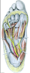
Foot - Layer 3
Adductor Hallucis

Transverse head: Plantr ligament of MTP joints
Oblique head: Base metatarsals 2 and 4
Both go to the lateral side of the base of the proximal phalanx

Foot - Layer 3
Flexor Digiti Minimi Brevis (*)

Base of the 5th Metatarsal to the base of the proximal phalanx

Foot - Layer 4
Plantar Insterossei

3 of them
Bases and medial side of metatarsals 3-5 (unipennate) to the medial side of the proximal phalanx of 3rd-5th digit
PAD - Plantar ADuct

Foot - Layer 4
Dorsal Interossei

4
Adjacent sides of metatarsals 1-5 (bipennate) to the 1st on medial side proximal phalanx of the 2nd digit and 2nd-4th on lateral side of proximal phalanx of 2nd-4th digits
DAB - Dorsal ABduct

Foot - Layer 4
Tendons

Peroneus Longus (*)
Tibialis Posterior (TP)

Foot - Layers 3 and 4


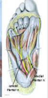
Plantar Nerves
What are they?

Terminal branches of Tibial nerve, deep to the flexor retinaculum. They enter the foot deep to Abductor Hallucis.