Reiner - Basal Ganglia Anatomy/Function Flashcards
(40 cards)
Late-stage Huntington’s is characterized by a loss of ENK+ and SP+ striatal neurons, and GPe neurons. What tx would be the best choice to combat the hypokinesia of late Huntington’s?
A lesion of the subthalamic nucleus
D2 type neurons are INH, and preferentially found on ENK+ striatal neurons. What would be the effect of a D2 antagonist on enkephalinergic neurons?
- INC activity and INC enkephalin production
- When a neuron is active, it is making more transmitter; will make less when it is inactive
- If you hit it with a D2 agonist, enkephalin and activity would go down
- NOTE: D1 type dopamine receptors are excitatory, and preferentially found on SP+ striatal neurons
Is subthalamic input to GPi INH or excitatory?
Excitatory
What is ballismus? What causes it?
- Ballismus: person afflicted w/unwanted movements
- Caused by destruction of the subthalamic nucleus, which leads to INC activity of motor thalamus neurons
Apomorphine (non-specific dopamine receptor agonist) produces hyperactivity. What would the effects of this drug be?
Diminished activity of GPi neurons
What is Haloperidol? How does it affect the basal ganglia?
- Haloperidol: dopamine (D2) receptor antagonist
- INC activity of GPi neurons
How does the profound loss of dopamine neurons acting on the basal ganglia motor circuit produce hypokinesia?
Via INC activity of subthalamic nucleus neurons
Huntington’s: enkephalinergic neurons lost before substance P neurons, leading to a progression from chorea to rigidity. What therapy would be desirable to combat the hyperkinesia of early Huntington’s?
Tx with a D2 receptor antagonist
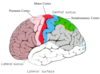
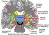
What are the parts of the basal ganglia? What other brain structures are associated with them?
- Caudate nucleus (yellow)
- Putamen (green)
- Globus pallidus (purple)
- Thalamus
- Subthalamic nucleus: interconnected with the basal ganglia
- Substantia nigra: in midbrain, interconnected with basal ganglia
1. Black bc neurons in substantia nigra contain the black pigment melanin
What are the main structures composing the basal ganglia? Where are they located anatomically?
- Caudate: medial, and located along the lateral wall of the lateral ventricle
-
Putamen: lateral and ventral to the caudate nucleus, and separated from it by the internal capsule
1. NOTE: caudate and putamen are part of the same structure, but separated by the internal capsule - Globus pallidus: external (GPe) and internal (GPi) parts

What are the 2 divisions of basal ganglia, and their functions (in general)?
- SOMATIC (dorsal) basal ganglia: caudate, putamen, globus pallidus -> movement control
- LIMBIC (ventral) basal ganglia: nucleus accumbens, olfactory tubercle, ventral pallidum -> motivation, reward, and affect
Describe the 3D relationship of the caudate and putamen.
- Head of the caudate (tail) and putamen (peach pit) are confluent, evidence they are part of same thing
- Fibers of internal capsule develop b/t putamen and head of caudate moving caudally
- Caudate nucleus has a long tail that sweeps posteriorly, bends and comes forward, and curls under main body of the caudate, ending rostrally at the amygdaloid nucleus (if one cuts slice through middle of caudate, head and tail will be in that slice)
- Thalamus is medial to the putamen

Why is the globus pallidus called this?
- Does not have many cells in it, so it looks pale when sliced and stained with a stain that reveals cells
- Globus name because it is rounded like a globe
- NOTE: also can be referred to just as the pallidus
What is another name for caudate + putamen?
Striatum (because many nerve fibers pass through them, giving them a striped appearance)
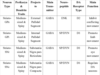
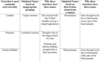
What are some outdated names for the basal ganglia that should NOT be used (table)?
I can’t imagine this is actually important
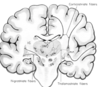
When stained with dopamine, what features of the basal ganglia will be enhanced?
- Dark region near bottom of image shows dopamine neurons of SUBSTANTIA NIGRA, at the base of the midbrain
- PUTAMEN, and head/tail of CAUDATE all brown bc substantia nigra has axons that travel to head and tail of caudate, and putamen and form dopaminergic terminals there
- Terminals present in all of those areas that are dark
- Globus pallidus does not receive many dopamine terminals, so it is pale
- NOTE: attached image shows rhesus monkey brain stained IHC for enzyme that reveals which neurons make dopamine
1. Note the cerebral peduncle, which is the midbrain continuation of the internal capsule

What are the two divisions of the substantia nigra? What is the significance of this division?
- PARS COMPACTA: dopaminergic neurons arranged in a compact layer in the upper part of the substantia nigra -> neurons contain the black pigment (melanin)
- PARS RETICULATA: net-like, meshwork of fibers below the pars compacta that contains few dopaminergic neurons -> resemble neurons in globus pallidus in their chemistry, shape, and function
- NOTE: image is close-up of substantia nigra in a rat

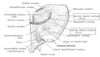
Where is the subthalamic nucleus located in the human brain?
- Above the cerebral peduncle, and below the bulk of the thalamus

What are the 2 types of neurons in the striatum?
-
Aspiny (A): about 5% of neurons in the striatum
1. Dendrites that do NOT possess stubby protrusions (spines) on them - Spiny (SN): 95% of neurons in striatum -> typically smaller than aspiny neurons

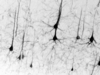
What is this? Why does it look like this?

- Close-up view of a spiny neuron (about 15 microns in width; aspiny are about 30 microns in width at their cell bodies): projection neuron of the striatum
- Dendrites w/stubby protrusions (spines) specialized for receiving terminals from o/brain regions -> neurons that need to integrate information from diverse sources possess dendritic spines
- Dendrites of some neurons are smooth, and receive their input on their smooth surface (aspiny)
What is the role of the spiny neurons in the striatum?
- Spiny neurons have a long axon that leaves the striatum (in contrast to the short axons of aspiny neurons that do NOT leave the striatum)
- Spiny neurons = projection neurons; aspiny neurons = local circuit/interneurons
- Whatever the striatum decides based on pattern of input it receives, it is spiny neurons that transmit that decision to other brain areas
- NOTE: there is further functional and neurochem diversity within these two types of neurons

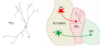
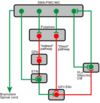
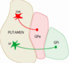
What are the 2 major subtypes of projection neurons in the striatum? Where do they project?
- All spiny (projection) neurons in the striatum use the nuerotransmitter, GABA
- 2 major subtypes based on different neuropeptides (adjunct NT’s that neurons often use, and can be neurochem signatures for defining subtypes):
1. Opioid neuropeptide, enkephalin (ENK): red in the illustration; nearly all project to GPe
2. Substance P (green): many project to GPi (there are add’l subtypes of striatal substance P neurons)
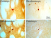
What does this image show?

- Dark labeling in GPi represents substance P-containing terminals of those substance P neurons in striatum that project to GPi (note: no noteworthy labeling of substance P terminals in GPe)
- Enkephalinergic neurons in caudate and putamen project to GPe, resulting in many terminals in GPe that can be visualized by enkephalin IHC (GPi largely devoid of enkephalin)
- NOTE: rhesus monkey basal ganglia
What does this image show?
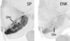
- Substance P image: substance P terminals located in upper shelf in substantia nigra where dopaminergic neurons are located (pars compacta) and in region of substantia nigra below dopaminergic neurons (pars reticulata)
1. Substance P neurons in striatum project to GPi, SNc, and SNr - Enkephalin neurons in striatum have only a hint of a projection to substantia nigra, and only to very most medial part -> bc it is minor, we will disregard it
- NOTE: rhesus monkey brain